[ANA] PE 7 (Identification + SQs)
1/78
There's no tags or description
Looks like no tags are added yet.
Name | Mastery | Learn | Test | Matching | Spaced | Call with Kai |
|---|
No analytics yet
Send a link to your students to track their progress
79 Terms
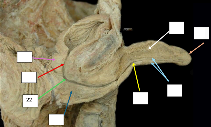
Clitoris
Blood Supply: Internal Pudendal a.
Origin: Internal Iliac a. (Anterior division)
Nerve Supply: Pudendal nerve (sensory fibers)
Lymph Drainage: Internal Iliac/Deep Inguinal nodes
Identify #1
Side Questions:
Blood Supply?
Origin
Nerve Supply?
Lymph Drainage?

Apex of the vestibule
Clitoris is found anteriorly by which structure?
Paired Corpora cavernosa
Ischiocavernosus muscle
Crura of the clitoris formed by
Paired corpora cavernosa that adjoin side by side below the pubic symphysis and enclosed in dense connective tissue
Body of the clitoris is formed by
Prepuce
Glans of the clitoris is partly covered by which structure
Labia Minora
Side Question:
Encloses which structure: Clitoris
Forms which structure anteriorly: Prepuce
Forms which posteriorly: Frenulum
Identify #2
Side Question:
Encloses which structure?
Forms which structure anteriorly?
Forms which posteriorly?

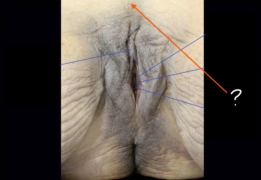
Labia Majora
Side Question:
Sensory Innervation
Anterior - Genital branch of the Genitofemoral n.
Posterior - Perineal branch of Pudendal n.
Ligament that terminates in this structure from the pelvic cavity: Round ligament of the Uterus
Identify #3
Side Question:
Sensory Innervation?
Anterior
Posterior
Ligament that terminates in this structure from the pelvic cavity?
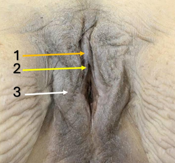
Labia Minora and Majora
Blood Supply: Internal Pudendal a.
Origin: Internal Iliac a.
Venous Drainage: External pudendal v. → Great Saphenous v.
Lymph Drainage: Superficial Inguinal nodes
Labia Minora and Majora
Blood Supply?
Venous Drainage?
Lymph Drainage?
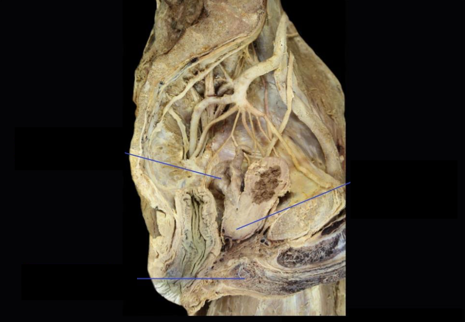
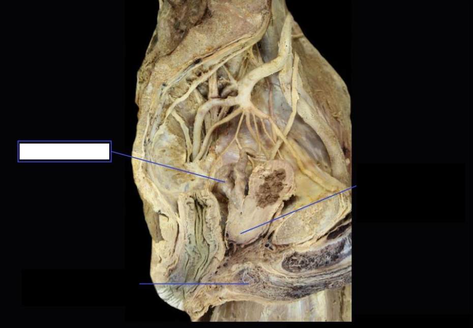
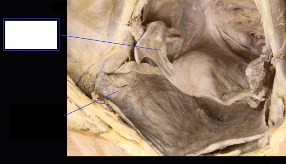
Body of the Uterus
Side Question:
What are the four primary supports of this structure?
Pelvic Diaphragm (Levator ani) - Main Support
*Cardinal Ligament (at the sides)
*Pubocervical Ligament (Anterior)
*Uterosacral Ligament (Posterior)
*Cervical Part
Lymph Drainage: External/Internal Iliac LN (Body and Cervix)
Identify
Side Question:
What are the four primary supports of this structure?
Lymph Drainage?

Round Ligament of the Uterus
Side Question:
Artery found in this ligament?
Sampson’s artery (The artery of the round ligament of the uterus)
Lymph Drainage: Superficial Inguinal LN
Identify
Side Question:
Artery found in this ligament?
Lymph Drainage?

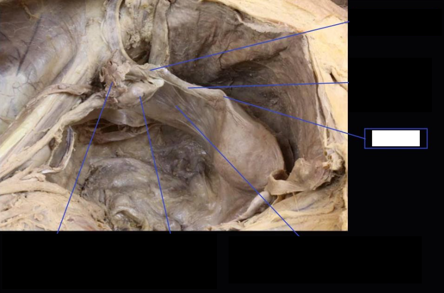
Broad Ligament of the Uterus
Side Question:
This structure is composed of the following:
Largest that supports the body and fundus of uterus: Mesometrium
Most superior that supports the fallopian tube: Mesosalpinx
Posterior and supports the ovary: Mesovarium
Identify
Side Question:
This structure is composed of the following extensions:
Largest that supports the body and fundus of uterus?
Most superior that supports the fallopian tube?
Posterior and supports the ovary?

Ampulla of the Fallopian Tube
Identify (Be Specific)

Fimbriae of the Fallopian Tube
Identify (Be specific)

Fallopian Tube
Blood Supply
Uterine a. (Origin: Internal Iliac a.)
Ovarian a. (Origin: Abdominal Aorta)
Venous Drainage: Uterine and Ovarian veins
Nerve Innervation: Inferior Hypogastric Plexus
Lymph Drainage: Internal Iliac and Para-aortic Nodes
Fallopian Tube
Blood Supply?
Venous Drainage?
Nerve Innervation?
Lymph Drainage?
Suspensory Ligament of the Ovary (IP Ligament)
Side Question:
This structure houses
Ovarian and lymphatic vessels
Autonomic nerves
This structure is attached to the following:
Fallopian tube
Ovary
Identify
Side Question:
This structure houses?
This structure is attached to the following (0.5 for each)?
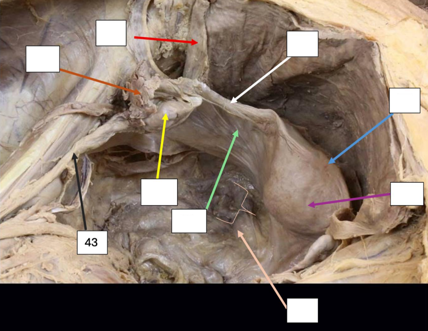
Ovary
Side Question:
Blood Supply: Ovarian a. (Origin: Abdominal Aorta)
Venous Drainage: Ovarian v. → R: IVC, L: Left Renal V.
Nerve Supply: Aortic Plexus
Lymph Drainage: Paraaortic nodes (LV1)
Ligaments attached to this structure:
Broad ligament (via mesovarium)
Suspensory ligament
True ligament of the ovary
Coverings:
Tunica Albuginea
Germinal Epithelium
Identify
Side Question:
Blood Supply?
Venous Drainage?
Nerve Supply?
Lymph Drainage?
Ligaments attached to this structure?
Coverings?

Rectouterine Pouch
Side Question:
This structure lies directly behind posterior fornix of the vagina
This structure is located between
Anterior: Uterus
Posterior: Rectum
Identify
Side Question:
This structure lies directly behind?
This structure is located between (anterior & posterior)?

Fundus of the Uterus
Side Question:
Blood Supply
Uterine a. (Origin: Internal Iliac a.)
Ovarian a. (Origin: Abdominal Aorta)
Venous Drainage: Uterine v. → Internal Iliac v.
Nerve Supply: Inferior Hypogastric Plexuses
Lymph Drainage: Paraaortic nodes (LV1)
Identify
Side Question:
Blood Supply?
Venous Drainage?
Nerve Supply?
Lymph Drainage?

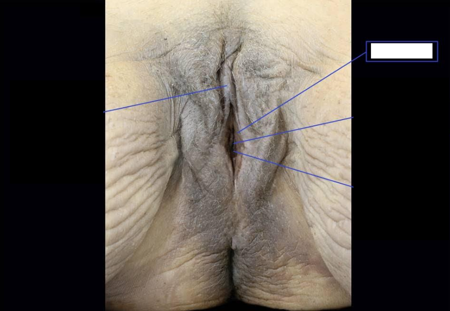
Vaginal Orifice
Side Question:
This structure is partially covered by the hymen
Structures anterior and posterior to this:
Anterior: Bartholin’s Glands, Urethra
Posterior: Anal Canal
This structure serves as an external opening into the vestibule
The size of this is reduced by the Bulbospongiosus
Identify
Side Question:
This structure is partially covered by?
Structures anterior and posterior to this?
This structure serves as an external opening into?
The size of this is reduced by which structure?

Body of Uterus
Identify
Fundus of Uterus
Identify

Uterine Cavity
Side Question:
This structure is lined by the endometrium
This structure is continuous with:
Superiorly: Fallopian Tubes (at Cornua)
Inferiorly: Cervical Canal (via Internal os)
Identify
Side Question:
This structure is lined by?
This structure is continuous with:
Superiorly?
Inferiorly?

Cervix/Cervical Canal
Side Question:
What do you call the folds of this structure: Plicae palmatae
Which opening connects this structure to the uterine cavity: Internal os
Which opening connects this structure to the vagina: External os
Specific part of this structure seen in the vagina lined by stratified squamous ep.: Ectocervix
Specific part of this structure that is lined with simple columnar ep.: Endocervix
Identify
Side Question:
What do you call the folds of this structure?
Which opening connects this structure to the uterine cavity?
Which opening connects this structure to the vagina?
Specific part of this structure seen in the vagina lined by stratified squamous ep.?
Specific part of this structure that is lined with simple columnar ep.?

Vaginal Canal
Side Question:
Supports of this structure
Upper 1/3: Levator ani, Cervical, Pubocervical, Uterosacral Ligaments
Middle 1/3: Urogenital Diaphragm
Lower 1/3: Perineal Body
This structure drains into which node? Internal Iliac LN
Identify
Side Question:
Supports of this structure
Upper 1/3
Middle 1/3
Lower 1/3
This structure drains into which node?
Vagina
Blood Supply: Vaginal a. (Origin: Internal Iliac a.)
Venous Drainage: Vaginal veins → Internal Iliac v.
Nerve Supply: Inferior Hypogastric Plexus
Lymph Drainage
Upper 1/3: External and Internal Iliac Nodes
Middle 1/3: Internal Iliac Nodes
Lower 1/3: Superficial Inguinal Nodes
Vagina
Blood Supply?
Nerve Supply?
Lymph Drainage?
Rectouterine Pouch
Identify

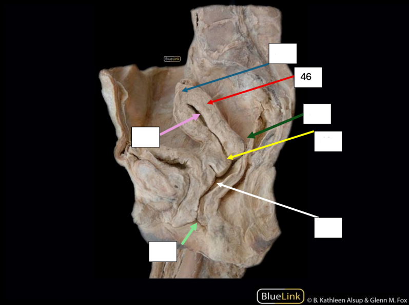
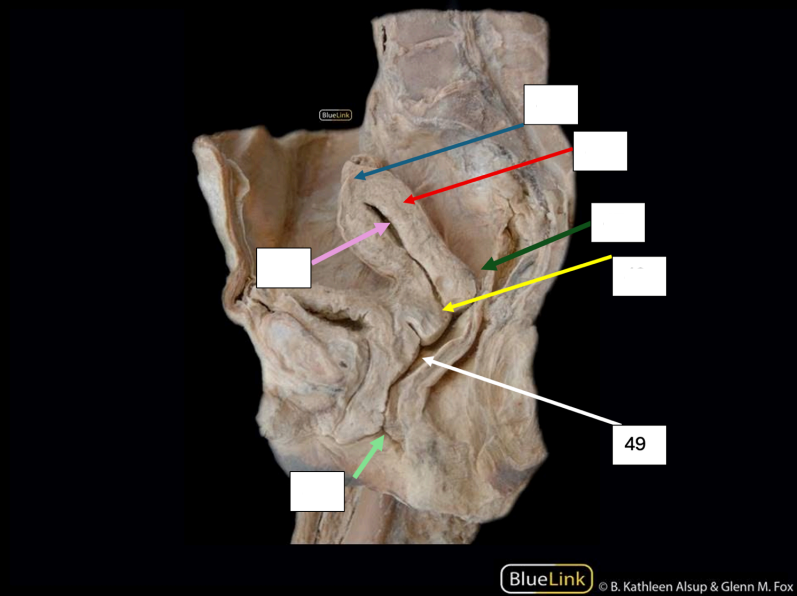
Internal Anal Sphincter
Side Question:
This structure is formed from the thickening of the smooth muscle of the circular muscle coat at the upper end of the anal canal.
Innervation: Inferior Hypogastric Plexus
Action of this structure during defecation: Relaxed
Identify
Side Question:
This structure is formed from?
Innervation?
Action of this structure during defecation?

Upper (Proximal 2/3) Anal Canal
Side Question:
This structure is derived from hindgut endoderm
This is lined by columnar epithelium
What do you call the vertical folds found in this structure: Anal columns (w/ Anal valves)
Blood Supply: Superior Rectal a. (Origin: Inferior mesenteric a.)
Venous Drainage: Superior Rectal v. → IMV → Portal Vein
Nerve Supply: Inferior Hypogastric Plexus
Lymph Drainage: Pararectal and Inferior Mesenteric nodes
Sensitive to stretch
Identify
Side Question:
This structure is derived from?
This is lined by which epithelium?
What do you call the vertical folds found in this structure?
Blood Supply?
Venous Drainage?
Nerve Supply?
Lymph Drainage?
Sensitive to?

Pectinate Line
Side Question:
This serves as a landmark that separates the upper and lower anal canal
Identify
Side Question:
This serves as a landmark that separates which structures?

Anal Columns
Side Question:
These are remains of proctodeal membrane
These are in between anal sinuses
This can be seen only in the upper anal canal
Identify
Side Question:
These are remains of?
These are in between which structure?
This can be seen only in which structure?

Lower (Distal 1/3) Anal Canal
Side Question:
This structure is derived from ectoderm of the proctodeum
This is lined by stratified squamous epithelium
Blood Supply: Inferior Rectal a. (Origin: Internal Pudendal a.)
Venous Drainage: Inferior Rectal v. → Internal Pudendal v. → Internal Iliac v.
Nerve Supply: Inferior Rectal nerve (somatic)
Lymph Drainage: Superficial Inguinal nodes
Sensitive to pain, temperature, touch, pressure
Identify
Side Question:
This structure is derived from?
This is lined by which epithelium?
What do you call the vertical folds found in this structure?
Blood Supply?
Venous Drainage?
Nerve Supply?
Lymph Drainage?
Sensitive to?

External Anal Sphincter
Side Question:
Innervation:
Inferior Rectal nerve
Perineal branch of S4
The deep part of this blends with the Puborectalis muscle to form a sling around the anorectal junction
Origin: Perineal body
Insertion: Coccyx
Identify
Side Question:
Innervation?
The deep part of this blends with which structure to form a sling around the anorectal junction?
Origin?
Insertion?
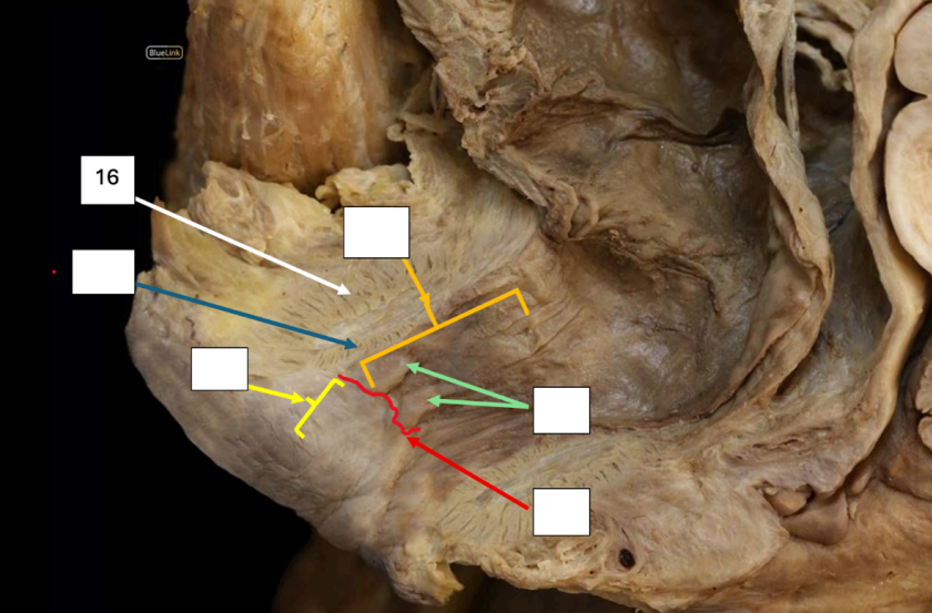
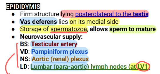
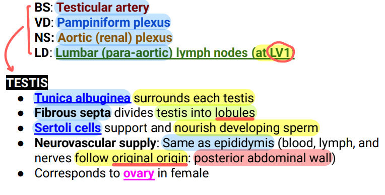
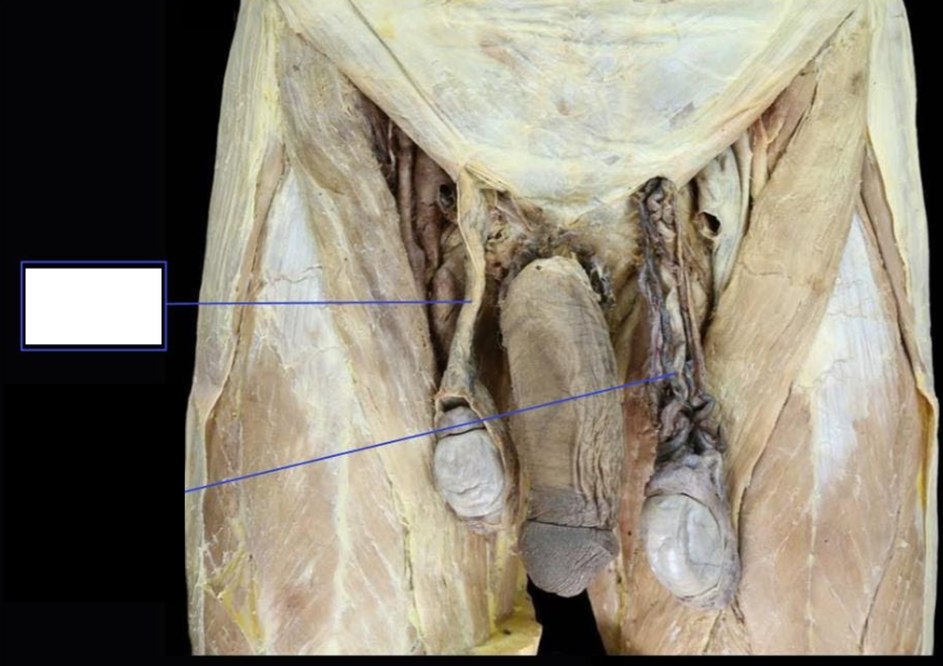
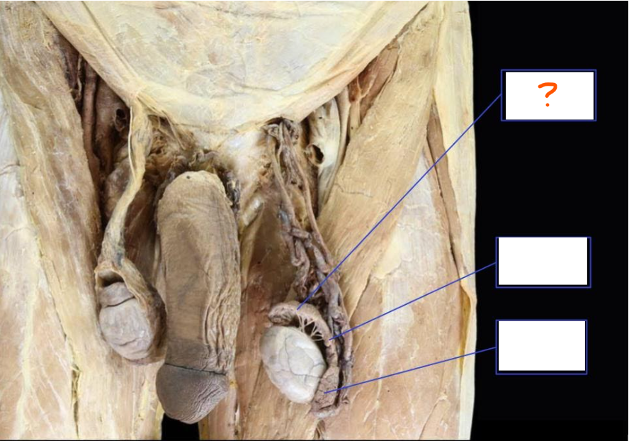
Epididymis
Identify
Side Question:
This structure lies in which position to the testis?
What structure lies on its medial side?
Blood Supply?
Venous Drainage?
Nerve Supply?
Lymph Drainage?

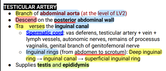
Testicular Artery
Identify
Side Question:
Origin?
Supplies which structures?

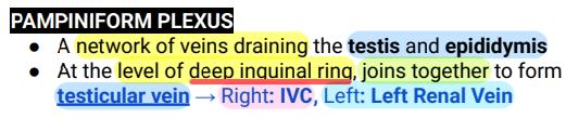
Pampiniform Plexus
Identify
Side Question:
This drains which structures?
At which level does this join together to form the testicular vein?
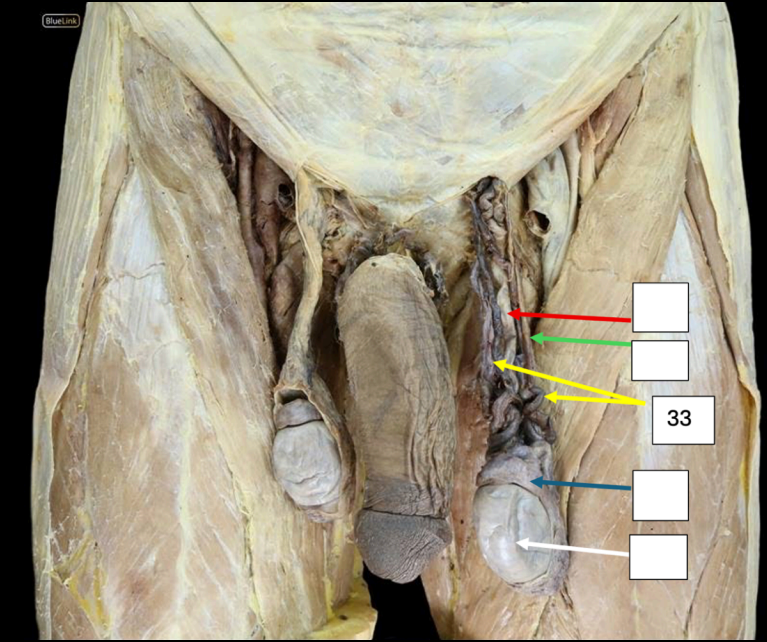
Vas deferens
Side Question:
This crosses which structure: Ureter
This conveys mature sperm from the epididymis to the ejaculatory duct
Its inferior end joins with which structure to form the ejaculatory duct: Ducts of the Seminal Vesicle
Blood Supply: Artery to the Ductus Deferens (Origin: Superior Vesical a.)
Near the testis: Spermatic Cord branches of Testicular artery
Distal Portion (Ampulla): Inferior vesical and middle rectal artery
Venous Drainage
Proximal: Pampiniform plexus → Testicular v.
Terminal: Vesicular Plexus / Prostatic Venous Plexus
Innervation: Inferior Hypogastric (Pelvic Plexus)
Lymph Drainage: External Iliac Nodes
Identify
Side Question:
This crosses which structure?
This conveys mature sperm from and to?
Its inferior end joins which structure to form the ejaculatory duct?
Blood Supply?
Venous Drainage?
Innervation?
Lymph Drainage?

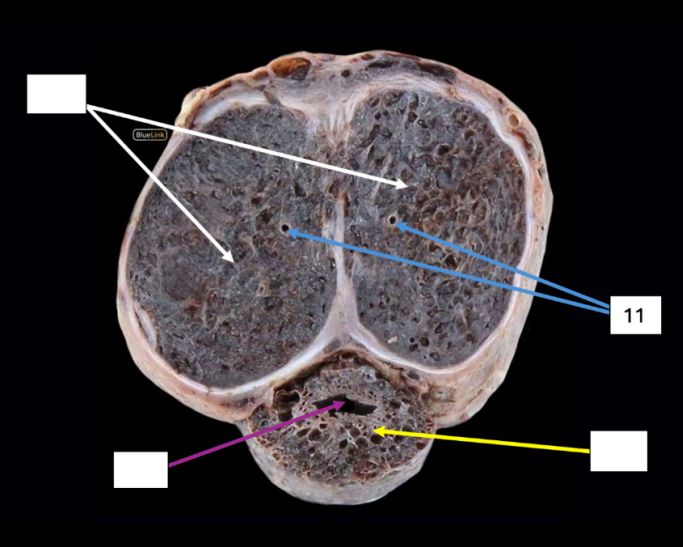
Testis
Identify
Side Question:
This is surrounded by which structure?
This is divided into lobules by which structure?
Blood Supply?
Venous Drainage?
Nerve Supply?
Lymph Drainage?

Deep Artery of Penis
Side Question:
Distribution: Crus, Corpora cavernosa
Origin: Internal Pudendal a.
Gives rise to Helicine arteries
Identify
Side Question:
Distribution?
Origin?
Gives rise to?
Penile/Spongy Urethra
Side Question:
Exits at the external meatus
Runs through and enclosed in a bulb by the corpus spongiosum
Held in place by Buck’s fascia
Lies within glans penis and dilated to form the Fossa terminalis (Navicular)
Identify
Side Question:
Exits at?
Runs through and enclosed in a bulb by which structure?
Held in place by which structure?
Lies within glans penis and dilated to form?

Corpus cavernosum
Side Question:
Each of these is surrounded by which structure: Tunica albuginea
This continues posteriorly to become which structure and is covered by which muscle: Crus of the Penis; Ischiocavernosus muscle
Together with Corpus spongiosum ventrally, this is enclosed by which structure: Buck’s/Deep Fascia
Identify
Side Question:
Each of these is surrounded by which structure?
This continues posteriorly to become which structure and is covered by which muscle?
Together with corpus spongiosum ventrally, this is enclosed by which structure?

Corpus spongiosum
Side Question:
This is found ventrally
The posterior portion of this is covered by Bulbosponsiosus muscle (serves as the insertion of this muscle from the perineal body)
Together with Corpus cavernosa dorsally, this is enclosed by Buck’s/Deep Fascia
This expands at its distal extremity to form the glans penis
Identify
Side Question:
This is found dorsally or ventrally?
The posterior portion of this is covered by?
Together with Corpus cavernosus dorsally, this is enclosed by which structure?
This expands at its distal extremity to form?

External Urethral Orifice
Side Question:
This is an opening in which corpus of the penis: Corpus spongiosum
This is found at the summit/tip of the glans of penis
What exits in this structure: Urine and semen
Identify
Side Question:
This is an opening in which corpus of the penis?
This is found at?
What exits in this structure?
Corona of Glans Penis
Side Question:
This is part of which erectile tissue? Corpus spongiosum
This serves as a junction between which structures? between glans and shaft
Identify
Side Question:
This is part of which erectile tissue?
This serves as a junction between which structures?

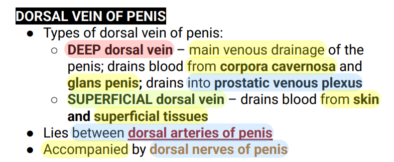
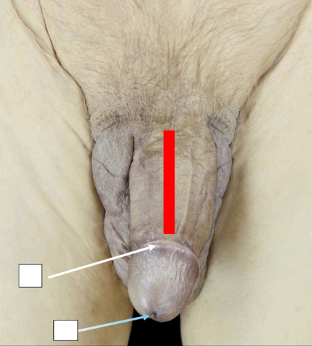
Dorsal Vein of Penis
Identify
Side Question:
The deep type of this structure drains blood from and into?
The superficial type of this structure drains blood from?
This lies between which structure?
This structure is accompanied by?
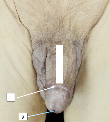
Frenulum
Side Question:
This connects the prepuce (foreskin) to the glans penis
Identify
Side Question:
This connects which structures?
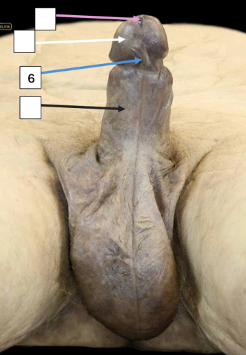
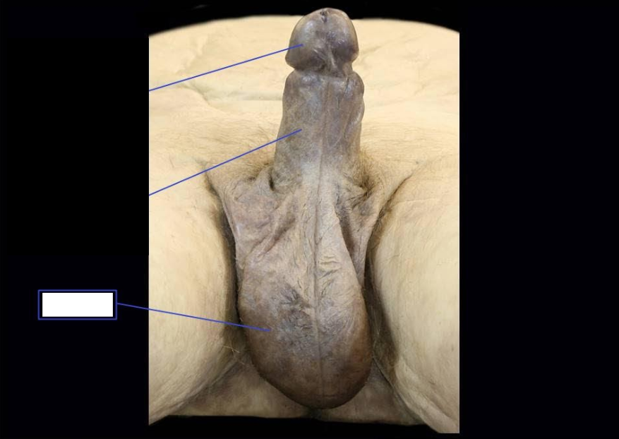
Glans Penis
Side Question:
This is a distal dilation of corpus spongiosum
What do you call the structure that covers this? Prepuce
This is perforated anteriorly by the external urethral meatus
Blood Supply: Both from the Internal Pudendal a.
Artery of the Bulb
Dorsal arteries of the Penis
Venous Drainage: Deep dorsal vein of the penis → Prostatic venous plexus
Lymph Drainage: Internal iliac nodes
Nerve Innervation: Dorsal nerve of the Penis from Pudendal nerve
Identify
Side Question:
This is a distal dilation of which structure?
What do you call the structure that covers this?
This is perforated anteriorly by?
Blood Supply?
Venous Drainage?
Lymph Drainage?
Nerve Innervation?

External Urethral Orifice
Identify

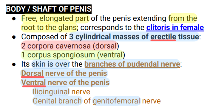
Penile Shaft / Body of Penis
Side Question:
Sensory innervation of the skin of this structure?
Ilioinguinal nerve
(Snell’s) Genital branch of the genitofemoral nerve
Innervation of the skin over the anterior part of the penis? Specific branch.
Dorsal nerve of the penis
Identify
Side Question:
Sensory innervation of the skin of this structure?
Innervation of the skin over the anterior part of the penis? Specific branch.

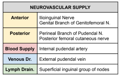
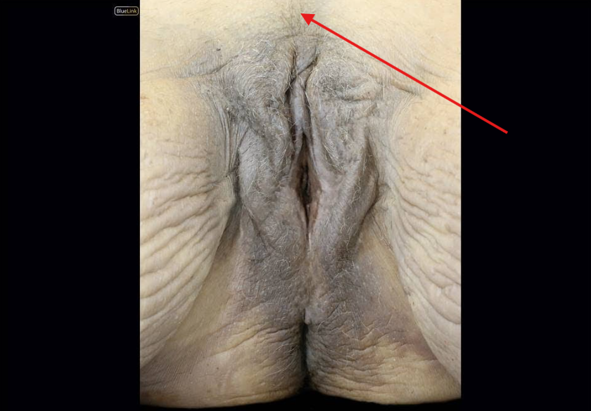
Mons Pubis
Side Question:
Neurovascular Supply?
Identify
Side Question:
Neurovascular Supply?
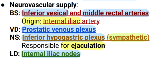
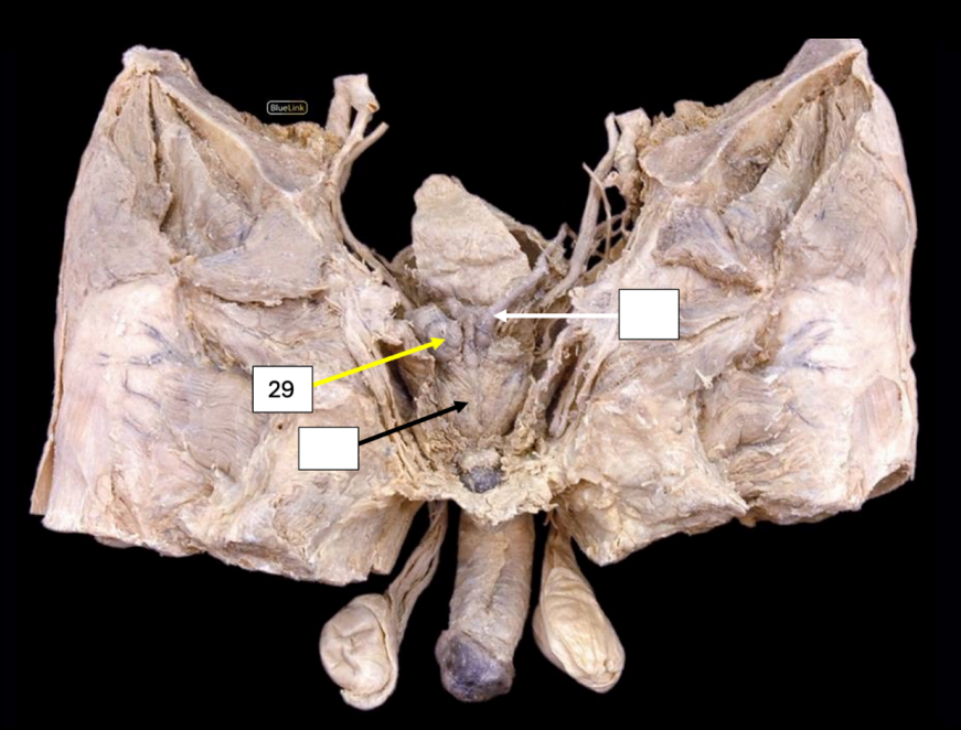
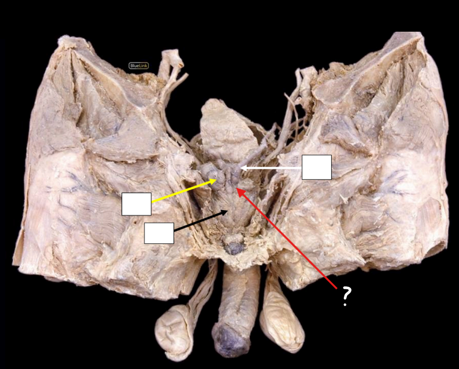
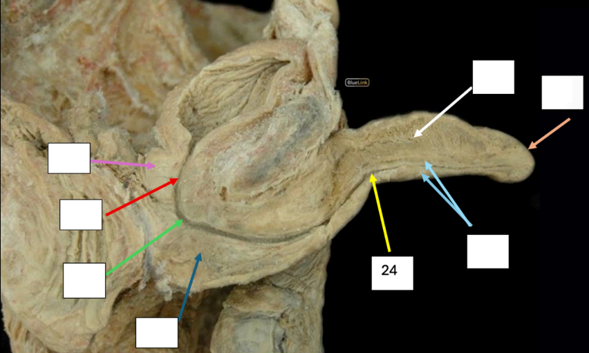
Prostate
*Prostatic venous plexus drains into Internal Iliac veins
Identify
Neurovascular Supply?

Vas Deferens
Identify
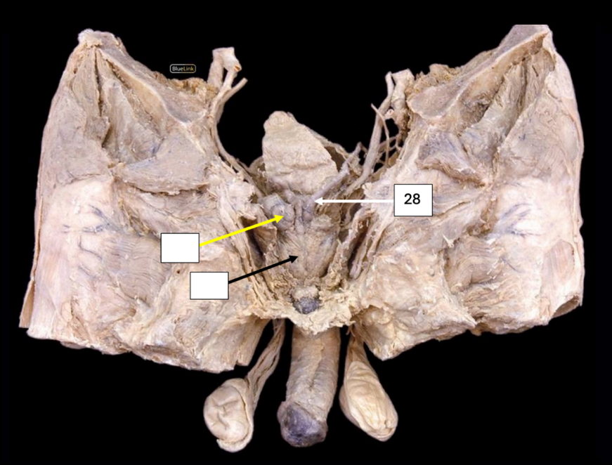
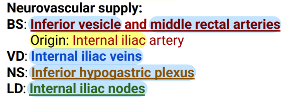
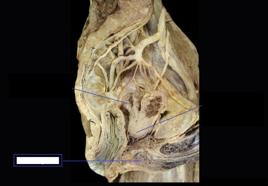
Seminal Vesicle
Side Question:
These lie at the base of the bladder
Produces citric acid and acid phosphatase
Structure found medially: vas deferens
Structure found posteriorly: rectum
Structure found inferiorly: joins with vas deferens to form ejaculatory duct
NVS?
Identify
Side Question:
These lie at the base of which structure?
Produces which secretion?
Structure found medially?
Structure found posteriorly?
Structure found inferiorly?
NVS?
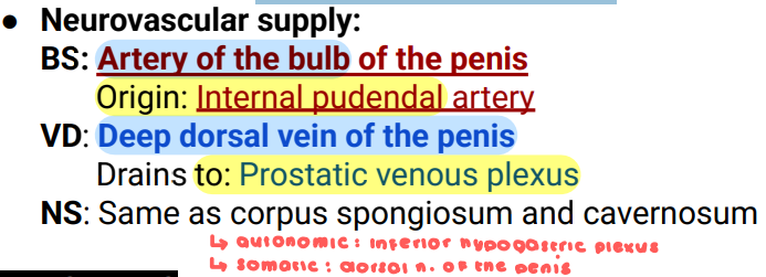
Bulb of Penis
Side Question:
This is the proximal (expanded) part of corpus spongiosum
Lies in the midline of the perineum
At the root of penis anterior to urogenital diaphragm
Continues as the body/shaft of the penis
Surrounded by bulbospongiosus muscle
NVS?
Identify
Side Question:
This is the proximal (expanded) part of?
Lies in the midline of?
At the root of penis anterior to which structure?
Continues as the?
Surrounded by what muscle?
NVS?
Prostate Gland
Side Question:
Lobe of this structure with no glandular tissue? Anterior lobe
Upper surface of the medial lobe of this is related to trigone of bladder
Its apex lies against which structure below and is closely related to?
Urogenital diaphragm; external urethral sphincter
Its base lies against which structure and is closely related to?
Bladder; internal urethral sphincter
Identify
Side Question:
Lobe of this structure with no glandular tissue?
Upper surface of the medial lobe of this is related to which structure?
Its apex lies against which structure below and is closely related to?
Its base lies against which structure and is closely related to?
neck of bladder
Base of the prostate is continuous with?
Urethra
Which structure enters the center of the base of prostate?
symphysis pubis; puboprostatic ligaments
Structure found anterior to the prostate gland and is attached by which ligament?
rectal ampulla
Structure found posterior to the prostate gland?
Levator ani muscles
Structure found laterally to the prostate gland?
Seminal Vesicle
Identify
Beginning of Ejaculatory Duct
Identify
Spongy (Penile) Urethra
Identify
Bulbospongiosus muscle
Side Question:
Origin: Perineal body
Insertion: Corpus Spongiosum
Innervation: Perineal branch of the Pudendal nerve
Action: Compresses the urethra/Sphincter of the vagina; Assists in erection
Identify
Side Question:
Origin?
Insertion?
Innervation?
Action?

Prostate (Posterior Lobe)
Side Question:
Situated behind the urethra and below the ejaculatory ducts
Identify (Be specific)
Side Question:
This is situated behind and below which structures?

Glans Penis
Identify

Prostatic Urethra
Side Question:
What drains in this structure? Seminal fluid
Identify
Side Question:
What drains in this structure?

Corpus Spongiosum
Identify
Side Question:
Blood Supply?
Venous Drainage?
Nerve Supply?

Membranous Urethra
Identify
Corpus Cavernosum
Identify
Side Question:
Blood Supply?
Venous Drainage?
Nerve Supply?

Ovarian Ligament
Identify
Isthmus of the Fallopian Tube
Identify
Vestibule
Identify
Mons Pubis
Identify
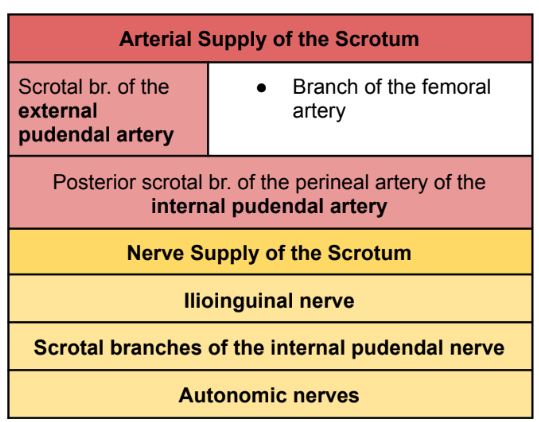
Scrotum
Side Question:
Wrinkling of this structure is caused by what muscle? Dartos muscle
The wrinkling of this structure is an extension or derivative of which abdominal wall layer: Camper’s fascia
If this structure had a superficial abscess, what group of nodes would enlarge? Inguinal group of nodes
Structure that superficially divides this into two: Median raphe
Identify
Side Question:
Wrinkling of this structure is caused by what muscle?
The wrinkling of this structure is an extension or derivative of which abdominal wall layer?
If this structure had a superficial abscess, what group of nodes would enlarge?
Structure that superficially divides this into two?
Note:
Innervation
Anterior scrotum
Ilioinguinal n.
Genital br. of the genitofemoral n.
Posterior scrotum
Pudendal n.
Lymph Drainage: Superficial Inguinal nodes
Neurovascular Supply of Scrotum
Spermatic Cord
Structure found in this?
Vas deferens
Testicular artery
Pampiniform plexus
Remains of processus vaginalis
Genital branch of genitofemoral nerve
Identify
Side Question:
Structure found in this?
Body of Epididymis
Identify (Be specific)

Head of Epididymis
Identify (Be specific)
Tail of Epididymis
Identify (Be specific)
