Muscle Physiology
1/22
Earn XP
Description and Tags
1.) Sorry for any spelling errors lol 2.) Notes recommended for flashcard mode only 3.) Any bolded card means it was not on the study guide but felt important to me
Name | Mastery | Learn | Test | Matching | Spaced |
|---|
No study sessions yet.
23 Terms
Draw the skeletal muscle structure from whole muscle to thick and thin filaments
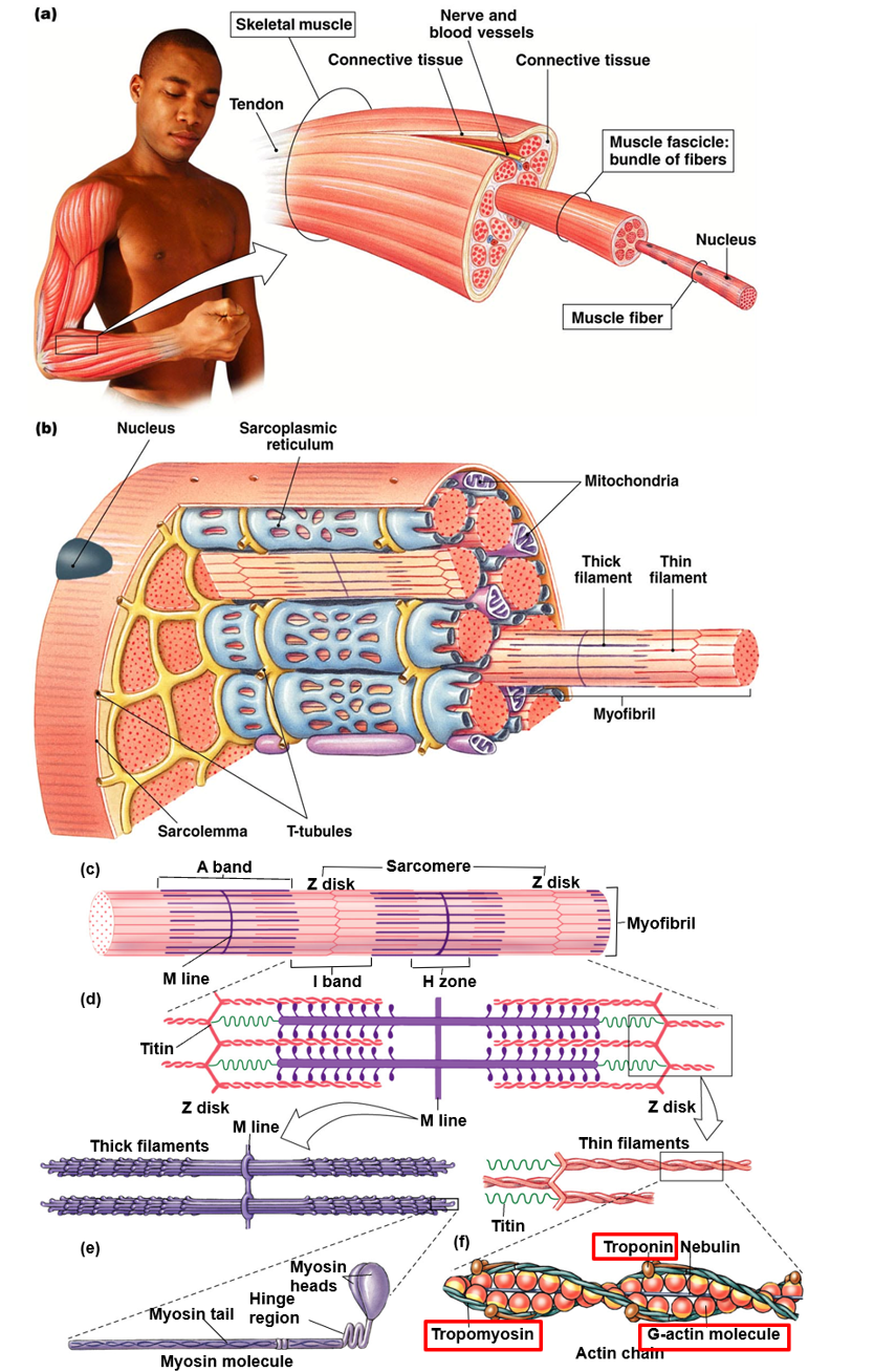
Sarcomere (Hint: 3)
Functional unit of skeletal muscle
Composed of thick filaments (myosin), thin filaments (actin)
From z disk to z disk
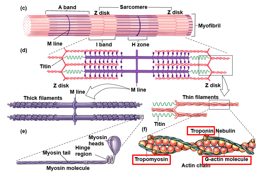
Thick Filaments (Hint: 3)
Bundles of proteins
Composed of only myosin
Anchored in place by titin (elastic) fibers

Thin Filaments (Hint: 2)
Made up of several structural proteins (major one is actin) anchored to z-lines
Polymer of myosin molecules, each of which has a flexible cross-bridge that binds ATP and actin

What two proteins have important roles in regulating contraction and relxation of skeletal muscle?
Troponin
Tropomyosin
Troponin
Calcium binds and induces a conformational change moving tropomyosin and exposing the myosin binding site on actin
Tropomyosin
Blocks the myosin binding site on the thin filament
Titin (Hint: 3)
Spring-like protein
Anchor thick filaments to z-disk
Key role in relaxation by springing the sarcomere back to resting length

When is muscle contraction triggered?
When a nerve impulse travels down a motor neuron until it reaches the neuromuscular junction
Neuromuscular Junction (Hint: 2)
Synapse between motor neuron and muscle fiber
Signals Ca2+
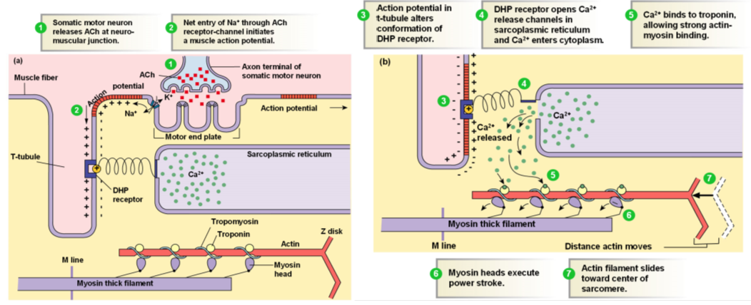
Describe the Excitation-Contraction Coupling Process (Hint: 7)
Somatic motor neuron releases Ach at neuromuscular junction
Net entry of Na+ through Ach receptor-channel initiates a muscle action potential
Action potential in t-tubule alters conformation of DHP receptor
DHP receptor opens Ca2+ release channels in sarcoplasmic reticulum and Ca2+ enters cytoplasm
Ca2+ binds to troponin, allowing strong actin-myosin binding
Myosin heads execute power stroke
Actin filament slides toward center of sarcomere
List the Process for Muscle Relaxation (Hint: 5)
Acetylcholinesterase removes Ach from synapse
Depolarization of motor end plate and muscle fiber action potentials cease
Calcium is pumped out of the myofibrils back into the sarcoplasmic reticulum via Ca2+-ATPase
Tropomyosin shifts back in position to block binding sites on actin
Filaments slide back into resting position (elastic elements- titin)
Describe Length-Tension Relationships (Hint: 2)
Most well known skeletal muscle graph
Structure of sarcomeres determines length of skeletal muscle

Length-Tension Relationship: Optimal Length (Hint: 2)
Sarcomere is in perfect position
Filaments have good overlap but still have plenty of space for filaments to slide
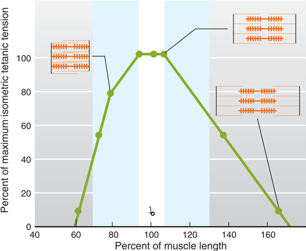
Length-Tension Relationship: Too Short (Hint: 2)
Z disks will physically hit thick filaments (why it goes to 0)
Not enough room for filaments to slide

Length-Tension Relationship: Too Long (Hint: 2)
Filaments have little to no overlap
Cannot cross bridge

List the 3 types of skeletal muscle fibers
Slow-oxidative (Type 1)
Fast-oxidative-glycolytic (Type 2A)
Fast-glycolytic (Type 2B)
Fast-Glycolytic Fibers (Type 2B) (Hint: 5)
White fibers
Largest in size
Most thick and thin filaments
Contract 2-3 times faster than slow-twitch (Faster uptake of Ca2+; Faster ATP splitting (more myosin ATPase))
Anaerobic/fatigue easily (power lifting; fast/delicate actions; Sprint)
Slow-Oxidative Fibers (Type 1) (Hint: 6)
Red fibers
Aerobic, less fatigue
More mitochondria (where we have oxidative phosphorylation done)
More capillaries (need to deliver more O2)
Abundant myoglobin (shuttles O2 from blood to mitochondria in muscles (gives red color))
Endurance activities; postural muscles (core and low back muscles)
Fast-Oxidative-Glycolytic Fibers (Type 2A) (Hint: 3)
Pink fibers
Intermediate speed
Anaerobic and aerobic- most adaptable to training
How does smooth muscle contraction differ from skeletal muscle?
Skeletal Muscle:
DHP
Calcium comes from sarcoplasmic reticulum
No CaM
No MLCK
Smooth Muscle
No DHP
Calcium comes from sarcoplasmic reticulum and extracellular sources
CaM
MLCK
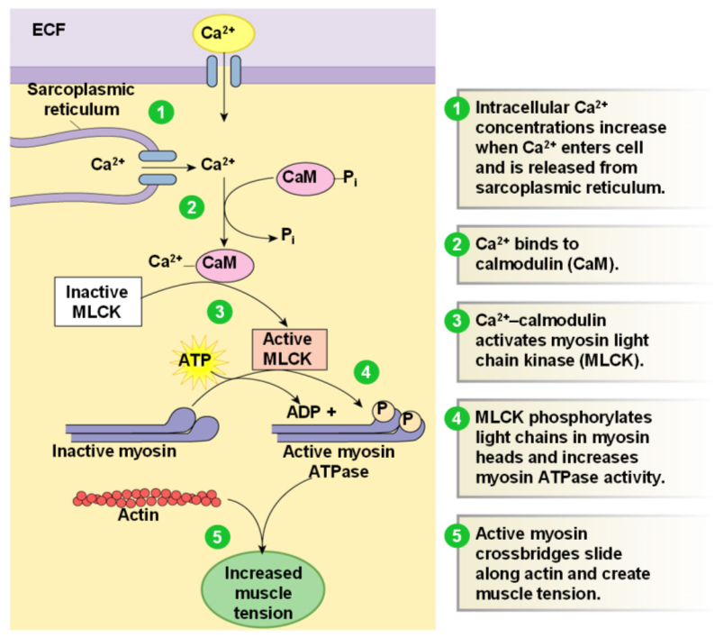
Describe the Smooth Muscle Contraction Process (Hint: 5)
Intracellular Ca2+ concentrations increase when Ca2+ enters cell and is released from sarcoplasmic reticulum
Initially Ca2+ comes from extracellular source
Ca2+ binds to calmodulin (CaM)
Ca2+-calmodulin activates myosin light chain kinase (MLCK)
MLCK only in smooth muscle; phosphorylates myosin n thick filament
Smooth muscle must be phosphorylated before it will bind to actin
MLCK phosphorylates light chains in myosin heads and increases myosin ATPase activity
To relax, myosin must be dephosphorylated
Active myosin cross bridges slide along actin and create muscle tension
Only in smooth muscle
Calmodulin (CaM)
A cytoplasmic protein not found on the thin filament