UNIT 4
1/688
Earn XP
Description and Tags
:)
Name | Mastery | Learn | Test | Matching | Spaced | Call with Kai |
|---|
No analytics yet
Send a link to your students to track their progress
689 Terms
What are the 3 components of the cardiovascular system?
Heart, vessels, and blood.
What is the role of the heart in the cardiovascular system?
Pump that creates pressure to move blood through the rest of the cardiovascular system
What are vessels in the cardiovascular system?
Tubes where blood flows through
Types of vessels?
arteries, arterioles, capillaries, venules, and veins.
What does blood carry throughout the body?
Oxygen, glucose, hormones, immune cells, proteins, wastes, and more.
What are the main functions of the cardiovascular system?
Distributes blood, delivers/exchanges nutrients/wastes/hormones, regulates heat, helps with clotting, and modulates inflammation.
What does the pulmonary circuit do?
Sends deoxygenated blood from the heart to the lungs to pick up oxygen and release carbon dioxide, then returns oxygenated blood to the heart.
What does the systemic circuit do?
Sends oxygen-rich blood from the heart to the body (except lungs) through arteries and collects waste to return to the lungs.
What type of blood do arteries carry and in which direction?
Most arteries carry oxygen-rich blood away from the heart. Except pulmonary arteries, they carry deoxygenated blood from the heart to the lungs.
What is the order of blood flow through vessels after arteries?
Arteries → arterioles → capillaries → venules → veins.
Where does exchange of nutrients, gases, and wastes occur?
In the capillaries.
What type of blood do veins carry and in which direction?
Most veins carry deoxygenated blood toward the heart, except for pulmonary veins, they carry oxygen rich blood from lungs to back to the heart
What separates the left and right sides of the heart?
The septum, a wall that divides the two sides.
How many chambers does the heart have? What are they?
Four chambers — 2 atria (top) and 2 ventricles (bottom).
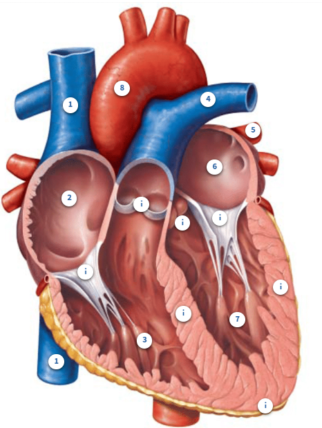
Anatomy of the heart
What is the superior vena cava?
The largest vein in the body that brings deoxygenated blood from the head, neck, chest, and arms to the right atrium.
Where does blood in the right atrium come from?
From the superior and inferior vena cava — returning from all body tissues except the lungs.
What does the right atrium do?
Pumps blood from every organ and tissue in the body except lungs to the right ventricle.
What does the right ventricle do?
Gets blood from the right atrium and sends it out of the heart through pulmonary arteries and into the pulmonary circuit.
Which side of the heart has lower oxygen and higher carbon dioxide?
Right
Why does blood go to the lungs from the right ventricle?
To drop off CO₂ and pick up oxygen.
Where do pulmonary arteries take blood?
Away from the heart and into the lungs.
How many pulmonary arteries are there and where do they go?
Two — one to the right lung and one to the left lung.
What do pulmonary veins do?
Bring oxygenated blood from the lungs back to the heart's left atrium
How many pulmonary veins are there?
Four — 2 right and 2 left.
Where does blood in the left atrium come from?
From the pulmonary veins (lungs).
Where does the left atrium sends blood?
to the left ventricle
What does the left ventricle do?
Pumps oxygen-rich blood to the systemic circuit through the aorta.
Which side of the heart has higher oxygen and lower carbon dioxide levels?
left
What is the aorta?
The largest artery in the body; carries blood from the left ventricle to the body (except lungs).
What does the pulmonary valve do?
Prevents blood from flowing back into the right ventricle from the pulmonary arteries
What does the aortic valve do?
Prevents blood from flowing back into the left ventricle from the aorta.
What does the left AV (atrioventricular) valve do?
Stops blood from flowing back into the left atrium from the left ventricle.
What does the right AV valve do?
Stops blood from flowing back into the right atrium from the right ventricle.
What is the interventricular septum?
The wall between the left and right ventricles, preventing blood from mixing.
Do the two atria have a septum?
yes, the interatrial septum
Why is the left ventricular myocardium wall thicker than the right?
Because it must pump blood forcefully throughout the whole body (against gravity).
What is the apex of the heart?
The bottom point of the heart where ventricular contraction starts all the way upwards.
Pathway of blood
Vena Cava (d) -> Right atrium -> Right ventricle -> Pulmonary artery-> Lungs -> pulmonary vein (o) -> left atrium -> left ventricle -> Systemic circuit
How many heart valves are there, and what are they called?
Four — aortic valve, pulmonary valve, right AV valve, and left AV valve.
Where are the aortic and pulmonary valves located?
At the exits of the ventricles.
Where are the AV (atrioventricular) valves located?
At the exits of the atria, leading into the ventricles.
What is the main function of heart valves?
To prevent backflow of blood into the wrong heart chamber.
What does the aortic valve do?
It sends blood from the left ventricle to the aorta and into systemic circulation.
What does the pulmonary valve do?
It sends blood from the right ventricle to the pulmonary arteries and into pulmonary circulation.
What are the “pockets” in the aortic and pulmonary valves called?
Cusps — they fill with blood and close the valve to stop backflow.
What do the cusps do when blood tries to go backward?
They fill up, expand, and close the valve like a parachute catching air.
What is another name for the aortic and pulmonary valves?
Semilunar valves — named for their crescent moon shape.
Where are the AV (atrioventricular) valves located?
Between the atria and the ventricles.
What do AV valves do?
They prevent backflow of blood from the ventricles to the atria.
What other names are used for the AV valves?
The right AV valve is also called the tricuspid valve, and the left AV valve is called the mitral valve
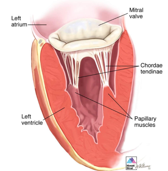
Anatomy of the left AV valve of the heart
:)
What are the two normal heart sounds called?
"Lub" and "Dub".
What creates the "lub" sound of the heart?
First sound which is created by the AV valves closing, which happens when the ventricles start to contract blood attempts to head back to the atria
What creates the "dub" sound of the heart?
Second sound from the aortic and pulmonary valves closing as the ventricles begin to relax and blood attempts to head back into the ventricles
What are heart murmurs?
Abnormal heart sounds due to valve issues
What is stenosis?
Valves do not open properly causing a high-pitched sound
What is valve regurgitation?
Valves don't open properly causing a swishing/whooshing sound
What might irregular heart contractions be called?
Arrhythmias — abnormal or irregular heartbeats.
What are the muscle cells of the heart called?
Cardiomyocytes (cardiac muscle cells)
What are skeletal muscle cells called?
Myofibers
What are 3 things needed for a skeletal muscle cell (myofiber) to contract and relax?
ATP, calcium (Ca²⁺), and electrical stimulation (action potential).
What are the two types of cardiomyocytes?
Contractile cells and nodal/conducting cells.
What is the function of contractile cardiomyocytes?
To pump blood through the heart.
What is the function of nodal/conducting cardiomyocytes?
To spread electrical signals through the heart.
Similarities between skeletal myofibers and cardiomyocytes
Striated muscle: Both have stripes due to the organization of thin/thick filaments
Calcium: Both need calcium to contract
Where do skeletal myofibers get their calcium from?
Sarcoplasmic reticulum
Where do Cardiomyocytes get their calcium from?
Sarcoplasmic reticulum and extracellular fluid
What's the sarcoplasmic reticulum?
An organelle inside of skeletal myofiber
What's the calcium’s role in skeletal myofibers?
Released from sarcoplasmic reticulum after action potential
What’s Calcium’s role in Cardiomyocytes?
calcium from extracellular fluid enters through ion channels and helps release calcium from the sarcoplasmic reticulum. AKA Calcium-induced calcium release.
ATP and mitochondria in skeletal myofibers
Requires ATP from mitochondria (fatigue can occur)
ATP and mitochondria in cardiomyocytes
Requires even more mitochondria than skeletal myofibers (fatigue cant occur)
Where does an action potential come from the skeletal myofiber?
From a motor neuron releasing a neurotransmitter
Do cardiomyocytes require outside neurons?
no, it can create its own action potential
Where does an action potential come from in the cardiomyocytes?
Nodal cells found within the heart
Shape of skeletal myofibers?
Cylindrical cells
Shape of cardiomyocytes?
Branched cells
How many nuclei does skeletal myofibers have?
multiple
How many nuclei do cardiomyocytes have?
one
Gap junctions in cardiomyocytes
Special channels that allow ions and small molecules to pass between cells. They allow direct communication between neighboring cells and enable ions to travel to cause depolarization in adjacent cells.
Intercalated discs in cardiomyocytes
Structures that lock cells together, preventing them from separating during heartbeats.
Where are gap junctions found?
At the intercalated discs.
Desmosomes
Special proteins that act as the glue that holds different cardiomyocytes together at the intercalated discs.
Are nodal/conducting cells self excitable?
Yes, they generate action potential to spread through the heart for contraction
Minimal action and myosin in nodal and conducting cells
Do not contract because they have minimal actin and myosin
What ion is important in cardiomyocyte action potentials?
Calcium (Ca²⁺).
What does the rise in calcium trigger in muscle cells?
It causes tropomyosin to move off myosin binding sites on actin, allowing contraction.
What is a typical resting heart rate?
Around 70–80 beats per minute (bpm).
What is the average heart rate for biological males?
About 70 bpm.
What is the average heart rate for biological females?
About 80 bpm (due to smaller heart size).
Why might trained athletes have slower heart rates (40–60 bpm)?
Their hearts are more efficient, pumping more forcefully, so they beat less often.
Where would you find nodal and conducting cells?
Sinoatrial node
Atrioventricular node
Atrioventricular bundle
Subendocardial branches
Where is the Sinoatrial node?
Upper Right atrium
What's considered the pacemaker of the heart?
The Sinoatrial node
What sets the heart rate?
Sinoatrial node
Is the sinoatrial node self excitable?
Yes
Where is Chloride more abundant?
Extracellular fluid
Where is Sodium more abundant?
Extracellular fluid
Where is Calcium more abundant?
Extracellular fluid