Fetal Heart Orientation and Views
1/79
There's no tags or description
Looks like no tags are added yet.
Name | Mastery | Learn | Test | Matching | Spaced | Call with Kai |
|---|
No analytics yet
Send a link to your students to track their progress
80 Terms
What percentage of cardiac anomalies can be detected with the 4-chamber view
50-60%
What percentage of cardiac anomalies can be detected when including great vessel imaging
Up to 85%
At what gestational age is the 4-chamber view obtainable in most cases
Around 18 weeks (~95%)
What is fetal lie
The orientation of the fetus in the uterus (longitudinal, transverse, oblique)
What is fetal presentation
The part of the fetus closest to the cervix
What are the types of fetal lie
Longitudinal, transverse, oblique
What are the types of fetal presentation
Vertex (head), breech (butt/feet), transverse
What landmark should the spine be assigned to
6 o'clock
What landmark represents the front of the fetus
12 o'clock
In vertex or transverse head right, how do the stomach and heart orient
Clockwise toward 9 o'clock
In breech or transverse head left, how do the stomach and heart orient
Counterclockwise toward 3 o'clock
What is levoposition
Heart located in the left chest (normal)
What is mesoposition
Heart located in the midline of the chest
What is dextroposition
Heart located in the right chest
What is the most common cause of dextroposition
Extrinsic mass effect (e.g., CPAM, pleural effusion, diaphragmatic hernia)
What is cardiac axis
The angle/orientation of the cardiac apex within the chest
What is the normal cardiac axis
45° ± 20° toward the left
What percentage of the chest should the heart occupy
About 1/3
What is levocardia
Apex points to the left (normal)
What is mesocardia
Apex points midline
What is dextrocardia
Apex points to the right
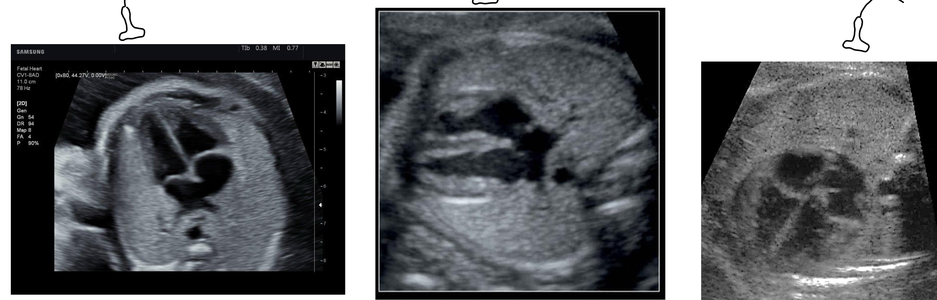
What 4 chamber views are these from left to right?
Apical
Subcostal
Basal
What are the three angles used to obtain the 4-chamber view
Apical, subcostal, basal
Which view is best for detecting septal defects (VSD, ASD)
Subcostal view
Why is the subcostal view best for septal defects
The ultrasound beam is perpendicular to the septum
How much of the chest should the heart occupy
1/3
What should be seen on either side of the chest in a proper view
Ribs
What should the cardiac axis be
~45° to the left
What should be true about the interventricular septum
It should be intact
How should the atria and ventricles appear
Symmetric
Which valve is more apical, tricuspid or mitral
Tricuspid
Where should the inferior pulmonary veins enter
Left atrium
Where is the aorta located relative to the left atrium
Posterior
What structure is seen in the right ventricle
Moderator band
Where does the foramen ovale flap open
Into the left atrium
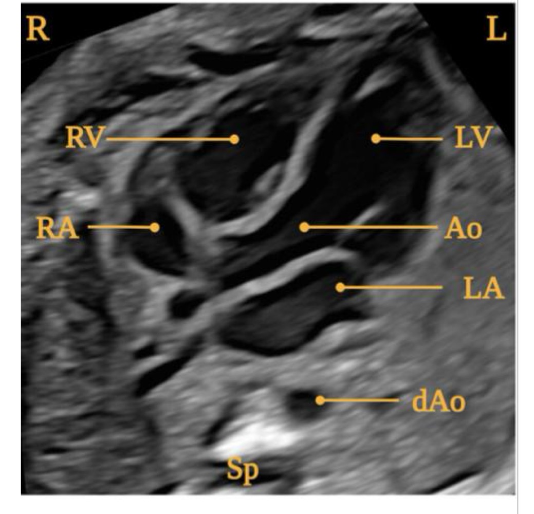
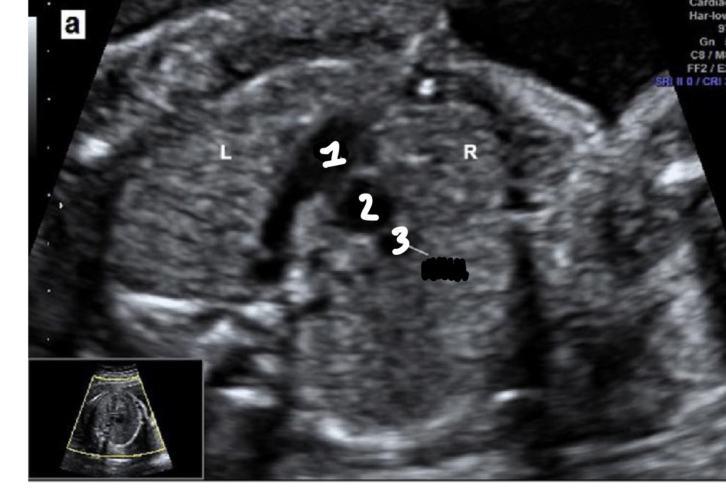
What view is this?
5 Chamber view
What additional structure is seen in the 5-chamber view
Aorta arising from the left ventricle
What is the purpose of the 5-chamber view
Evaluate aortic root and interventricular septum
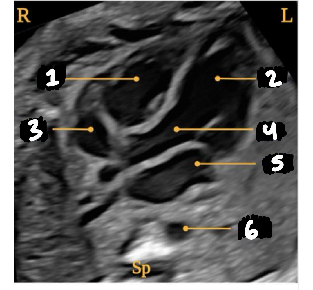
Right ventricle
Left ventricle
Right atrium
Aorta
Left atrium
Descending aorta
What does LVOT stand for
Left ventricular outflow tract
What vessel arises from the left ventricle in LVOT
Aorta
What is the "ballerina foot" sign associated with
LVOT view
What abnormality can be assessed in LVOT
Ventricular septal defect (VSD)
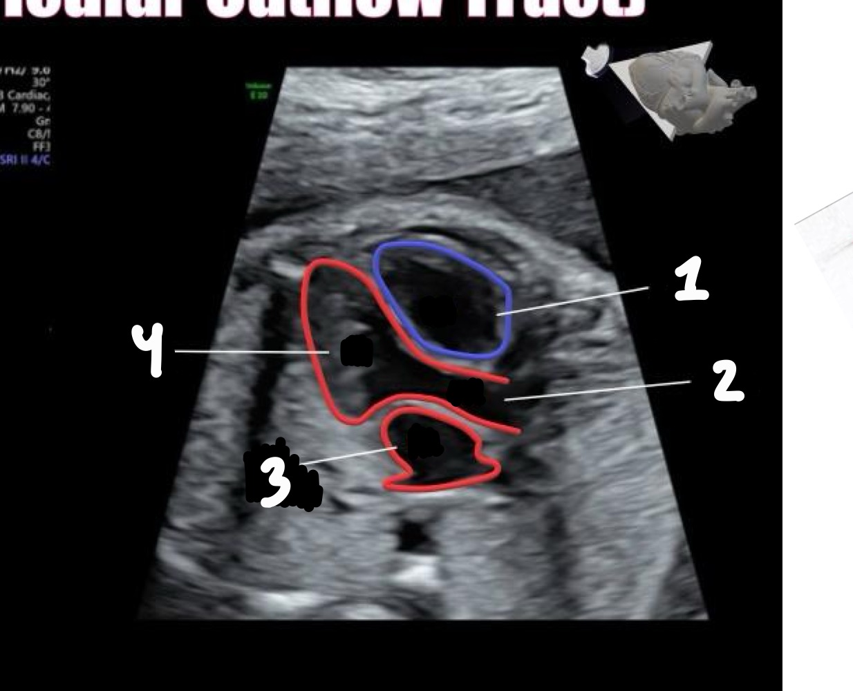
Right ventricle
Aorta
Left atrium
Left ventricle
What does RVOT stand for
Right ventricular outflow tract
What vessel arises from the right ventricle
Pulmonary artery
What structures are seen after the pulmonary artery in RVOT
Ductus arteriosus and right pulmonary artery
What are common descriptors for RVOT appearance
Snail or walking man
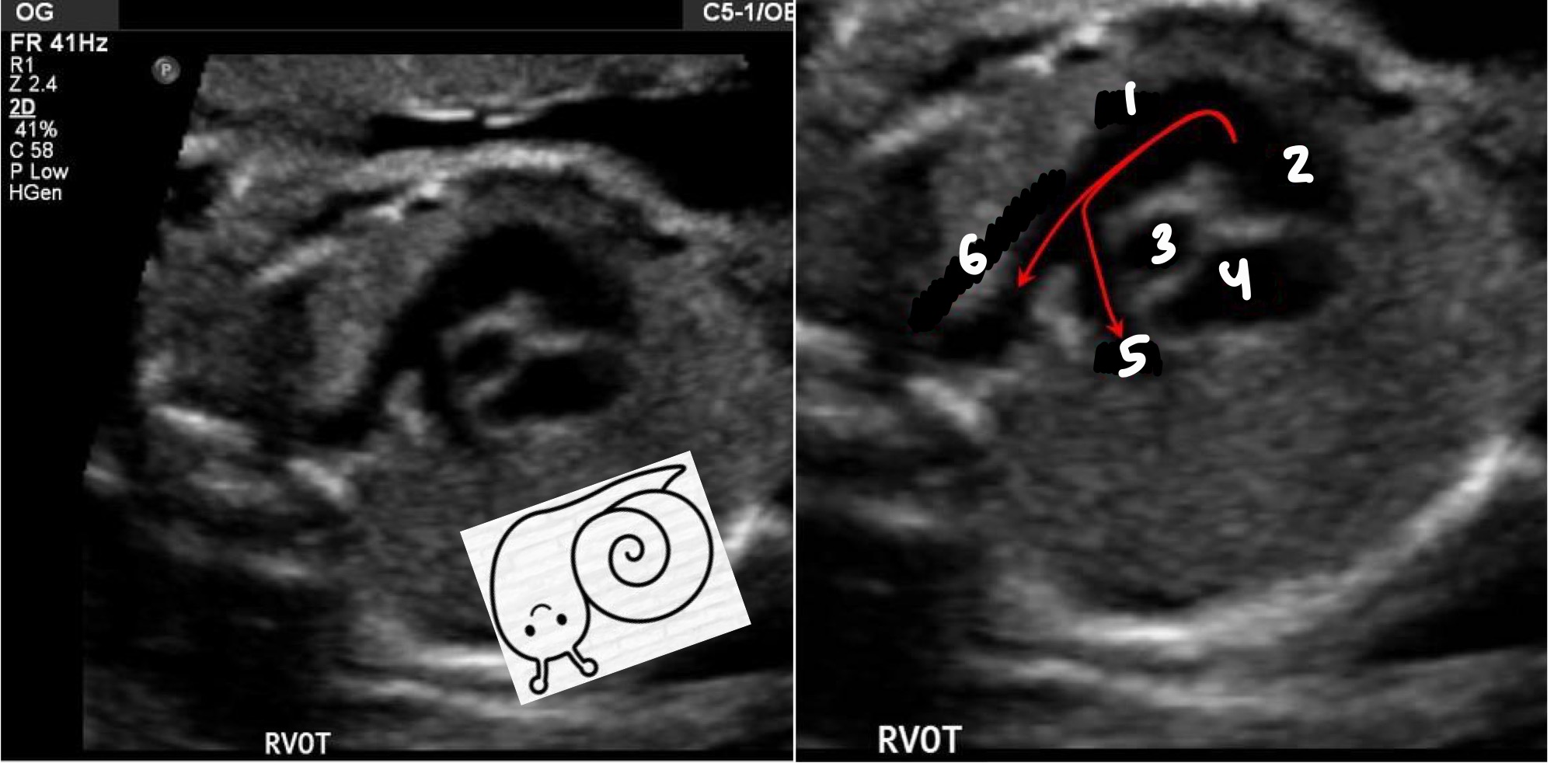
MPA
Right ventricle
Aorta
Right atrium
Right pulmonary artery
Ductus Arteriosus
How does the pulmonary artery compare in size to the aorta
Equal or slightly larger
What happens to the great vessels during development
They spiral 180°
What is the normal relationship between the pulmonary artery and aorta
Pulmonary artery crosses over the aorta
In LVOT, what direction does the aorta travel
Left to right
In RVOT, what direction does the pulmonary artery travel
Right to left
What direction should blood flow in both the aorta and pulmonary artery
Away from the transducer toward the spine
How should normal blood flow appear
Smooth and laminar
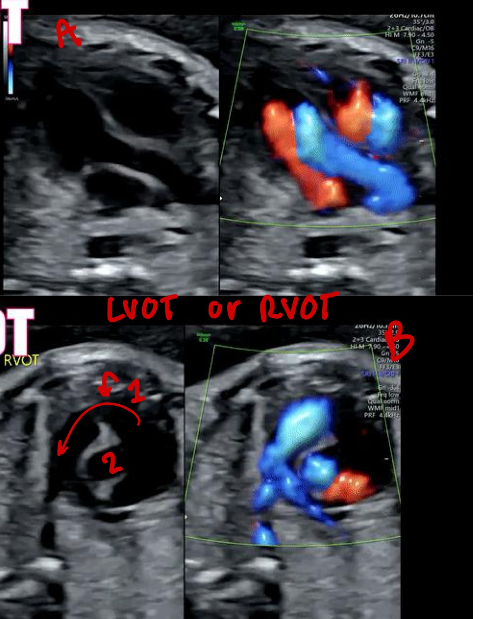
Pulmonary artery
Aorta
A= LVOT
B= RVOT
What is the shape of the aortic arch
Candy cane (tight curve)
What branches arise from the aortic arch IN ORDER
Brachiocephalic, left common carotid, left subclavian
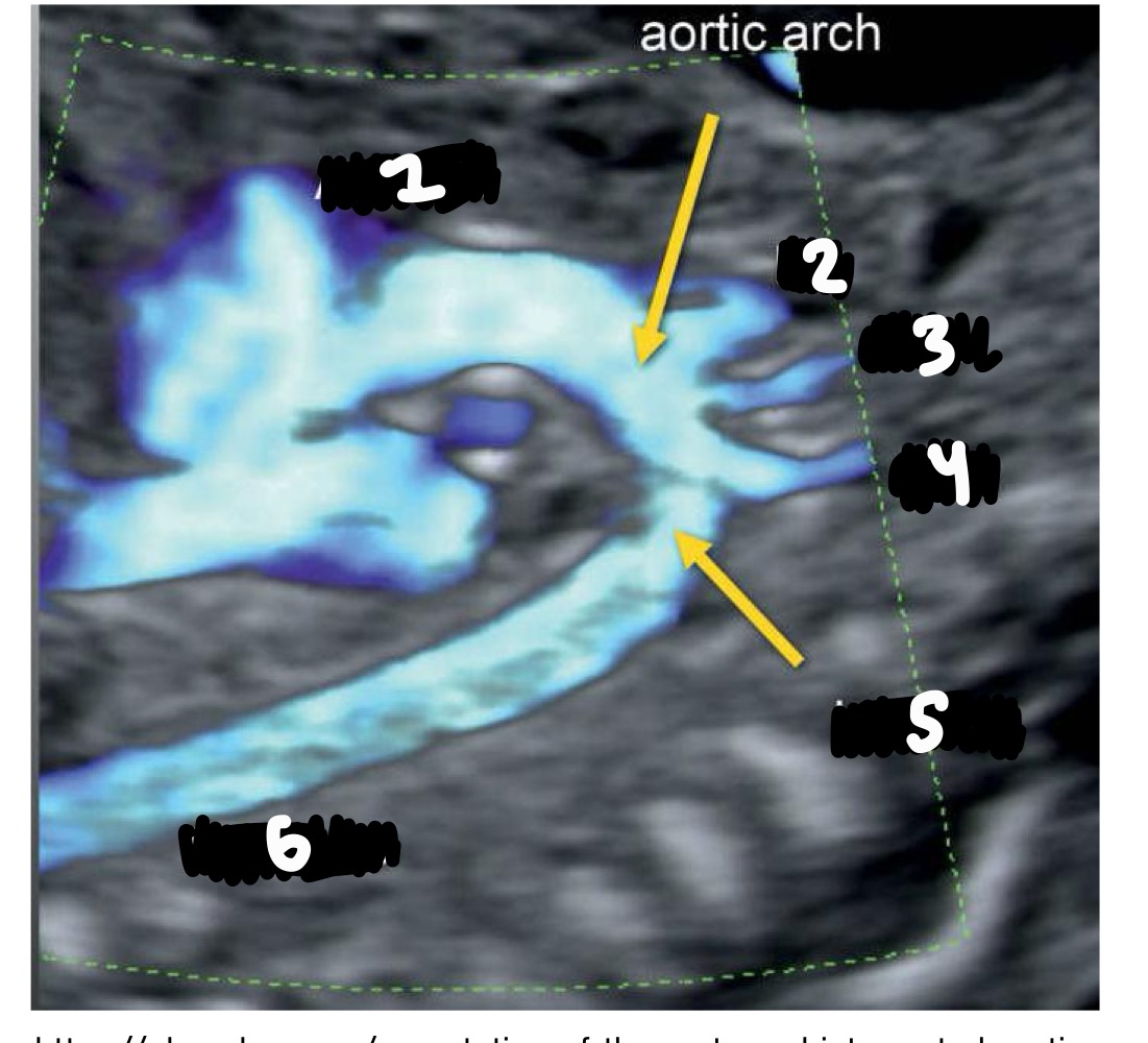
Ascending aorta
Innominate/brachiocephalic artery
Left common carotid
Left subclavian artery
Isthmus
Descending AO
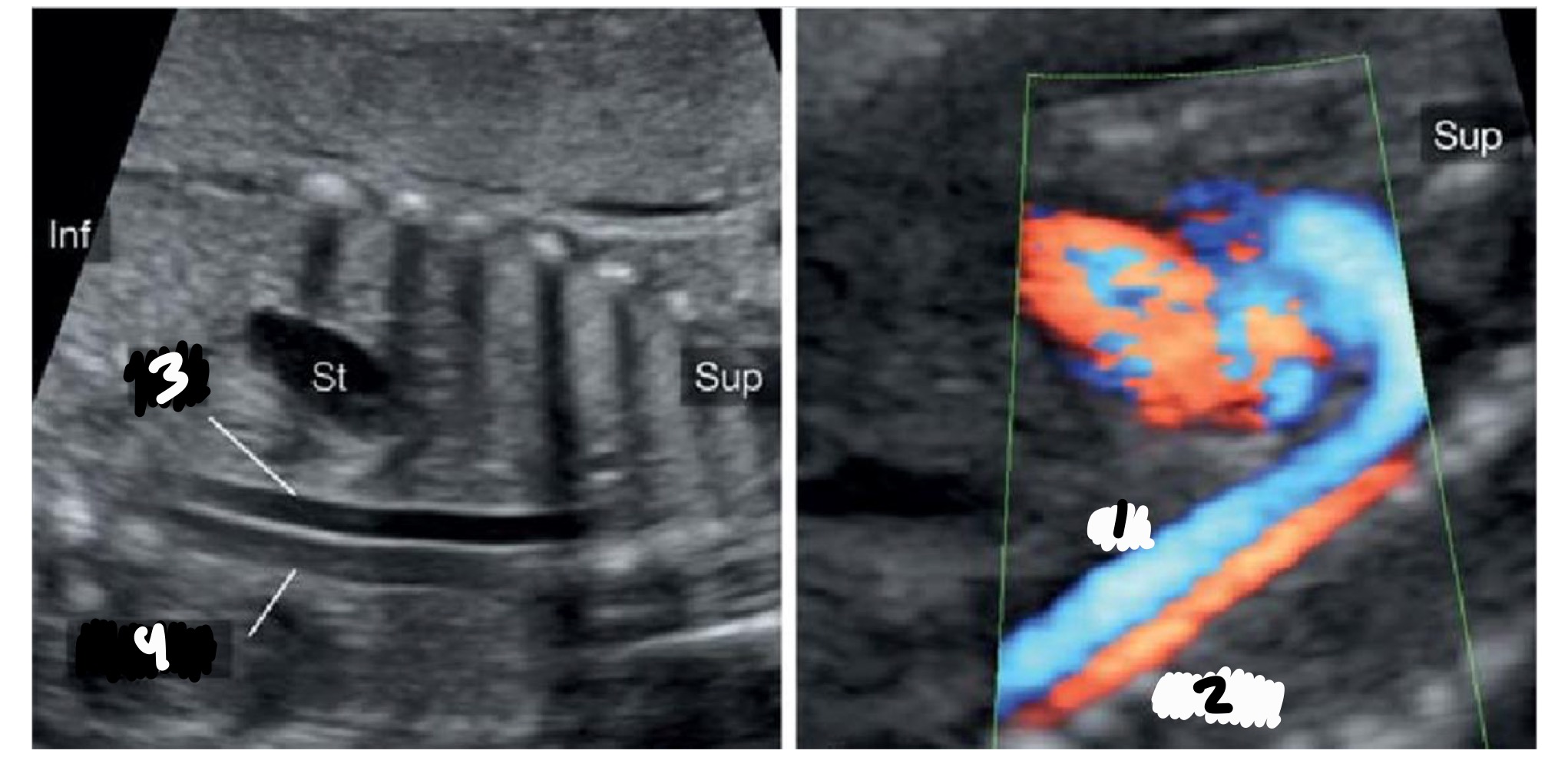
What is the shape of the ductal arch
Hockey stick (wide curve)
How many branches does the ductal arch have
None
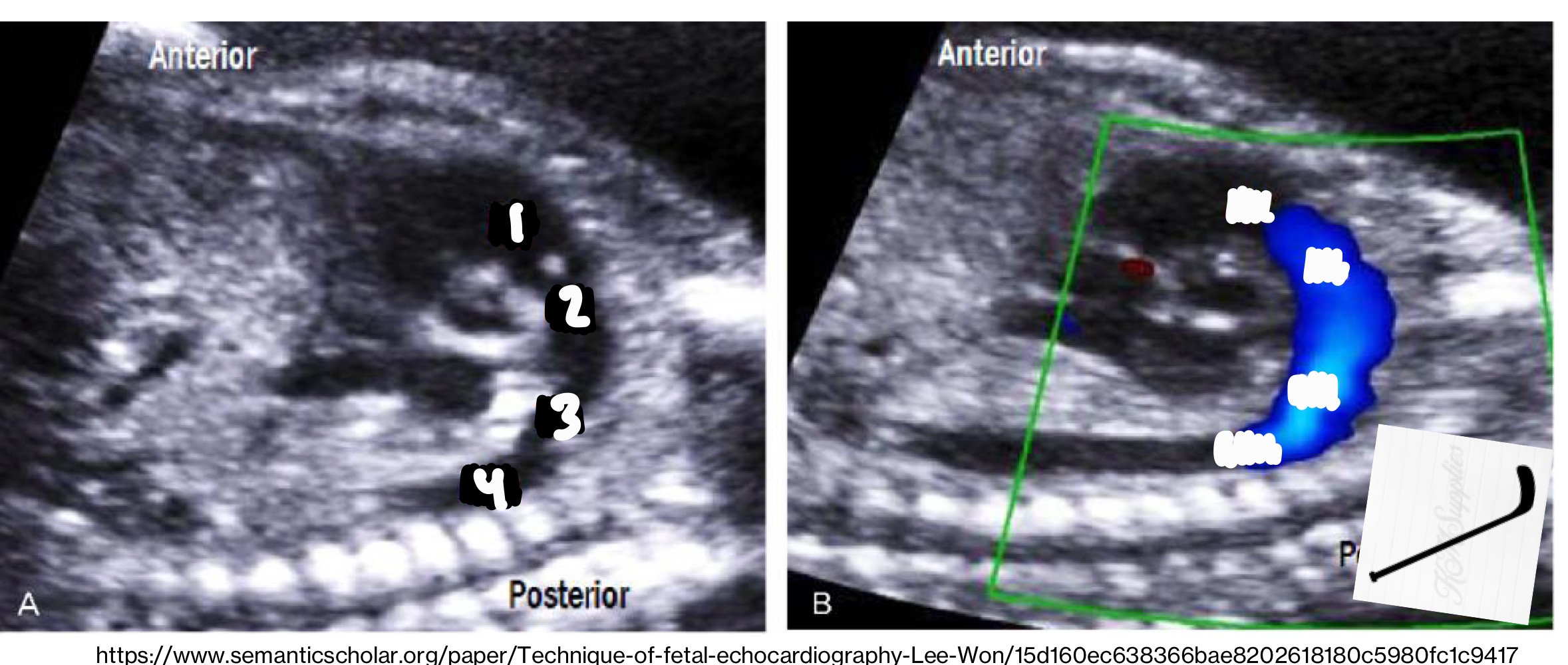
Right ventricle
Pulmonary artery
Ductus arteriosis
Descending aorta
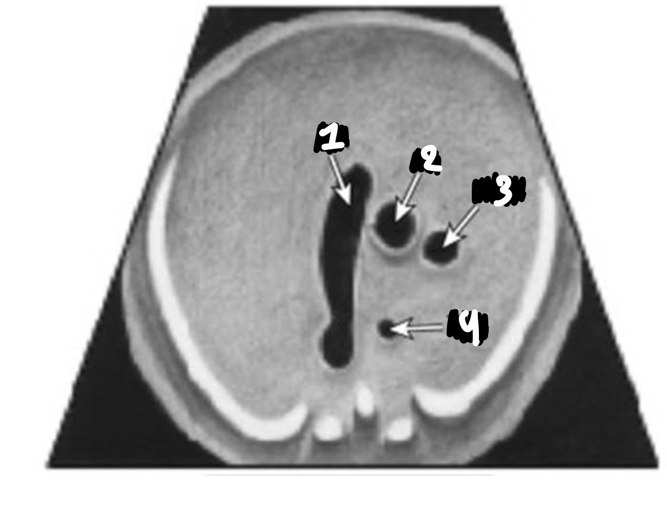
What vessels are seen in the 3 vessel view
SVC, aorta, pulmonary artery
Which vessel is smallest in 3VV
SVC
Which vessel is largest in 3VV
Pulmonary artery
How should the pulmonary artery compare to the aorta
Equal or larger
Pulmonary artery
Aorta
SVC
Pulmonary artery
Aorta
SVC
Trachea
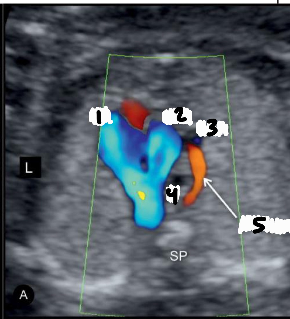
Pulmonary artery
Ascending aorta
SVC
Trachea
Azygous
What structure connects the aorta and pulmonary artery in 3VTV
Ductus arteriosus
Where is the trachea located relative to the aorta
To the right
What direction should flow be in 3VTV
Away from the transducer
What vessels are seen entering the right atrium in the bicaval view
SVC and IVC

SVC
Right Atrium
IVC
How should the SVC and IVC compare in size
Similar caliber
What does the azygous vein drain
Thoracic and abdominal walls
Aorta
Azygous vein
Aorta
Azygous vein
Where does the azygous vein empty
Into the SVC
Where might the azygous vein be seen
Parallel to the aorta or joining SVC in 3VT