3.1 Exchange surface
1/81
There's no tags or description
Looks like no tags are added yet.
Name | Mastery | Learn | Test | Matching | Spaced | Call with Kai |
|---|
No analytics yet
Send a link to your students to track their progress
82 Terms
spec point -
the need for specialised exchange surfaces
To include
SA:V ratio
metabolic activity
single-celled and multicellular organisms
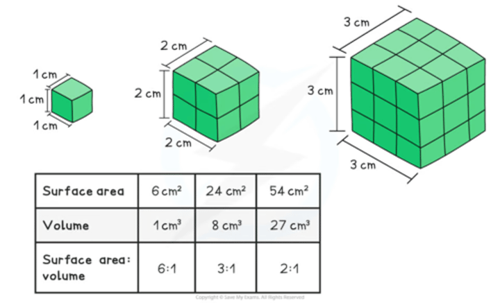
ratio = surface area / volume
as organisms increase in size what happens to their SA:V ratio
decreases
(less SA for absorption of ____)
(greater volume= longer diffusion distance)
larger surface area allows for....
small volume means...
max. absorption of nutrients and gases and secretion of waste products
means diffusion distance to all organelles is short
As the size of an organism increases, it’s surface area : volume ratio decreases. Notice for this particular shape the distance between the surface and the centre increases with size.
2 reasons for the need for a specialised system for gas exchange + explain them
supply of oxygen -organisms require ATP for biochemical processes = survive
removal of co2- co2 is toxic waste product of aerobic respiration =, if it accumulates in cells/tissue pH may be altered
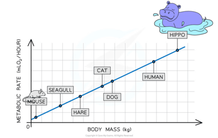
metabolic rate of an organism is ....
amount energy expended by that organism within given period of time
(it increases with body mass)
basal metabolic rate (BMR) is...
metabolic rate of an organism when at rest
-significantly lower when organism is actively moving
metabolic rate of organisms can be measured/estimated using different methods and apparatus :
-
-
-
-o2 consumption- respirometers
-co2 production - co2 probe
-health production- calorimeter
-how does body mass affect metabolic rate
-how does metabolic rate+ body mass compare to the BMR
-greater mass of organism = higher metabolic rate
-BMR per unit of body mass is higher on smaller animals than larger
smaller animals have greater SA:V ratio so lose more heat = have to use more energy to maintain their body temp
features of effective exchange surface
-large SA
-short diffusion distance
-good blood supply
-ventilation mechanism
spec point-
the features of an efficient exchange surface
To include:
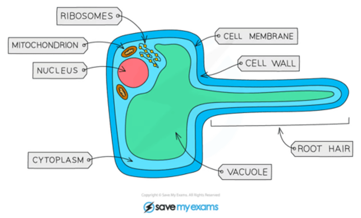
• increased surface area - root hair cells
• thin layer - alveoli
• good blood supply/ventilation to maintain gradient - gills/alveolus.
name the exchange surface in plants and how it increases its SA
root hair cells
-root hair that increases SA so rate of water uptake by osmosis is greater
-the exchange of o2 and co2 occurs btw the...
-o2 + co2 exchanged in a process of.....
-alveoli and capillaries in lungs
-simple diffusion (passive)
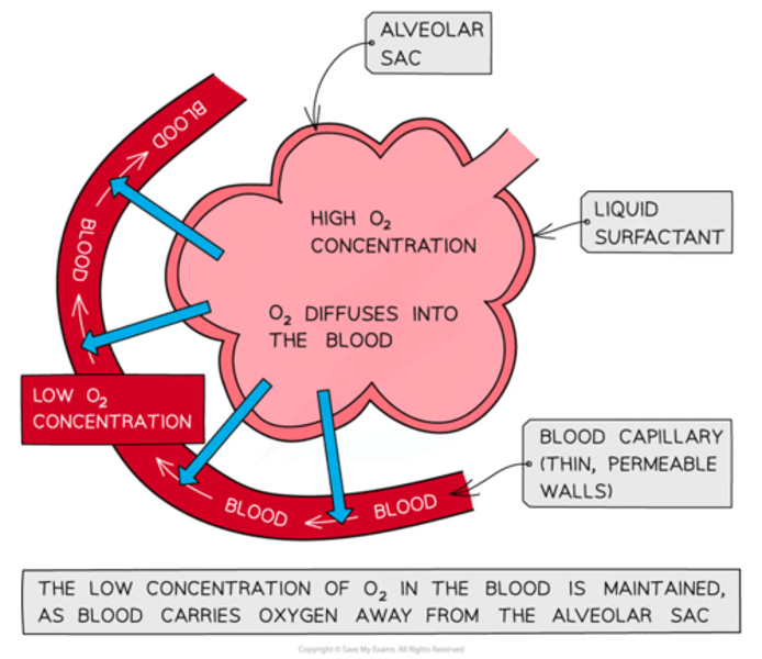
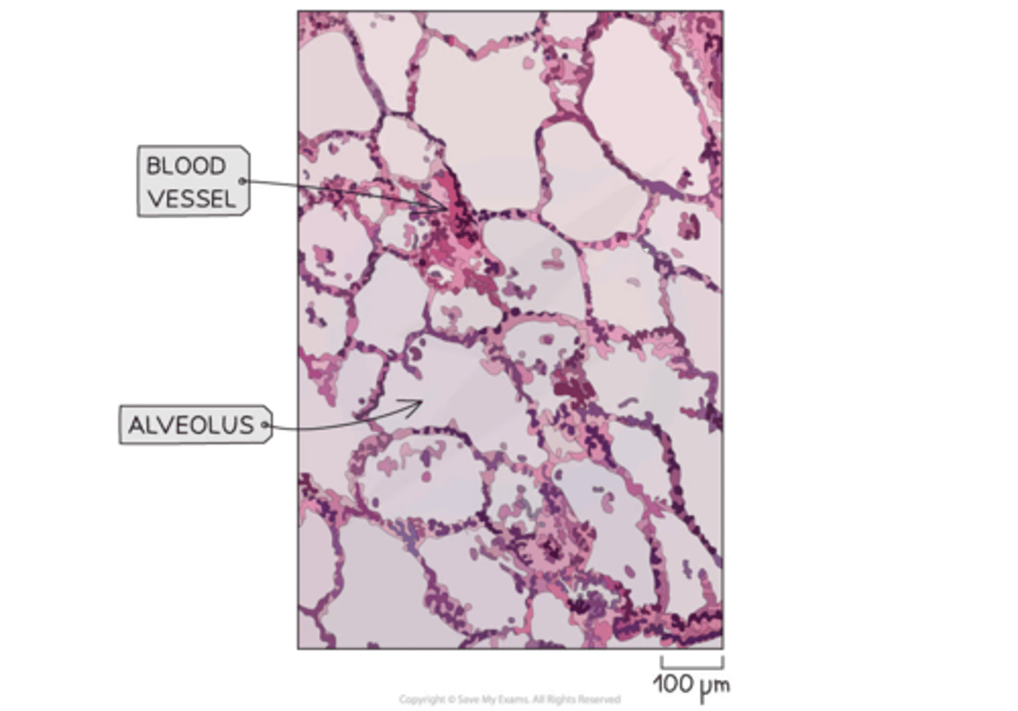
basic explanation for what goes in the alveoli/sort of why too
-air in alveoli contains high conc of o2- diffuses into blood capillaries + carried away (a.res.)
-blood in capps = low conc of o2 + high co2 conc - co2 diffuses into alevoli and is exhaled
explain why alveoli are good for effective gas exchange
-one cell thick alveoli walls + flattened cells = very short diffusion distance = quick g.e
-large no. of alveoli increasing SA available for diffusion
-extensive capillary network which are one cell thick/flattened = short gas diffuse distance + constant flow maintains the concentration gradient for gas exchange
-alveoli have surfactant
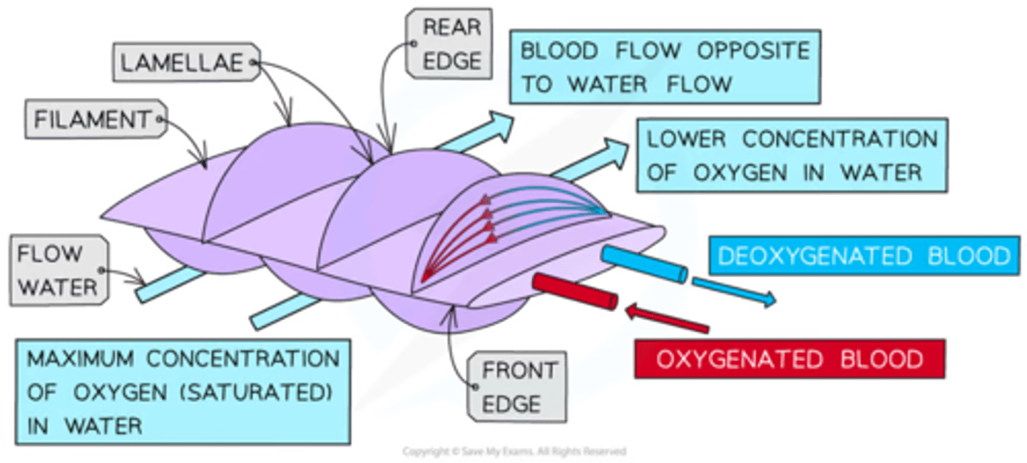
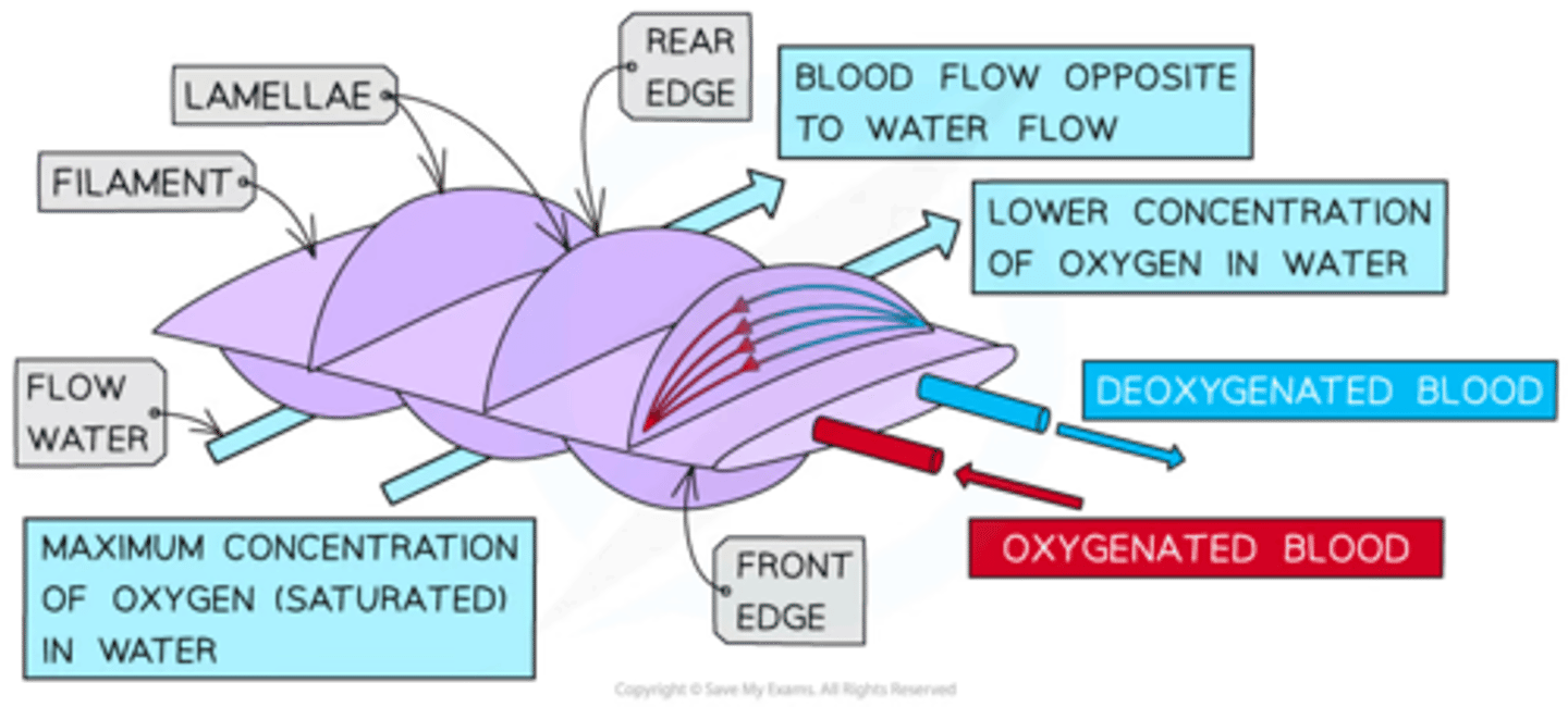
how are fish gills adapted to directly extract o2 from water
(named system)
extensive capillary network- ensures blood flows in opposite direction to flow of water - it is a counter-current system
what does the counter current system ensure
the conc gradient is maintained along whole length of the capillary
explain how counter current works
-water.... where its found /enters
-conc
-water does what ...how much ....where conc are
Water with highest o2 content is found next to blood that is oxygenated
-There is still a difference in conc so diffusion of o2 into the blood still occurs
Water continues to supply the blood with o2 along the whole gill arch + ends w/ water w/ the lowest o2 conc adjacent to the most deoxygenated blood with continued diffusion occurring
Image showing the structure of fish gills and the counter-current system within gills
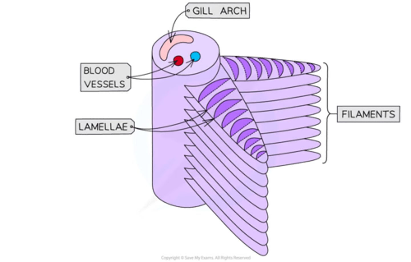
Image showing the structure of fish gills and the counter-current system within gills
what else helps to maintain a concentration gradient across an exchange surface
ventilation mechanism
ventilation (mass flow of gases) in lungs help to ensure that there is....
always a higher conc of o2 in the alveoli than in the blood
the movement involved in breathing causes....
what it does to the air
the air in the alveoli to change.
Breathing removes air with low amounts of oxygen and high amounts of carbon dioxide and replaces it with air that has high amounts of oxygen and low amounts of carbon dioxide
spec point
the structures and functions of the components of the mammalian gaseous exchange system
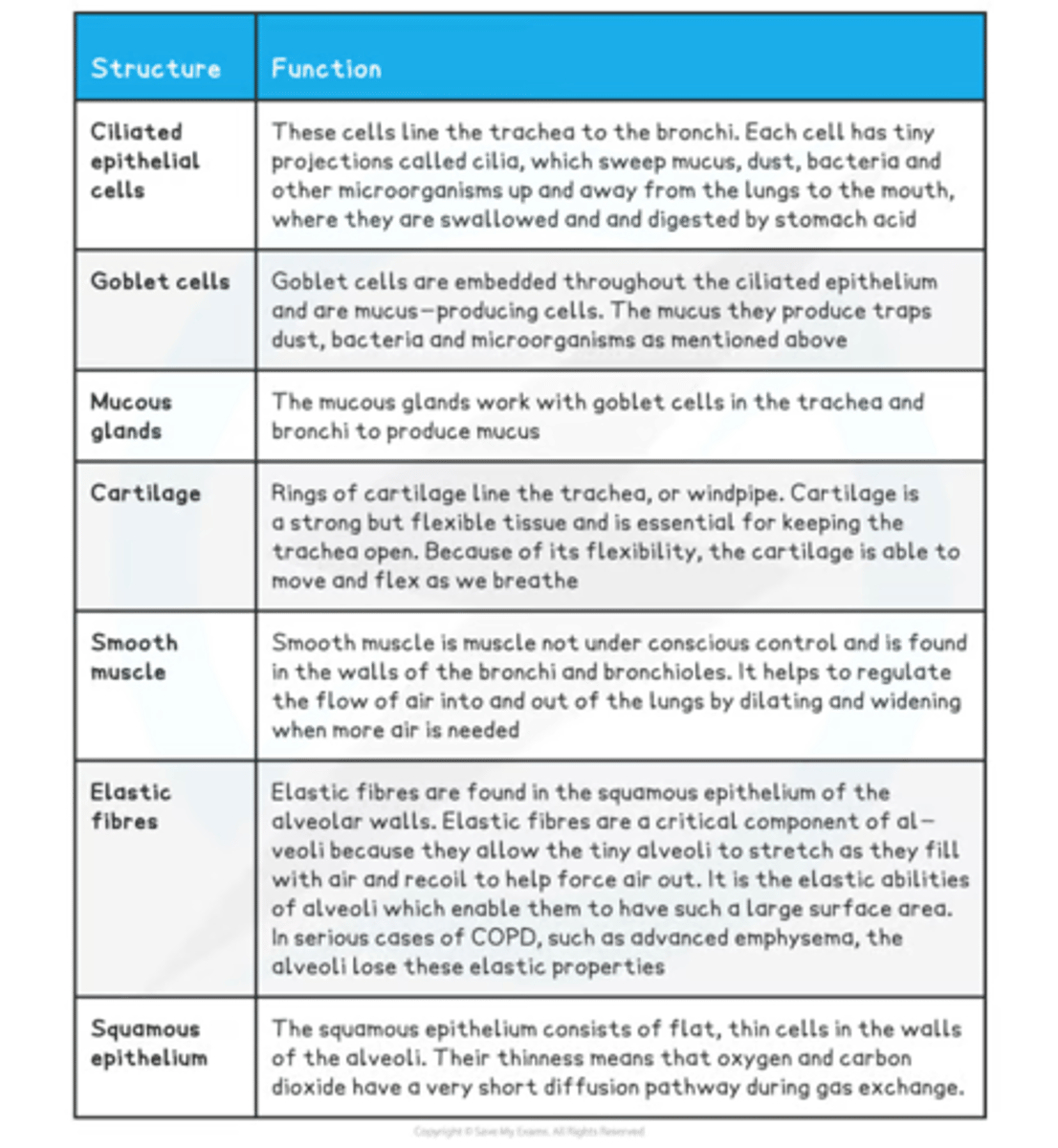
To include the distribution and functions of cartilage, ciliated epithelium, goblet cells, smooth muscle and elastic fibres in the trachea, bronchi, bronchioles and alveoli.
-what 2 cells and one type of gland that plays vital role in maintaining health of gas exchange system
-what are the tissues of the gas exchange system (play a role in it)
-
-
-
-
-ciliated epithelial cells
-goblet cells
-mucous glands
-cartilage
-smooth muscle
-elastic fibres
-squamous epithelial tissue
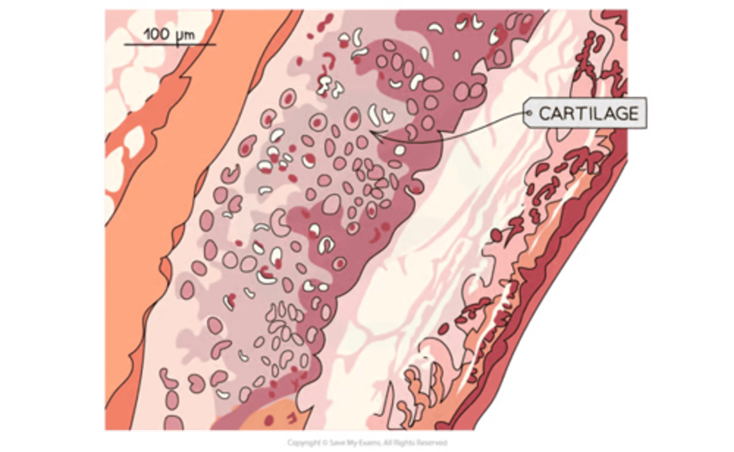
-what is cartilage
-one place it is found
-how it helps there
strong + flexible tissue
-in the rings along the trachea (tracheal rings)
-support the trachea + ensure it stays open while allowing it to move + flex while we breathe
-ciliated epithelium is ______ _____
-found (where)
-each cell has.......
-specialised tissue
-along the trachea down to the bronchi
-each cell has small projections of cilia which sweep mucus, dust + bacteria upwards, away from lungs
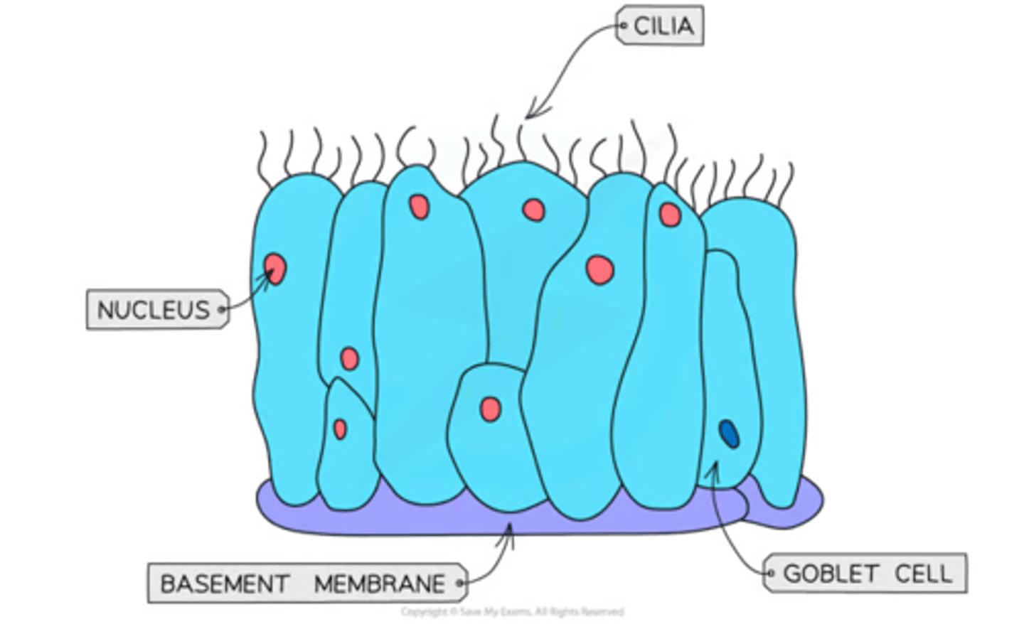
goblet cells can be found....
they are _______-_______ cells that.....(role)
(explanation of role/what happens, movement of mucus)
-scattered throughout the ciliated epithelium in the trachea
-mucus-producing cells secrete viscous mucus = traps dust, bacteria + other microorganisms + prevent them from reaching lungs
-mucus then swept along cilia of ciliated epithelium upwards + is swallowed
(then destroyed by HCL)
where is the squamous epithelium found
what does it form.... name a sig. characteristic of it
-in alveoli as a thin lining = gas exchange
-forms the structure of the alveolar wall + so is very thin + permeable for the easy diffusion of gases
smooth muscle can be found...
it helps do what......
-throughout the walls of the bronchi and bronchioles
-regulate flow of air into the lungs by dilating when more air is needed and constricting when less is needed
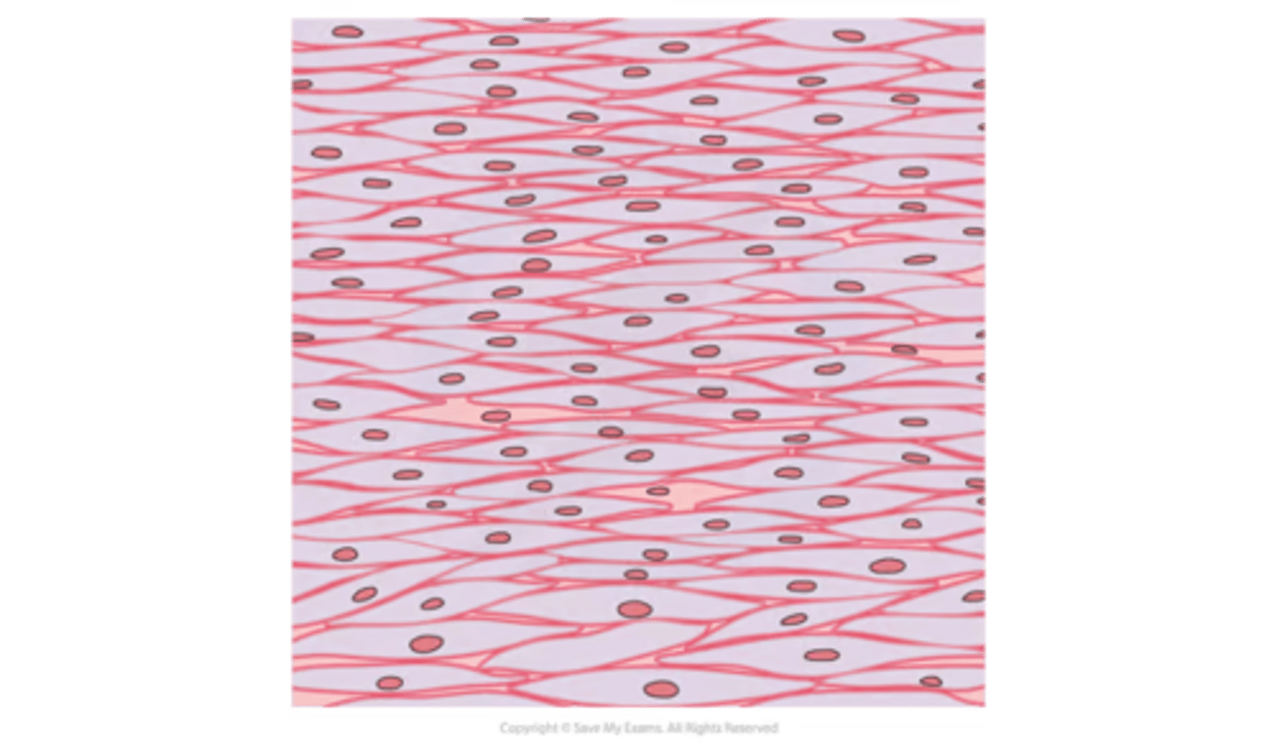
elastic fibres are present in....
why are they so important
how do they effect expiration
all lung tissue
-enable lung to stretch + recoil
-the ability to recoil is what makes expiration a passive process
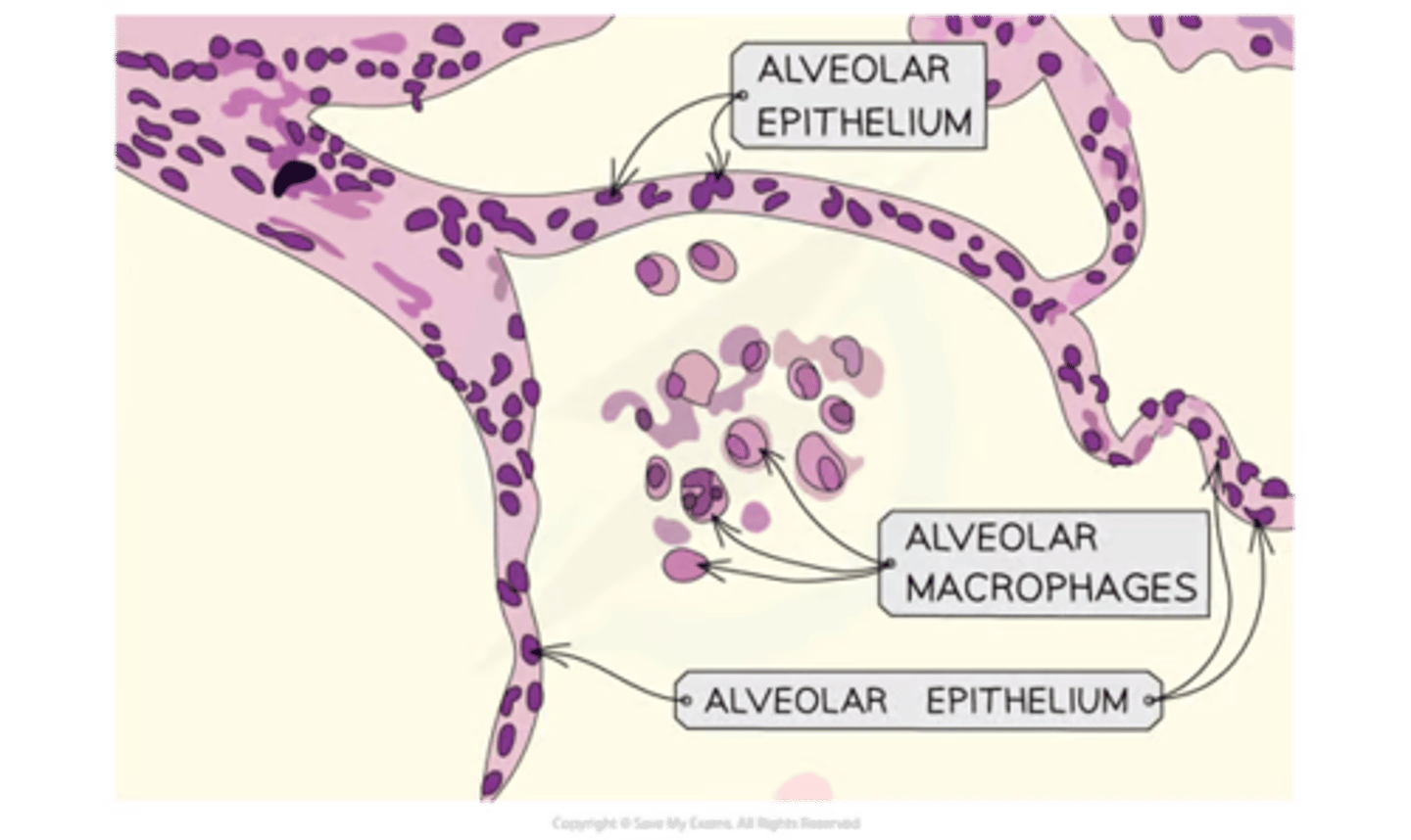
each alveolus is surrounded by an extensive network of capillaries
-co2 moves
-o2 goes
-diameter of capps
-this ensures.....
co2 out of capps into alveoli to be exhaled
o2 from alveoli into capps = carried around body
3-4 um - only wide enough for 1 rbc to travel at one time
-ensures sufficient time for gas exchange
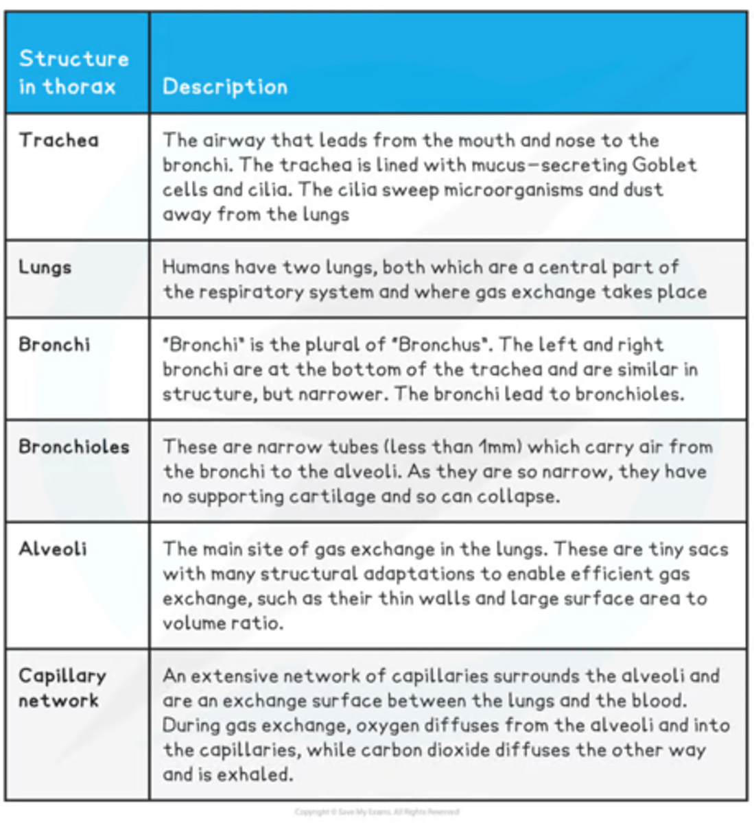
summary table of structures + function in gas exchange system
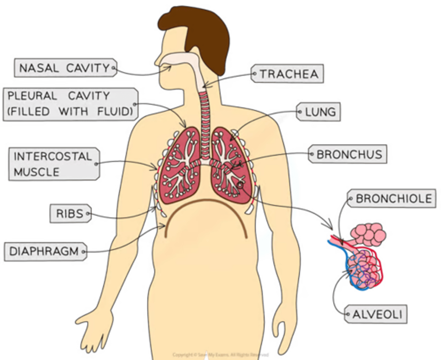
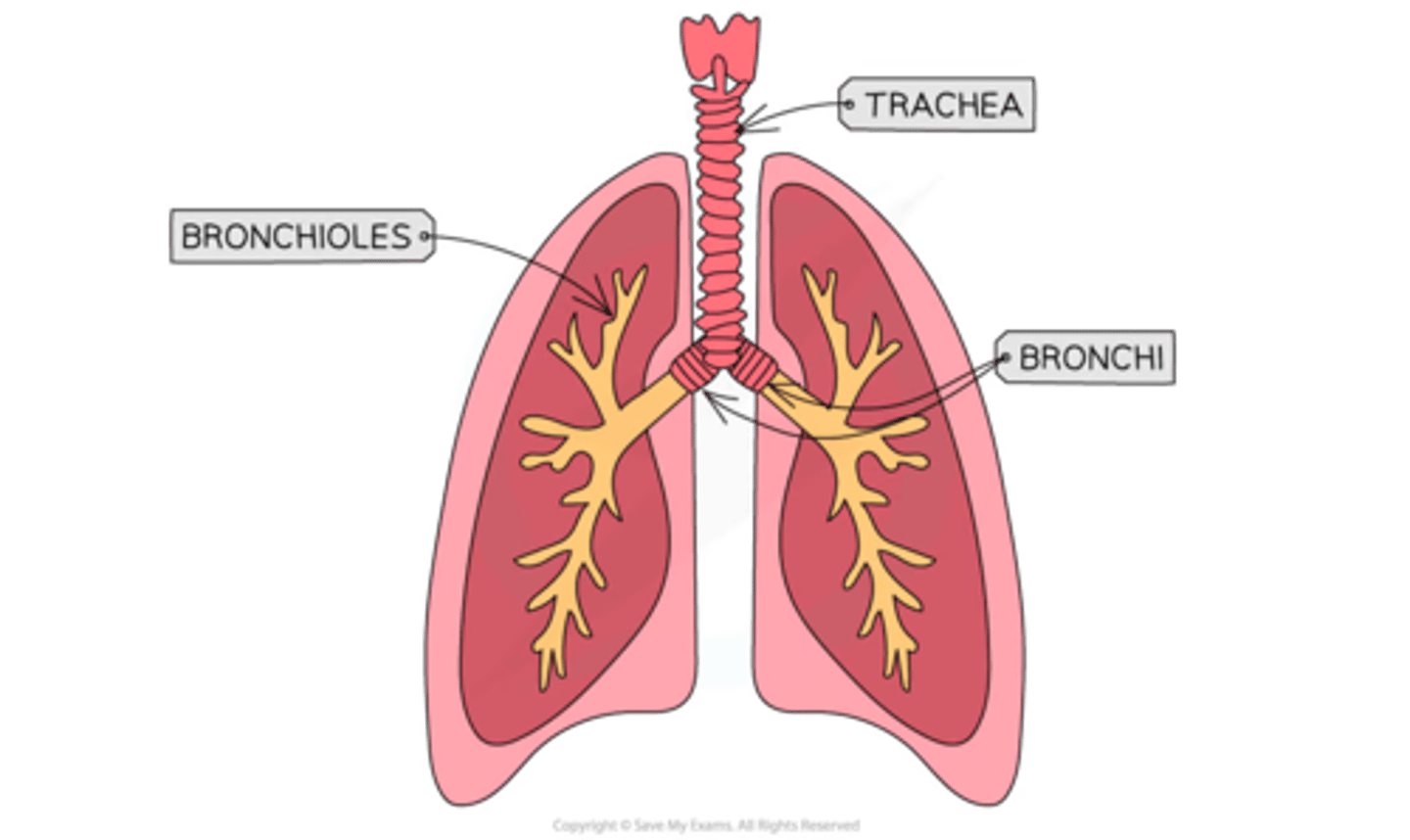
name all the components of the gas exchange system
+ where are all of them located
trachea
bronchi
bronchioles
alveoli
capillary network
thorax- they're thorax's structures
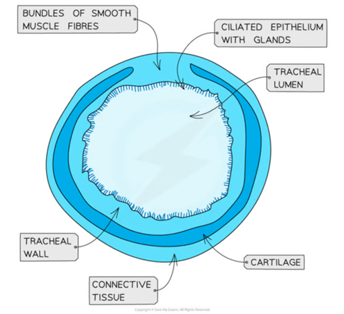
-the trachea is....
-_________ ensures air channel remain open
-why are they that shape
-what is it lined with
-what is inside the trachea + what it helps w/
-the wall of trachea contains...
-channel that allows air to travel to lungs
-C-shaped rings of cartilage
C-shaped bc prevents any friction from rubbing with oesophagus (close behind)
-lined w/ ciliated epithelium
-substantial covering of mucus inside the trachea (by goblet cells + mucous glands)to trap dust/bacteria + prevent entry into lungs
-wall contains smooth muscle + elastic fibres
bronchi structure compared to trachea
-similar but thinner walls + smaller diameter
-not c-shaped cartilage but can form full rings + irregular blocks
bronchioles
-what are they....
-do they have cartilage
-lined w/... yet not containing.....
-size / relevant size + location link
-larger bronchioles possess...
-smallest bronchioles do not have...
-narrow self-supporting tubes w/ thin walls
-usually no cartilage
-lined w/ ciliated epithelium
-don't have any goblet cells
-vary in size, smaller closer to alveoli
-larger ones possess elastic fibres + smooth muscle adjusting size of airway
-smallest ones don't have smooth muscle but do have elastic fibres
-groups of alveoli are located
-alveolar wall consists of a...
-sig. thing they have + location
-fluid
ends of bronchioles
single layer of epithelium
elastic fibres in the extracellular matrix
-capp network
-surfactant-watery fluid facilitating diffusion of gases
summary table of thorax structures + descriptions
spec point-
the mechanism of ventilation in mammals
to include the function of the rib cage, intercostal muscles (internal and external) and diaphragm
what 2 things ensures that there is always a higher concentration of oxygen in the alveoli than in the blood
ventilation
continuous flow of blood in capps
passage of air through
1.
2.
3.
4.
5.
1. nose/mouth
2. trachea (windpipe)
3. bronchi
4. bronchioles
5. alveoli
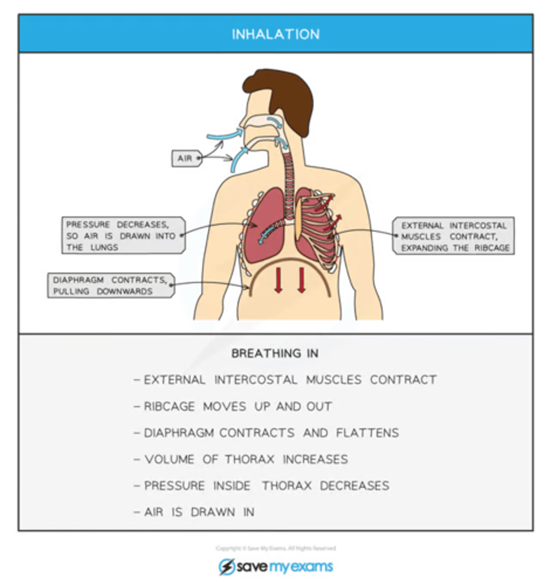
the breathing-in process causes...
as a result....
the volume in chest to increase and the air pressure in the lungs to decrease
as a result air moves down the pressure gradient and rushes into the lungs
mechanism when at rest for breathing-in
diaphragm contracts and flattens, increasing chest volume
mechanism for breathing-in when exercising
additionally to flattening of diaphragm the external intercostal muscles contract, causing the ribcage to move upwards and downwards
The process of breathing in (inhalation)
spec point -
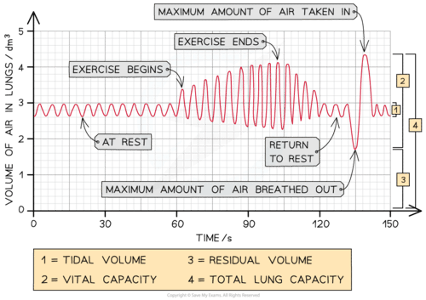
the relationship between vital capacity, tidal volume, breathing rate and oxygen uptake
to include analysis and interpretation of primary and secondary data e.g. from a data logger or spirometer.
what are the four main ways breathing cat)n be scientifically measured (what are each)-
-
-
-
-
vital capacity - the maximum volume of air that can be breathed in or out in one breath
tidal volume- the volume of air that is breathed in or out during normal breathing (rest)
breathing rate- no. of breaths taken in 1 minute
oxygen uptake- this is volume of oxygen used up by someone in a given time
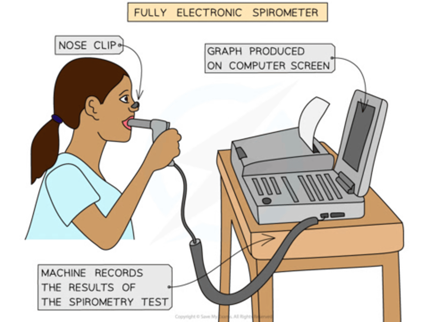
The breathing measurements described above can all be made using a piece of apparatus known as a
spirometer
how does the spirometer work
-The person breathes in/out through the spirometer
-co2 is absorbed from the exhaled air by soda lime to stop the conc. of co2 in the re-breathed air from getting too high, as this can cause respiratory distress
-As subject breathes through the spirometer, a trace is drawn on a rotating drum of paper or a graph is formed digitally, which can be viewed on a computer
-From this trace, the subject's vital capacity, tidal volume and breathing rate can all be calculated
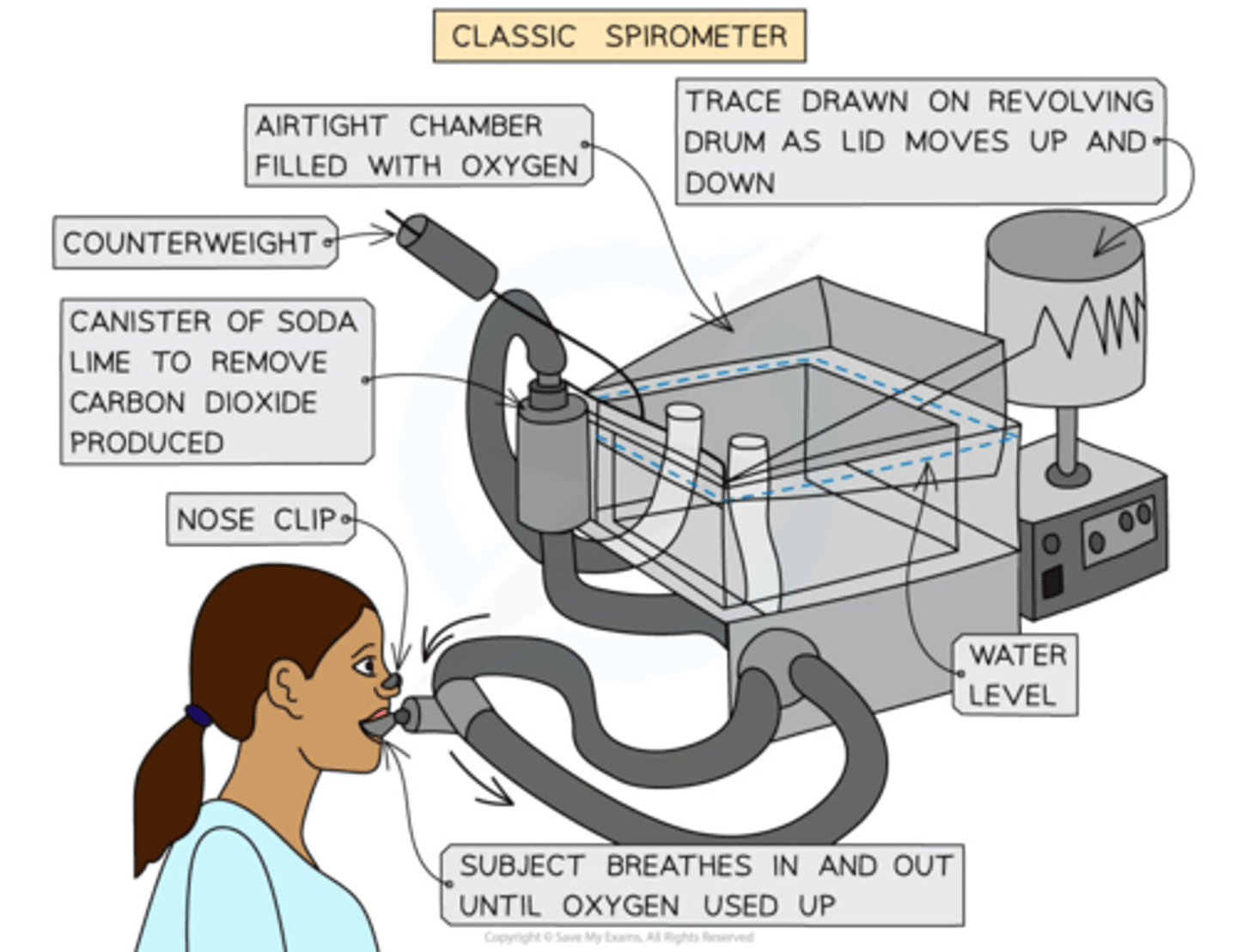
how can oxygen uptake be calculated by using spirometer
co2 is removed from the exhaled air, meaning that the total volume of air available in the spirometer gradually decreases, as oxygen is extracted from it by the subject's breathing
This change in volume is used as a measure of oxygen uptake
A small amount of air, known as the ______ ______ is always retained in the lungs
residual volume
Analysing Data from a Spirometer
The results from a spirometer (either in the form of a trace drawn on graph paper or a digital graph created by a computer) can be used to calculate vital capacity, tidal volume and breathing rate.
The changes in the volume of air present in the lungs are shown here. Note the residual volume, this is the volume of air left in the lungs after as much air has been breathed out as possible.

Worked example
From the spirometer data in the image on this slide calculate
-the breathing rate during the first minute
-then the breathing rate during the second minute.
quite easy, don't overthink it

answer
Step One: Count the number of breaths in the first 60 seconds.
Step Two: Give appropriate units.
Step Three: Count the number of breaths in the second 60 seconds.
Step Four: Give appropriate units.
Step One
One breath is shown by the trace going up and then down, so there are 12 breaths in the first 60 seconds.
Step Two
Breathing rate should be given in breaths min⁻¹ (breaths per minute), so the breathing rate during the first minute = 12 breaths min⁻¹.
Step Three
There are 14 breaths in the second 60 seconds.
Step Four
The breathing rate during the second minute = 14 breaths min⁻¹
Worked example
Calculate the tidal volume during rest and the peak tidal volume during exercise.
Step One: For the 'at rest' phase of the trace, measure the difference between the top and bottom of the trace in terms of the volume of air in the lungs.
Step Two: At the peak tidal volume during exercise, measure the difference between the top and bottom of the trace in terms of the volume of air in the lungs.
Step One
During rest, the tidal volume = 3 dm³ - 2.6 dm³
= 0.4 dm³
Step Two
The peak tidal volume during exercise occurs right at the end of the exercise period (at around 100 seconds):
= 4.1 dm³ - 2.3 dm³
= 1.8 dm³
spec point-
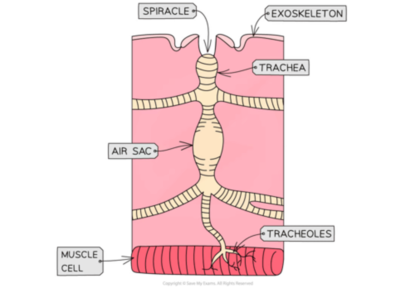
the mechanisms of ventilation and gas exchange in bony fish and insects
To include:
• bony fish – changes in volume of the buccal cavity and the functions of the operculum, gill filaments and gill lamellae (gill plates); countercurrent flow
• insects – spiracles, trachea, thoracic and abdominal movement to change body volume, exchange with tracheal fluid.
all insects possess a .... (means they can't do something ) therefore they have....
rigid exoskeleton with a waxy coating that is impermeable to gases
have evolved a breathing system that delivers o2 directly to all organs and tissue of their bodies
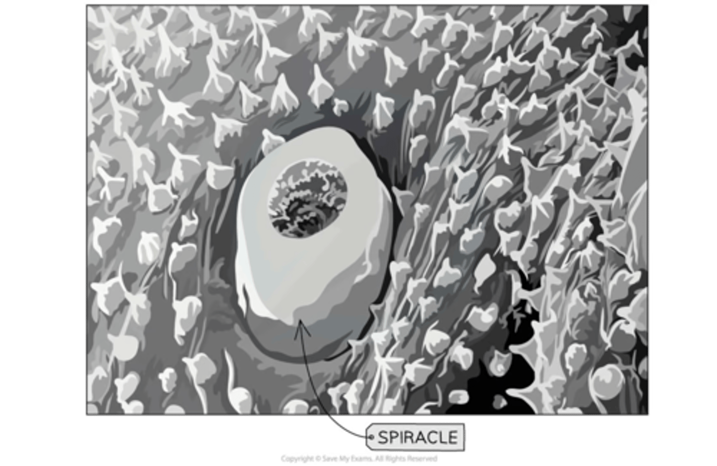
what is one component/element of insects that lead to gas exchange/ventilation + explain it
(s________)
spiracle
an opening in the exoskeleton of an insect which has valves
-allows air to enter insect and flow into system of tracheae
another element of insects that lead to gas exchange/ventilation + explain it
(T______)
tracheae - are tubes within insect respiratory system which lead to tracheoles (narrower tubes)
a large no. of tracheoles run btw cells and into the muscle fibres- site of gas exchange
insect's tracheal system (described above in 3 cards)
-what is the site of gas exchange in it?
tracheoles
very active/flying insects need more rapid supply + intake of o2 so what do they do + how
create....
-
-
-
create a mass flow of air into tracheal system by:
-closing the spiracles
-using abdominal muscles to create a pumping movement for ventilation
-also during flight the fluid at narrow ends of the tracheoles is drawn into repairing muscle so gas diffuses across quicker (due to diffusion distance being shorter)
what are fish adapted to do and why
directly extract o2 from water
as o2 dissolves less readily in water
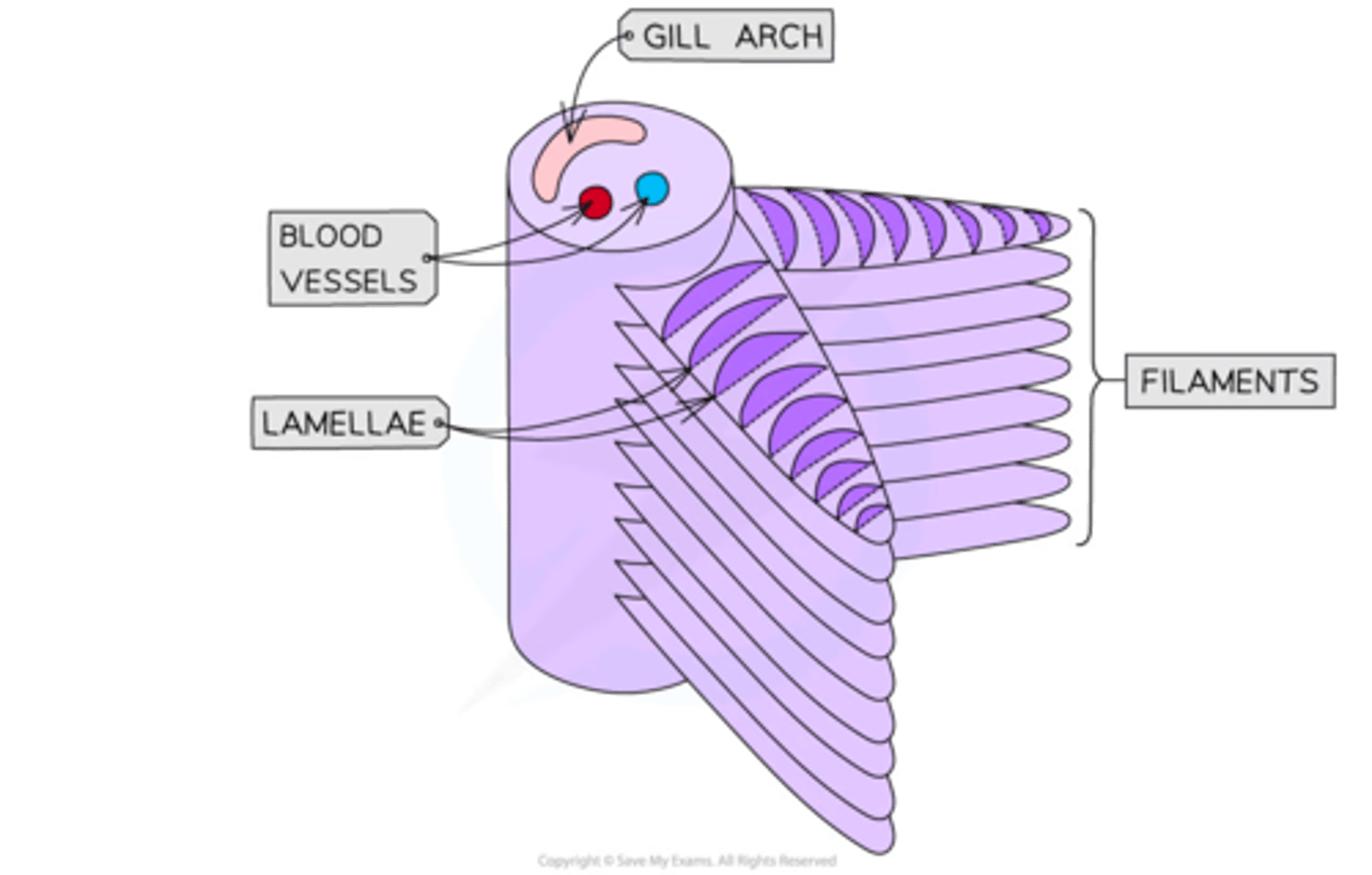
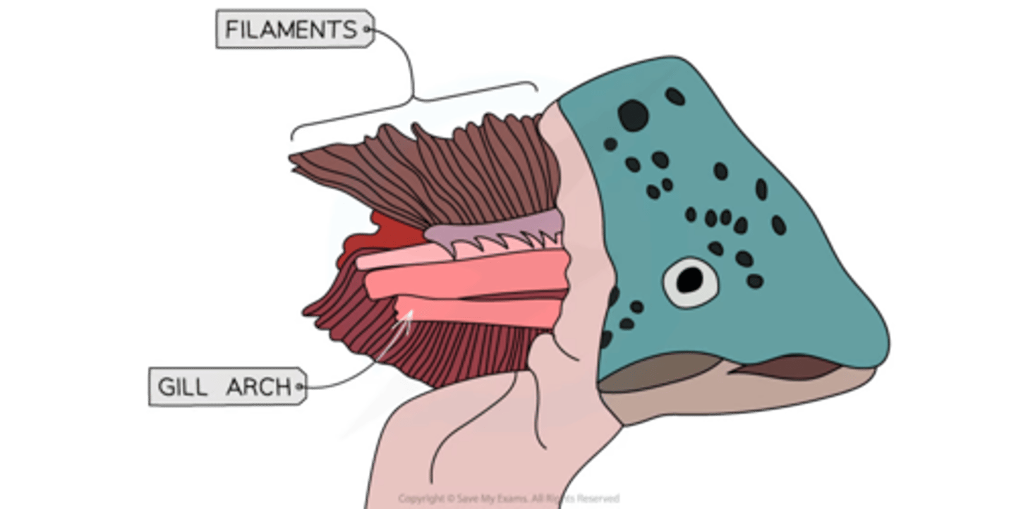
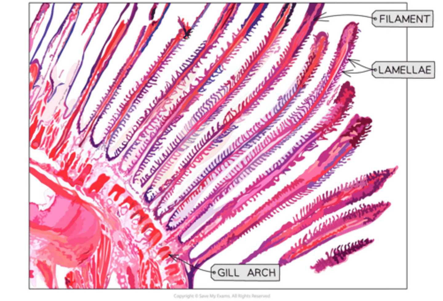
list the structure of fish gills in bony fish:
-
-
-
-
-series of gills each side of head
-each gill arch is attached to 2 stacks of filaments
-on the surface if each filament there are rows of lamellae
-the lamellae surface consists of a single layer of flattened cells that cover a vast network of capillaries
mechanism for gills of fish gas exchange
-
-
-
-the capillary system within the lamellae ensures that the blood flow is in the opposite direction to the flow of water - it is a counter-current system (C.C.S)
-the c.c.s. ensures the conc gradient is maintained along the whole length of the capillary
-the water with the lowest o2 conc is found adjacent to the most deoxygenated blood
Image showing the structure of fish gills and the counter-current system within gills
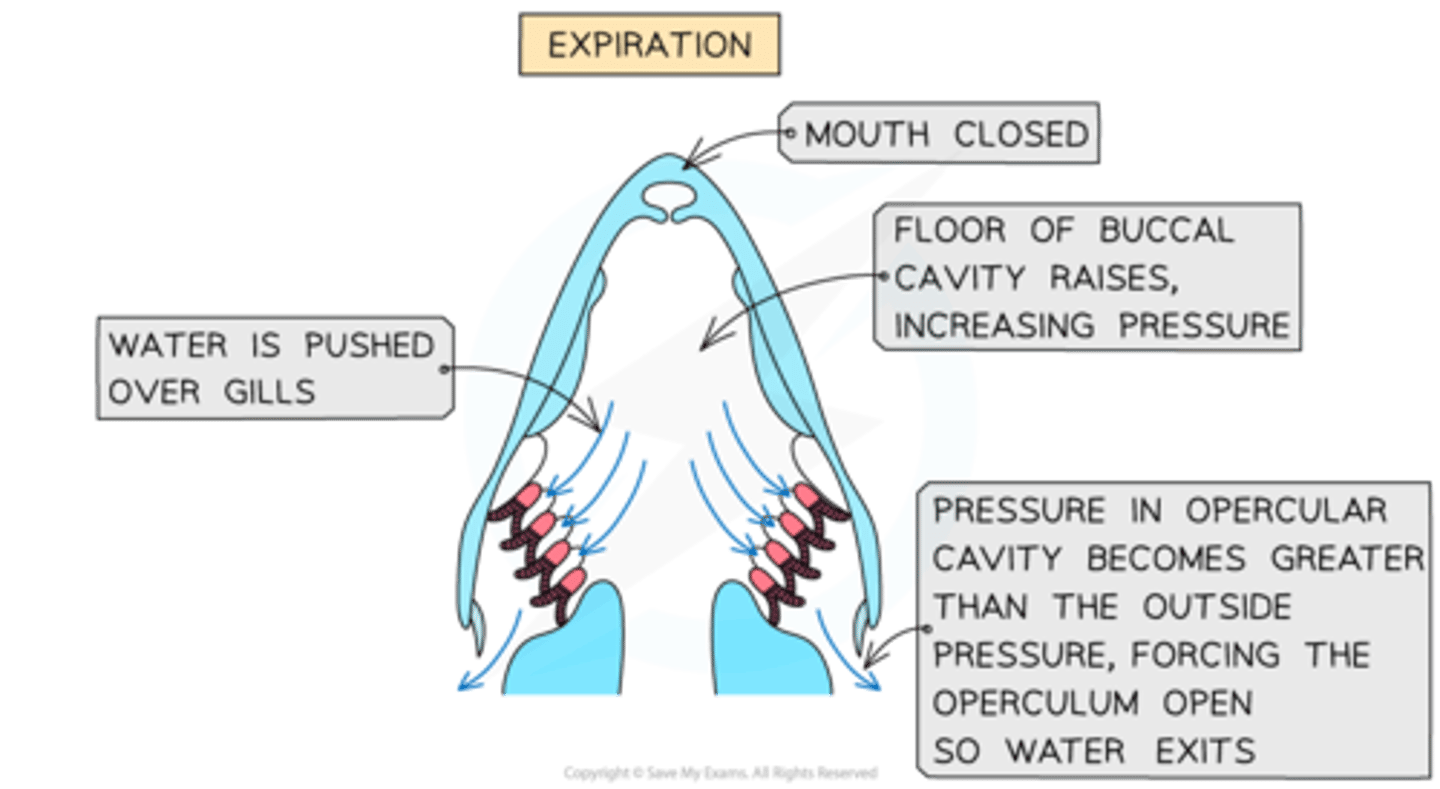
ventilation mechanism in fish
-what does it do......to ensure.......
-pushes water over the surface of gills
-ensure they are constantly supplied with water rich in o2 - maintain conc gradient
ventilation mechanism in fish
**when fish open their mouth they do what....
-this causes
lower the floor of the buccal cavity
-causes a decrease in pressure within cavity
ventilation mechanism in fish
what happens after fish decreases pressure by opening mouth + lowering floor of buccal cavity
-the pressure is higher outside the mouth of fish and so water flows into buccal cavity
ventilation mechanism in fish what follows the
-decrease pressure: buccal + mouth + lower floor
+
-water flowing into buccal by lower pressure
water flows from buccal cavity y(high pressure) into gill cavity (low pressure)
final step of ventilation mechanism in fish
**
as water enters pressure begins to build up in gill cavity + causes the operculum (flap of tissue covering gills) to be forced open + water exit the fish
-the operculum is pulled shit when the floor of buccal cavity is lowered at the star of the next cycle !
The pressure differences created by the opening of the mouth causes water to be constantly pushed across the surface of the gills
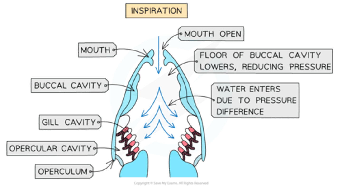
The pressure differences created by the opening of the mouth causes water to be constantly pushed across the surface of the gills
rough method that is holistic for dissection fish/insect gas exchange
think sequence
what you use each tool for
what you must avoid and thus do
-wear lab coat, gloves, glasses to avoid contamination with bio. material
-place specimen on dissecting board
-use scissors/scalpel/forceps (tools) to access desired structure
-scalpel: cut away from body + fingers far to reduce harm chance
-scissors: for cutting large sections of tissue
-scalpel- finer precise cut
-use pins to move other section of specimen aside to leave desired structure exposed
limitations of dissection
-
-
-
-hard to see some of smaller finer structures within organs
-specimens don't reflect how the tissue would look in living organism
-only single specimen dissected then anomalies found within that specimen may be ignored
the key structures that can be seen from dissection of mammalian lungs
-
-
-
trachea
bronchi
bronchioles
the key structures that can be seen from a dissection of fish gills
-
-
gill arch
filaments
what is different about insect tracheal system dissection
small size so specialised equipment and skills are sometimes required
+
microscopes to observe them + stain it
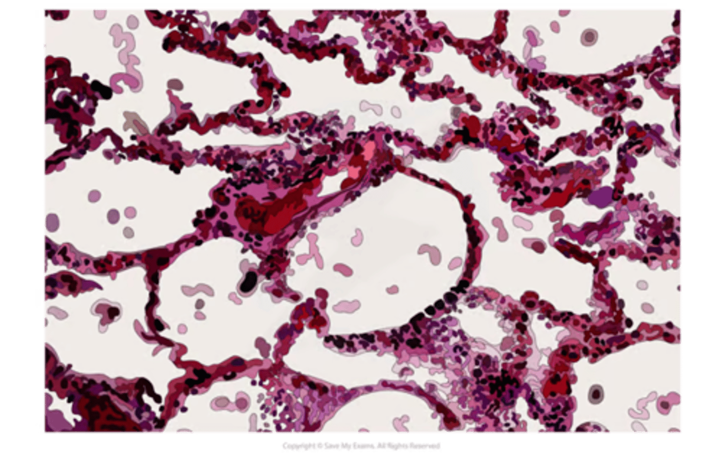
what to expect in lung dissection - mammalian gas exchange (down microscope)
-alveoli are different sizes and shapes bc no longer inflated
-the nuclei are shown as dark dots
-blood vessels can be found btw alveoli
-sometimes wbcs present in tissue
what to expect in fish dissection (microscope)
-gill arch (resembles backbone for gills)
-different filaments are shown w/ many of lamellae visible
what to expect in insect dissection down microscope
-electron microscopes = clear images of spiracle structure found on surface of insects