Anatomy Lab - Bones
1/208
There's no tags or description
Looks like no tags are added yet.
Name | Mastery | Learn | Test | Matching | Spaced | Call with Kai |
|---|
No analytics yet
Send a link to your students to track their progress
209 Terms
two types of osseous tissue
compact bone and spongy bone
types of bone classification
long bones, short bones, flat bones, irregular bones
long bones (classification)
cylindrical; shaft with two ends (femur, phalanges)
short bones (classification)
cube shaped (tarsals, carpals)
flat bones (classification)
thin and curved (some skull, scapula, ribs)
irregular bones (classification)
complex shape (vertebra, facial bones)
spongy bone
A
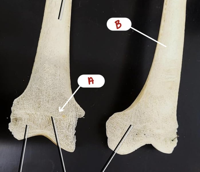
compact bone
B

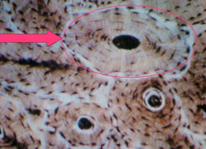
osteocyte in lacuna
(name the structure indicated by the pointer)
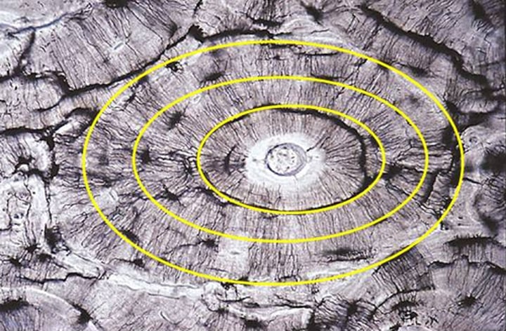
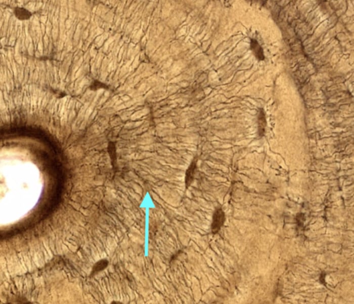
lamella
(what is indicated by the lines?)
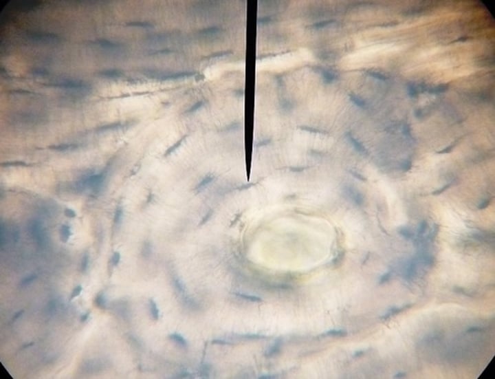
central canal (bone slide)
(dark spot in the middle)
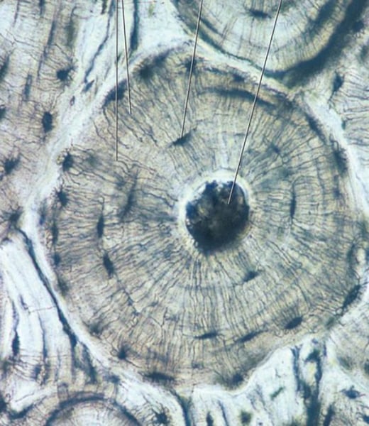
osteon
(whole structure)
canaliculi
striations between lamella
bone formation zones (5 total)
resting zone, proliferation zone, hypertrophic zone, calcification zone, ossification zone
resting zone
A

proliferation zone
B

hypertrophic zone
C

calcification zone
D

ossification zone
E
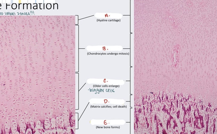
what happens in the proliferation zone?
chondrocytes undergo mitosis
what happens in the hypertrophic zone?
older cells enlarge
what happens in the ossification zone?
new bone forms
what is found in the resting zone?
hyaline cartilage
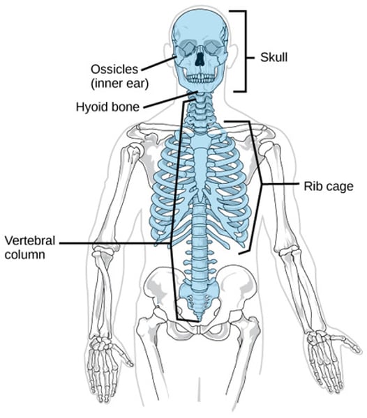
axial skeleton
includes skull, vertebral column, and thoracic cage
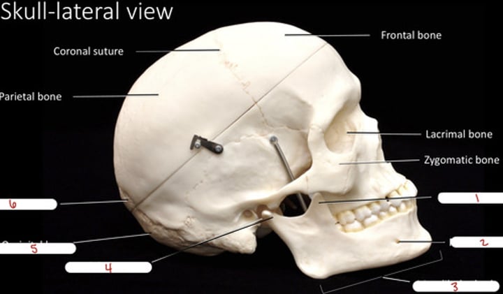
coronal suture
1

parietal bone
2

temporal bone
3

squamous suture
4

external acoustic meatus
5/6

mastoid process
7

frontal bone
8

sphenoid bone
9

nasal bone
10

lacrimal bone
11

zygomatic bone
12

maxilla
13
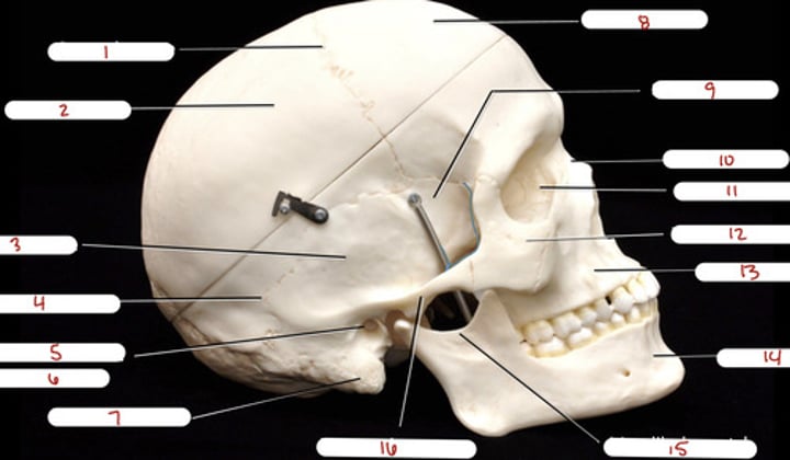
mandible
14

mandibular notch
15

zygomatic process
16

coronoid process
1

mental foramen
2
mandibular body
3

condylar process
4

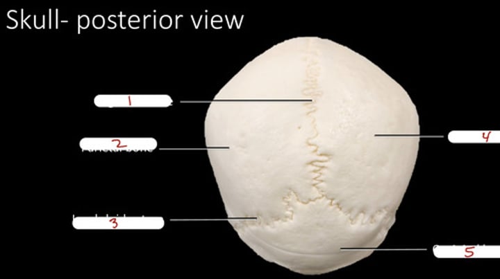
occipital bone
5

lambdoid suture
6

sagittal suture
1

parietal bone (posterior view)
2

lambdoid suture (posterior view)
3

parietal bone
4

occipital bone (posterior view)
5
nasal bone (anterior view)
1

superior orbital fissure
2

zygomatic bone (anterior view)
3

inferior nasal concha
4

maxilla (anterior view)
5

frontal bone (anterior view)
6

supraorbital foramen
7

sphenoid bone (anterior view)
8

middle nasal concha
9

infraorbital foramen
10
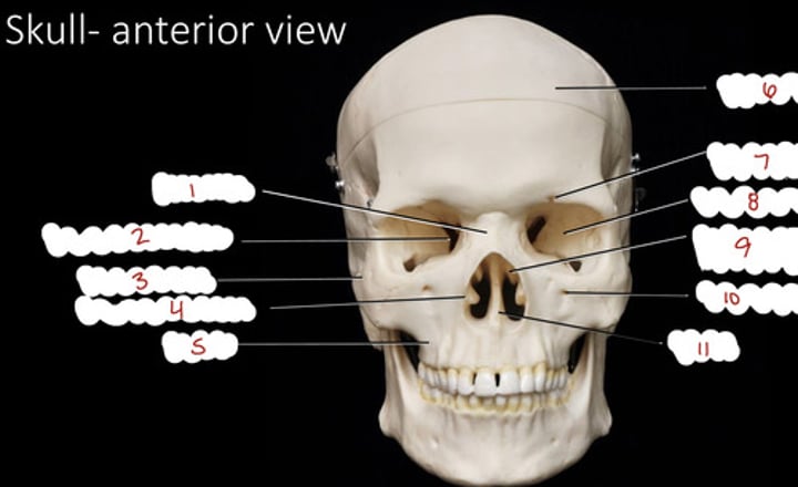
vomer
11

ethmoid bone (crista galli)
1

sphenoid (lesser wing)
2

foramen ovale
3
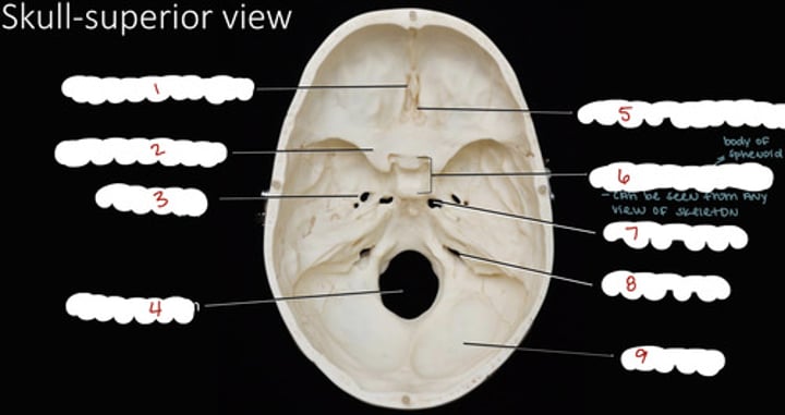
foramen magnum
4

ethmoid bone (cribriform plate)
5

sphenoid - sella turcica
6

foramen lacerum
7

jugular foramen
8

occipital bone (superior view)
9

sphenoid - greater wing
1
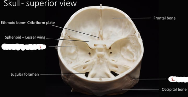
lamboid suture (superior view)
2

incisive fossa
1

median palatine suture
2

vomer (inferior view)
3

styloid process (skull)
4

foramen magnum (inferior view)
5

occipital bone (inferior view)
6

occipital condyle
7

carotid canal
8

foramen lacerum (inferior view)
9
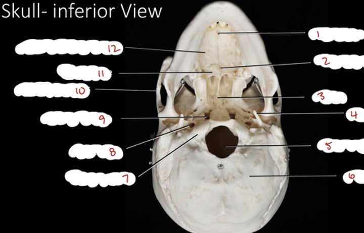
zygomatic process (inferior view)
10

palatine bone
11

maxilla - palatine process
12

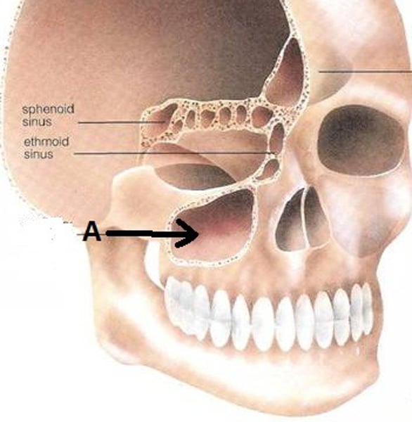
frontal sinus
cavity within frontal bone
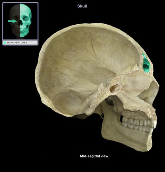
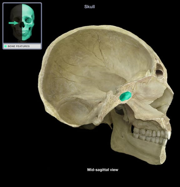
sphenoid sinus
cavity within sphenoid bone
maxillary sinus
cavity within maxilla
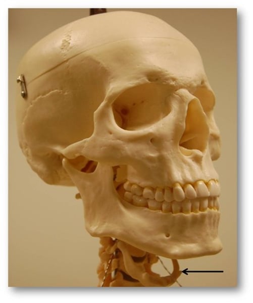
hyoid bone
(held together with tendons and ligaments)
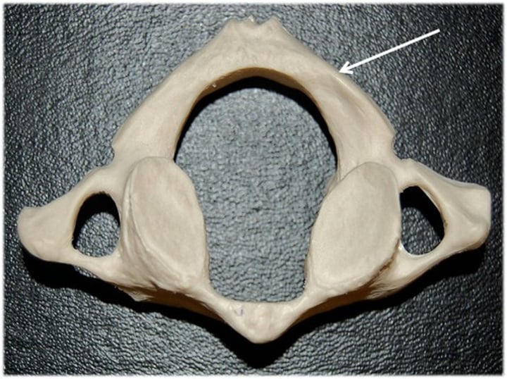
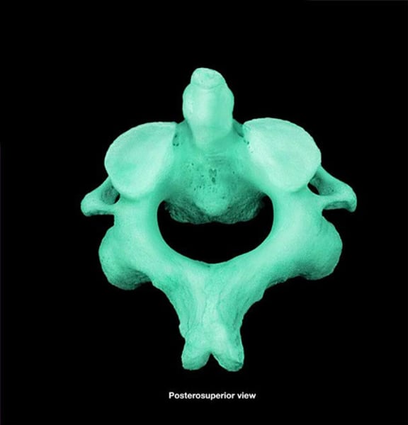
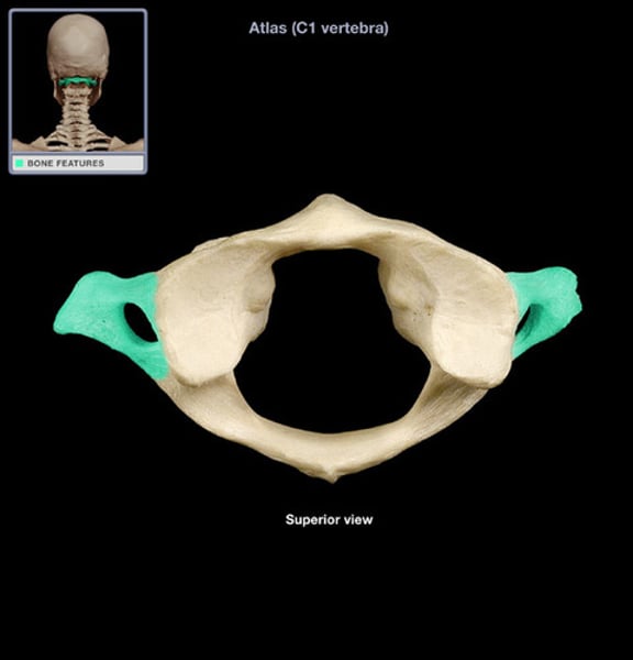
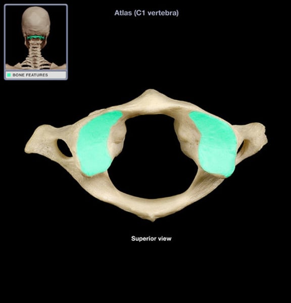
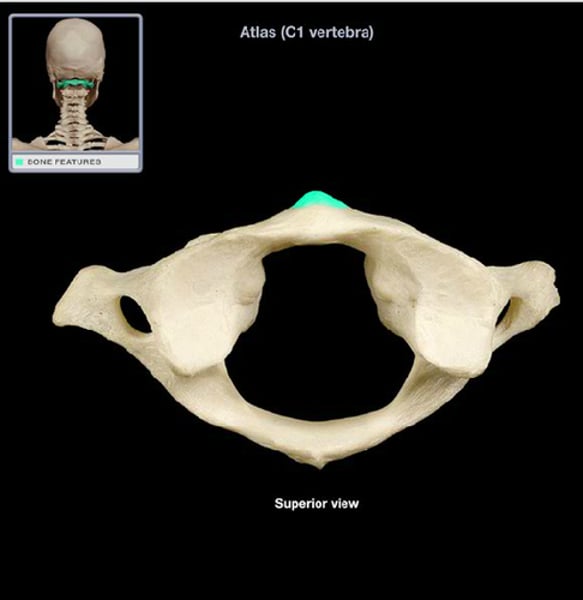
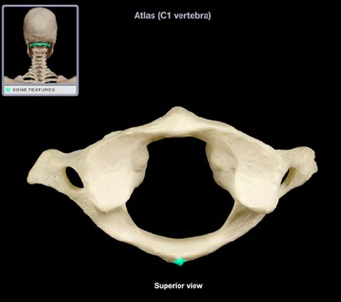
atlas
(C1; top vertebra)
axis
(C2; under atlas)
atlas transverse foramen
(highlighted area)
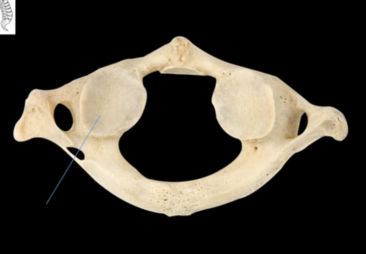
atlas superior articular facet
(highlighted area)
atlas anterior tubercle
(highlighted area)
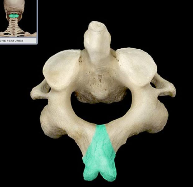
atlas posterior tubercle
(highlighted area)
atlas inferior articular facet
(where arrow is)
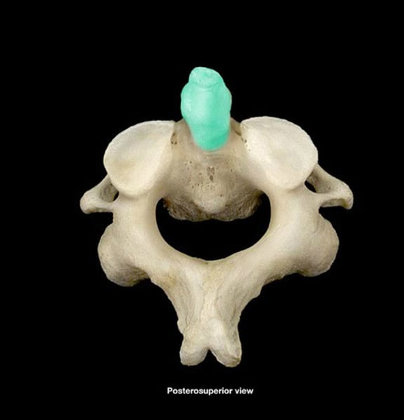
axis dens (odontoid process)
(highlighted area)
axis spinous process
(highlighted area)
cervical vertebrae (C1-C7)
(has 3 foramen)
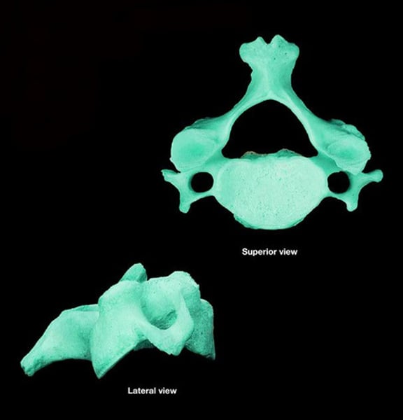
spinous process (cervical vertebra)
1
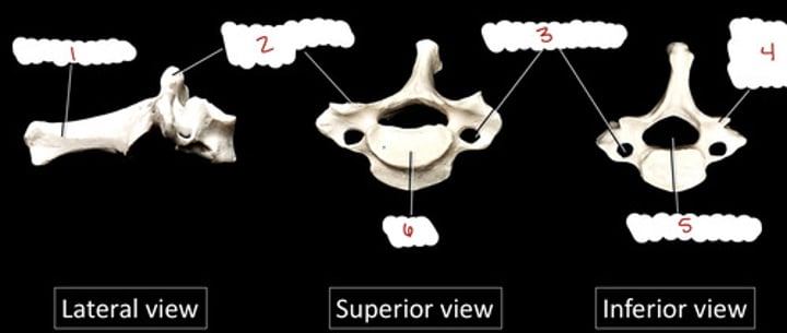
superior articular process (cervical)
2
