midertnm study (real class)
1/55
Earn XP
Description and Tags
MMMMMADFS
Name | Mastery | Learn | Test | Matching | Spaced | Call with Kai |
|---|
No analytics yet
Send a link to your students to track their progress
56 Terms
Golgi Apparatus
Cell Organelle: Manipulates products from the rough Endoplasmic Reticulum (the one with ribosomes on it), and produces new organelles called lysosomes
Rough Endoplasmic Reticulum
Cell Organelle: Studded with ribosomes. The ribosomes function is making proteins.
Smooth Endoplasmic Reticulum
Cell Organelle: Does not contain ribosomes, and functions in making lipids.
Cytoplasm
Cell Organelle: The fluid inside the cell. Contains a network of channels and support structures called the cytoskeleton.
Centrosome
Cell Organelle: Important in producing a structure called the mitotic spindle that helps to separate the chromosomes during mitosis. Consists of 2 hollow cylinders called centrioles, constructed from tubular proteins
Cilia
Important in cell movements. Moves substances across cells. Cells can have many.
Flagellum
Long protein structure that moves the cell. Cells can only have one.
Mitochondrion
Cell Organelle: Composed of two separate bilayer membranes. Along the inner membrane are various molecules that work together to produce ATP, the cell's major energy currency.
Peroxisome
Cell Organelle: Membrane-bound organelle that contains an abundance of enzymes for detoxifying harmful substances and lipid metabolism. Differs from lysosomes.
Nucleus
Cell Organelle: Control center of the cell; contains genetic material that determines the entire structure and function of the cell.
Interphase
Mitosis: prepatory phase consisting of a G1, S, and G2 stage.
G1: cell produces copies of it organelles, centrioles replicate (lasts 8-12 hrs)
S: cell replicates DNA and ends up with 2 sets of identical chromosomes (lasts 6-8 hrs)
G2: Centrioles complete replication and protein synthesis (lasts 2-5 hrs)
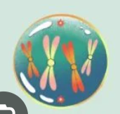
Prophase
Mitosis: Genetic material of the cell forms tightly coiled chromosomes, and the nuclear membrane begins to break up. Microtubules form the mitotic spindle at opposite ends of the cell.
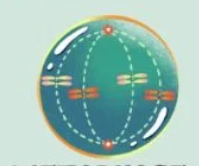
Metaphase
Mitosis: Chromosomes line up at the center of the cell, the area called the metaphase plate.
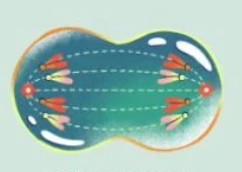
Anaphase
Mitosis: Spindle fibers shorten and centromeres divide, separating the pair of chromosomes. The chromosomes move to opposite sides of the cell.
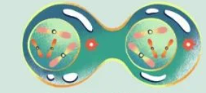
Telophase
Mitosis: The final stage, the nuclear membrane and nucleolus begin to reappear, and the mitotic spindle breaks up and the chromosomes uncoil. Two daughter cells are present.
Interphase Prophase Metaphase Anaphase Telophase
Type the order of mitosis stages (no commas, only spaces)
Facilitated diffusion
When non-lipid-soluble substances get in and out of the cell via a protein channel
Active transport
When substances are moved against their concentration gradients by carrier proteins. Costs ATP.
Osmosis
When water moves through a semipermeable membrane, from a higher water concentration from a lower water concentration.
Facilitated diffusion
When non-lipid-soluble substances get in and out of the cell via a protein channel
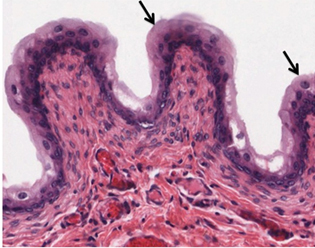
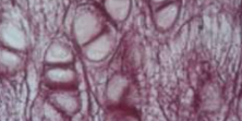
Epidermis
Layers of the Integument: Epithelium composed of multiple layers of cells. The basal layer consists of cuboidal cells, whereas the outer layers are squamous (flat, scale-like), keratinized cells, so the whole epithelium is often described as being keratinized stratified squamous epithelium.
Dermis
Layers of the Integument: Made of dense, irregular connective tissue that houses blood vessels, hair follicles, sweat glands and other structures.
Hypodermis
Composed mainly of loose connective and fatty tissues, located under the dermis.
First-Degree Burn
Superficial burn that affects only the epidermis. They typically heal on their own within a few days. Ex. mild sunburns
Second-Degree Burn
Burn that goes deeper and affects the epidermis and a portion of the dermis. Results in swelling and painful blistering of the skin.
Third-Degree Burn
Burn that fully extends into the epidermis and dermis, destroying the tissue and affecting the nerve endings and sensory function. These are serious burns that appear white, red, or black; they require medical attention and will heal slowly without it.
Fourth-Degree Burn
Burn affecting the underlying muscle and bone.
Transitional Epithelium
Epithelium: Special case found in the urinary bladder. Looks like stratified squamous epithelium, but instead of cells being rounder in deeper layers and flattened at the top, cells are still rounder in the deeper layers, but also are rounded at the superficial layers.
Pseudostratified columnar epithelium
Epithelium: Special case that looks stratified (more than one layer), but isnt. Cells are found at various levels, but there is still only one layer.
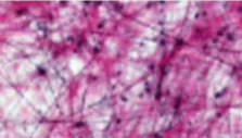
Areolar connective tissue
Connective tissue: part of the loose connective category; contains fibroblasts, collagen, elastic and reticular fibers. Lines organs, surrounds muscle fibers, blood, and lymphatic vessels.
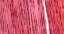
Dense connective tissue
Connective tissue: Contains fibroblasts and collagen fibers. Ligaments and tendons have this tissue where the fibers run parallel along the lines of force.
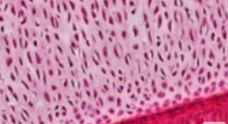
Hyaline Cartilage
Cartilage: The most abundant cartilage in the body. Contains collagen fibers distributed throughout the matrix along with chondrocytes in lacunae. The matrix is smooth. Found at the ends of bones, in the nose, ribs, and serves as a template for bone formation.
Fibrocartilage
Cartilage: Contains chondrocytes in lacunae with thick collagen fibers distributed throughout the matrix. Found in intervertebral disks, pubis symphysis, menisci of the knee and in the small disks in the temporomandibular joints.
Elastic Cartilage
Contains chondrocytes in a matrix containing elastic and collagen fibers. Found in the ears, epiglottis, and eustachian tubes.
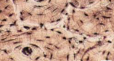
Bone
Connective tissue: Contains osteocytes in lacunae suspended in a hydroxyapatite matrix. The most rigid and strongest connective tissue. Contains good blood supply which promotes healing
Hyaline Cartilage
Cartilage: The most abundant cartilage in the body. Contains collagen fibers distributed throughout the matrix along with chondrocytes in lacunae. The matrix is smooth. Found at the ends of bones, in the nose, ribs, and serves as a template for bone formation.
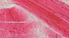
Skeletal Muscle
Muscle: Attaches to bones by way of ligaments and contracts to produce movement. Can also produce heat. Consists of long cells (myocytes) that give it a red color. Contains lines called striations produced by the overlapping contractile protein filaments. It’s cells contain multiple nuclei.
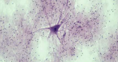
Nervous Tissue
Tissue: Consists of nerve cells called neurons and supportive cells called neuroglia. Processes and carries info throughout the nervous system.
Scar
Collagen-rich skin formed after the process of wound healing that differs from normal skin. Occurs in cases in which there is repair of skin damage, but the skin fails to regenerate the original skin structure.
Keloid
Raised or hypertrophic scar, occurs from an overproduction of scar tissue because the process of collagen formation does not stop when the wound is healed.
Callus
Basal stem cells in the stratum basale are triggered to divide more often to increase the thickness of the skin at the point of abrasion to protect the rest of the body from further damage.
Eccrine Sweat Gland
Gland that produces a hypotonic sweat for thermoregulation, found all over the skin’s surface, but especially abundant on the palms of the hand, the soles of the feet, and the forehead. Coils from the dermis to the surface.
Apocrine Sweat Gland
Sweat gland associated with hair follicles in densely hairy areas, such as armpits and genital regions. Larger than eccrine sweat glands and lie deeper in the dermis, sometimes reaching into the hypodermis. In addition to water and salts, includes organic compounds that make the sweat thicker and subject to bacterial decomposition.
Nail
An accessory structure of the integumentary system. Contains keratinized cells that are pushed from the root to the distal portions.
Glycolysis
During the energy consuming phase, 2 ATP is consumed, transferring 2 phosphates to the glucose molecule. The glucose molecule splits into 2 3-carbon compounds, each with a phosphate. During the second phase, an additional phosphate is added to each of the 3-carbon compounds. During the energy-releasing phase, the phosphates are removed from both 3-carbon compounds, and used to produce 4 ATP molecules
2
How many ATP is consumed during the energy consuming phase of Glycolysis?
4
How much ATP is made from the energy-releasing phase of glycolysis?
2
What is the net gain of ATP from glycolysis? (Per glucose molecule)
Krebs Cycle
Each pyruvate that is generated by glycolysis is converted into a 2-carbon acetyl-CoA molecule. Acetyl CoA is systematically processed through the cycle, producing high-energy NADH, FADH2, and ATP molecules.
3
How many NADH is made from the Krebs Cycle?
1
How many FADH2 is made from the Krebs Cycle?
1
How much ATP does the Krebs Cycle produce (Per turn/Acetyl Coenzyme A)
Electron Transport Chain
A series of electron carriers and ion pumps that are used to pump H+ ions out of the inner mitochondrial matrix to power a protein to phosphorylate ADP.
Phosphocreatine system
The body's fastest, anaerobic energy pathway, providing immediate fuel for high-intensity, short-duration activities (0–15 seconds). It functions by breaking down _______________ to rapidly regenerate adenosine triphosphate (ATP), the cellular currency for muscle contraction, allowing for maximum power output.
What enters the Krebs Cycle?
Acetyl Coenzyme A