NEURO2020 WEEK 5
1/32
There's no tags or description
Looks like no tags are added yet.
Name | Mastery | Learn | Test | Matching | Spaced | Call with Kai |
|---|
No analytics yet
Send a link to your students to track their progress
33 Terms
LIGHT
Electromagnetic radiation
Wavelength = colour
Amplitude = brightness
Humans respond to approx. 400-700nm
EYES
Light enters through pupil
Refracted by cornea
Fine-tuned by lens
Image lands at retina
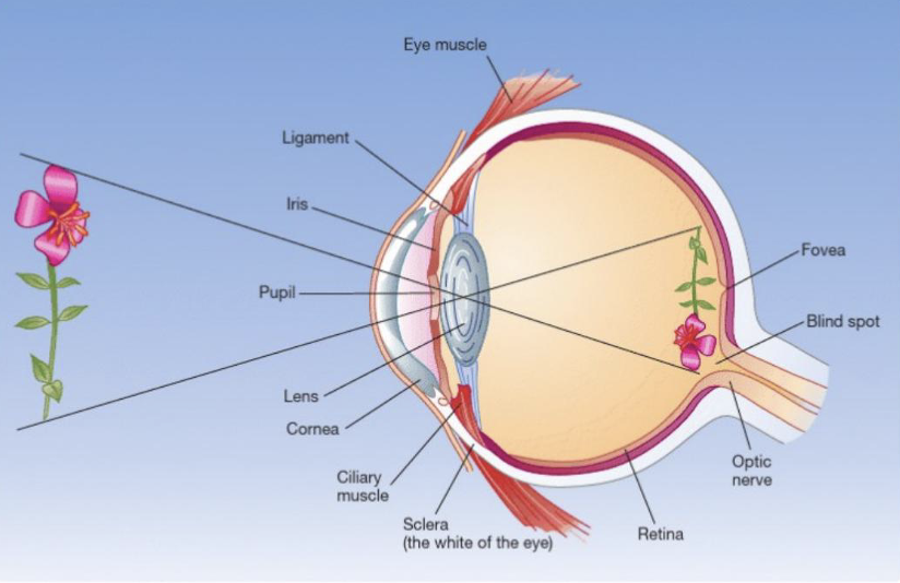
FOVEA
small central depression where fine-detail vision is sharpest
Layers are cleared aside for maximum acuity

EYE PLACEMENT
Laterally placed eyes
Usually prey
Wider view
Detect predator sneaking up
Frontal face eyes
Binocular disparity
Can break camouflage
Usually predator
Cyclopean eye
Combining both images
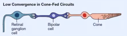
CONES
densely packed in the fovea
Short, medium, long wavelength cones
High visual acuity
Not sensitive to dim lights
Colour vision
Low convergence
One/two cones connected to the retinal ganglion cell
Precise spatial information
Photopic system
Peak sensitivity of 550nm
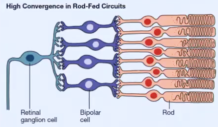
RODS
Non-existent in fovea
Many in periphery
Achromatic
Very sensitive to dim lights
Lower acuity
High convergence
Many rods connected to retinal ganglion cell
Vague spatial information
Scotopic system
Peak sensitivity of 500nm
BLIND SPOT
Area with no photoreceptors
Occurs where optic nerve exits the eye
Brain does perceptual filling in
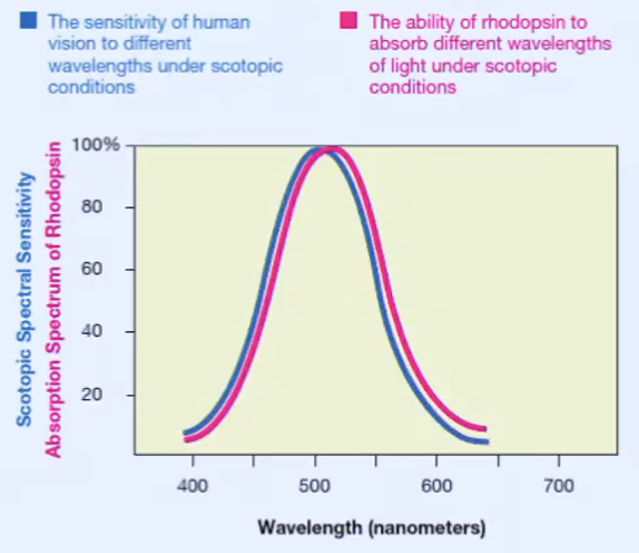
SCOTOPIC SPECTRAL SENSITIVITY & BEHAVIOUR
Behavioural measures of human sensitivity to different wavelengths closely matches the ability of rhodopsin (rod photoreceptor) to absorb those wavelengths of light.
Visual transduction is conversion of light energy into neural signals by the receptors.
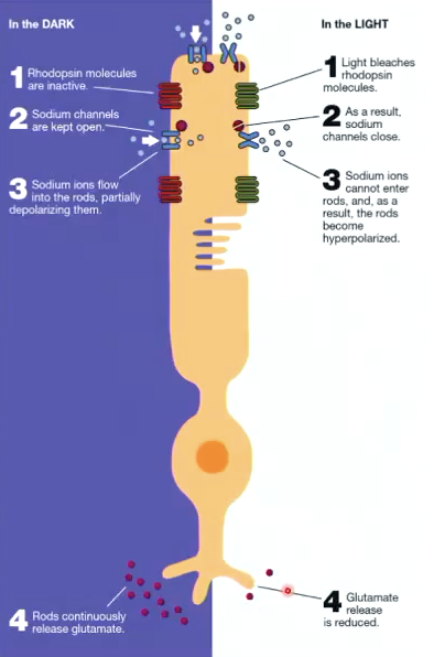
ROD LIGHT ABORPTION
In the dark:
Rhodopsin molecules are inactive
Na+ channels kept open
Na+ ions flow into the rods, partially depolarising them.
Steady flow of excitatory glutamate being emitted from rod
In the light:
Light bleaches rhodopsin molecules
Na+ channels close
Na+ ions can’t enter rods and rods become hyperpolarised
Flow of excitatory glutamate is reduced.
*therefore, the presence of light is signalled by a decrease in activity
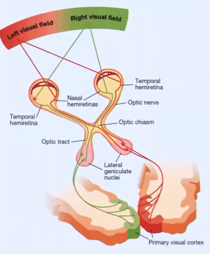
RETINA-GENICULATE-STRIATE PATHWAY: HEMI DECUSSATION
Anything in right visual field casts an image to the left side of the retina in the left and right eye
Vice versa
Information in the right visual field is transmitted to the left visual cortex
Vice versa
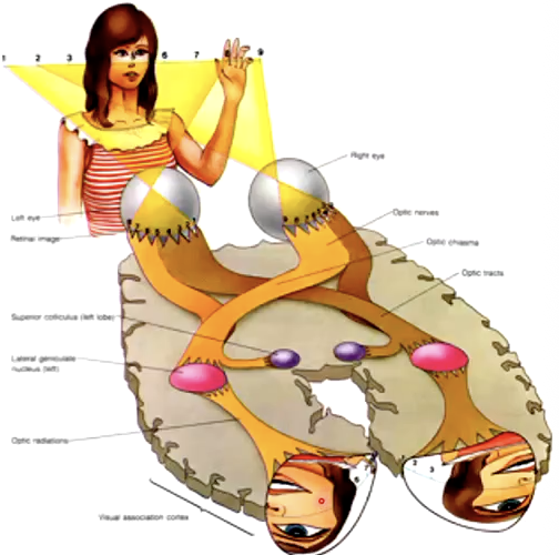
RETINA-GENICULATE-STRIATE PATHWAY: RETINOTOPIC MAPPING
Adjacent regions process features that fall on adjacent regions on the retina in the visual cortex
The order of stimulation on the retina is maintained all the way through the pathway to the visual cortex
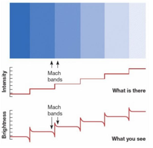
LATERAL INHIBITION
Mechanism by which neighbouring photoreceptors mutually suppress each other's activity
Bright-side receptor sends strong inhibition to its dim neighbour
Dim-side receptor can only send weak inhibition back
MACH BANDS
Bright side looks brighter at border, dark side looks darker in adjacent uniform rectangles
RECEPTIVE FIELD
Region in space/on the retina, that when stimulated, affects the behaviour of a neuron that is connected to it
RESPONSES OF ON-CENTRE + OFF-CENTRE CELLS
Responses of on-centre cell:
There is an 'on' response when light is shone anywhere in the centre of the field
There is an 'off' response when a spot of light is shone anywhere in the periphery of the field
*off-centre process is the opposite
*if both off and on regions were illuminated together, there was little reaction
SIMPLE CELLS
Simple cells form multiple aligned centre-surround receptive fields to create orientation specific receptive fields.
Respond most vigorously when static bars with an approximate orientation falls onto the 'on' subfield of the receptive field
Regions don't need to be uniform

SPATIAL FREQUENCY - SIMPLE CELLS
Low spatial frequency:
Activates simple cells with widely separated subfields
High spatial frequency:
Activates simple cells with less separated subfields
COMPLEX CELLS
Larger receptive fields
They respond to oriented contour anywhere within their receptive field
Receive input from both eyes although prefer one eye over the other
Respond more vigorously when both eyes stimulated simultaneously
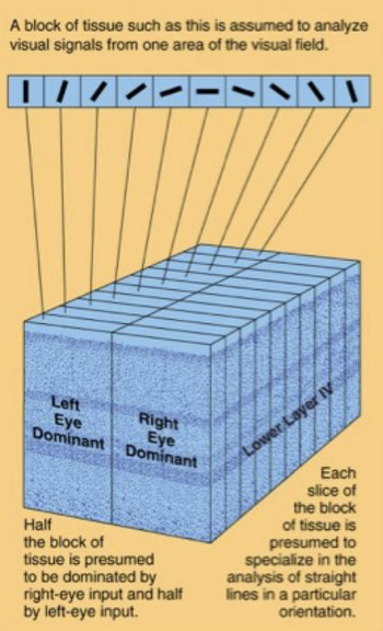
ORGANISATION OF PRIMARY VISUAL CORTEX (V1)
V1 is organised into functional columns perpendicular to cortical surface
Columns alternate in eye dominance across the cortical surface
Hypercolumn:
A full set of orientation columns for both eyes
Within column cells share:
Same retinal location
Same eye dominance
Same preferred orientation
SCOTOMAS & CORTICAL FILLING-IN
Damage to V1 causes scotoma (cortical blind spot)
Brain fills it in automatically
BLIND SIGHT
Patients with large V1 scotomas report no conscious awareness of objects in blind field yet can still interact with objects there
Incomplete damage leaving residual processing
Parallel visual pathways that reach secondary visual cortex without passing through V1
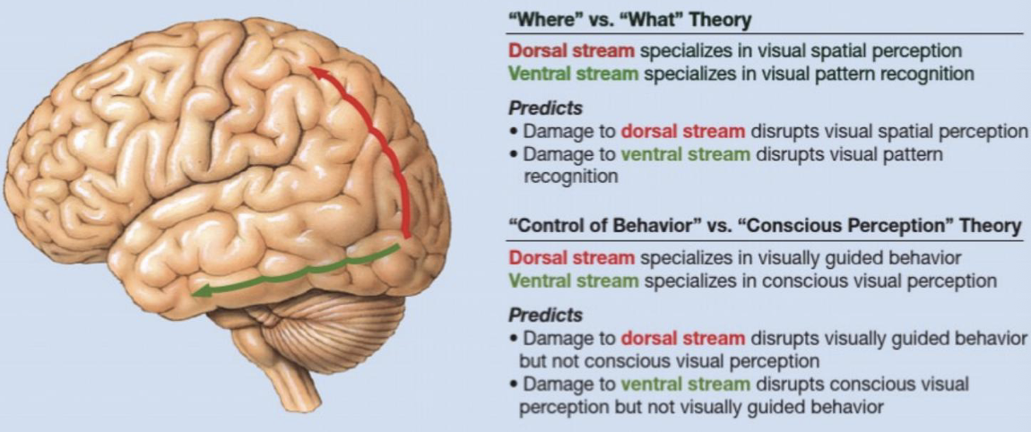
DORSAL STREAM (PARIETAL CORTEX)
Specialises in visual spatial perception
Specialises in visually guided behaviour
Reaching/intercepting/ interacting with objects
Damage impairs motor interaction but not verbal description

VENTRAL STREAM (INFEROTEMPORAL CORTEX)
Specialises in visual pattern recognition
Specialises in conscious visual perception
Damage impairs description/recognition, but not motor interaction
PULFRICH ILLUSION
Illusion where a 2D moving object is perceived as 3D (moving in depth) dye to a delay in signal processing between eyes.
EX. ball swinging left to right it perceived as swinging in a circle
PULFRICH ILLUSION COMPONENTS
Stereoscopic Vision
Simple harmonic motion of the pendulum
The filter/lens
STEREOSCOPIC VISION
2 eyes get slightly different images of the world
brain combines these 2 images to get a single perception
important for depth perception
FIXATION:
fixate → eyes focus and align → image falls on fovea
PERIPHERAL:
image falls beside fovea, on retina.
UNCROSSED DISPARITY
Focusing/fixating on an image BEHIND what you were initially fixated on. Object appears further away in depth
farther than fixation point
image appears further to left in space to left eye
image appears further to right in space to right eye
CROSSED DISPARITY
Focusing/fixating on an image IN FRONT what you were initially fixated on. Object appears nearer in depth
nearer than fixation point
image appears further to right in space to left eye
image appears further to left in space to right eye
SIMPLE HARMONIC MOTION
Ball accelerates as travelling down due to gravity. At bottom it is maximum gravity. Ball slows down as it travels back up, due to going against gravity.
DIMMING FILTER/LENS
Signal transduction from retina to brain is faster for brighter images
filter over your eyes dims the image that falls on your retina
filter in ONE eye, means that signals from one eye are slightly delayed
COMBINATION: FILTER OVER LEFT EYE
ball appears further away on the same side as filtered eye
delay in filtered eye causes ball to always be perceived as behind
ball appears to move in clockwise direction
LEFT TO RIGHT MOTION
uncrossed disparity
further away in depth
illusory backward arc
RIGHT TO LEFT MOTION
crossed disparity
nearer in depth
illusory forward arc
COMBINATION: FILTER OVER RIGHT EYE
ball appears further away on the same side as filtered eye
delay in filtered eye causes ball to always be perceived as behind
ball appears to move in anti-clockwise direction
LEFT TO RIGHT MOTION
crossed disparity
nearer away in depth
illusory forward arc
RIGHT TO LEFT MOTION
uncrossed disparity
further in depth
illusory backward arc
MONOCULAR DEPTH CUES (ONE EYE)
OCCLUSION - chair blocks persons leg therefore, person behind chair
RETINAL SIZE - object gets ‘bigger’ when closer
AMES WINDOW - using angles and shading
LINEAR PERSPECTIVE - parallel lines converge in the distance
TEXTURE GRADIENT - more detailed closer to
SHADING - light generally comes from above (convex vs concave)
LIGHT SCATTER - distant objects appear hazier & bluer
MOTION PARALLEX - objects further away appear to move slower