L10. Malignant Tumors of the Musculoskeletal System
0.0(0)
Card Sorting
1/71
Earn XP
Description and Tags
Last updated 3:27 AM on 11/21/22
Name | Mastery | Learn | Test | Matching | Spaced | Call with Kai |
|---|
No analytics yet
Send a link to your students to track their progress
72 Terms
1
New cards
Soft tissue sarcomas (STS) are __________ and ___________ diseases.
rare and heterogenous
13k diagnoses; >5000 deaths
13k diagnoses; >5000 deaths
2
New cards
What is crucial in STS diagnosis?
tissue diagnosis
pathology should be reviewed by an experienced pathologist
pathology should be reviewed by an experienced pathologist
3
New cards
STS account for ______% of adult malignancies and ____% of pediatric malignancies.
1%
15%
15%
4
New cards
When is the peak incidence of STS and are males or females more likely to be affected?
15
males
males
5
New cards
What are the two most common STS?
Osteosarcoma (56%)
Ewing's Sarcomas (34%)
Ewing's Sarcomas (34%)
6
New cards
What STS is most common in children?
Rhabdomyosarcoma (RMS)
7
New cards
_____% of STS involve the limbs.
75%
although they can occur anywhere with connective tissues
although they can occur anywhere with connective tissues
8
New cards
Is there screening for STS?
no because very rare
9
New cards
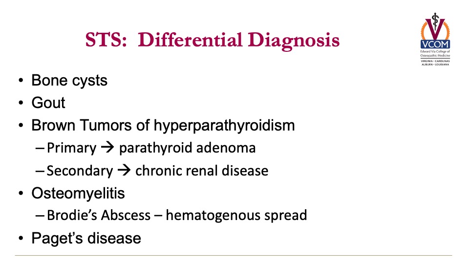
What are 8 other differential diagnoses for STS? How can you differentiate?
1. endochonomas (tend to be benign)
2. osteoid osteoma (proximal femur & relieved with NSAIDs)
3. myositis ossificans (post-traumatic)
4. bone cysts
5. gout
6. brown tumors of hyperparathyroidism
7. osteomyelitis
8. paget's disease
2. osteoid osteoma (proximal femur & relieved with NSAIDs)
3. myositis ossificans (post-traumatic)
4. bone cysts
5. gout
6. brown tumors of hyperparathyroidism
7. osteomyelitis
8. paget's disease
10
New cards
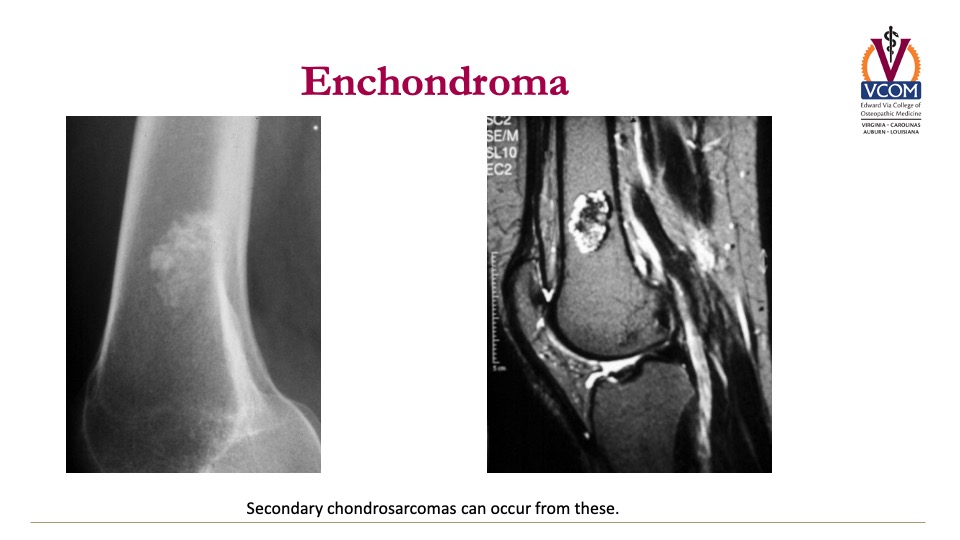
What is the appearance of an enchondroma?
Left side is a radiograph
Right is a MRI
Right is a MRI
11
New cards
What secondary STS can occur from enchondromas?
secondary chondrosarcomas
12
New cards
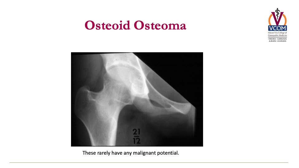
What is the appearance of an osteoid osteoma?
these rarely have any malignant potential
13
New cards
Primary brown tumors of hyperparathyroidism are caused by what?
parathyroid adenoma
14
New cards
Secondary brown tumors of hyperparathyroidism are caused by what?
chronic renal disease
15
New cards
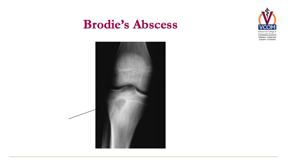
What is seen on imaging for osteomyelitis?
brodie's abscess
16
New cards
What is Paget's Disease and how can it be differentiated from STS?
Disorder of bone metabolism resulting in benign growths
May cause pain or nerve impingement & is often found incidentally on radiographs
Alkaline phosphatase is highly elevated
May cause pain or nerve impingement & is often found incidentally on radiographs
Alkaline phosphatase is highly elevated
17
New cards
What is the treatment of Paget's Disease?
IV bisphosphonates
**Zoledronic acid**
**Zoledronic acid**
18
New cards
If symptoms worsen in Paget's disease, you should reimage due to what concern?
secondary sarcoma (due to increased cell turnover)
19
New cards
STS: differential diagnosis
20
New cards
What are the risk factors for STS?
1. prior radiation therapy to the area affected
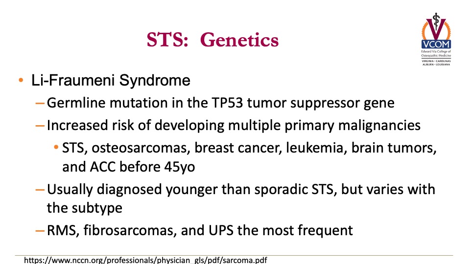
2. genetic cancer syndrome
- Li-Fraumeni syndrome
- Familial adenomatous polyposis (FAP)
- Carney-Stratakis Syndrome
- Hereditary retinoblastoma
- Neurofibromatosis
2. genetic cancer syndrome
- Li-Fraumeni syndrome
- Familial adenomatous polyposis (FAP)
- Carney-Stratakis Syndrome
- Hereditary retinoblastoma
- Neurofibromatosis
21
New cards
What mutation causes Li-Fraumeni Syndreom?
germline mutation in TP53 tumor suppressor gene
22
New cards
Li-Fraumeni syndrome increases your risk of developing multiple primary malignancies, such as?
STS, osteosarcomas, breast cancer, leukemia, brain cancers, and ACC before 45 years old
23
New cards
Familial adenomatous polyposis (FAP) is an inherited autosomal _____________ colorectal cancer syndrome.
dominant
24
New cards
What mutation causes FAP?
FAP?
germline mutations in the adenomatous polyposis coli (APC) gene on chromosome 5q21
germline mutations in the adenomatous polyposis coli (APC) gene on chromosome 5q21
25
New cards
What is FAP characterized by?
adenomatous colon polyps that progress to CRC by 35-40 year old
26
New cards
What is Gardner's syndrome?
Variant of FAP with extracolonic manifestations such as osteomas,
skin cysts, congenital hypertrophy of the retinal pigmented epithelium, and desmoid tumors (aggressive fibromatosis)
skin cysts, congenital hypertrophy of the retinal pigmented epithelium, and desmoid tumors (aggressive fibromatosis)
27
New cards
What is Carney-Stratakis Syndrome? What is the inheritence?
autosomal dominant
GISTs and paragangliomas
GISTs and paragangliomas
28
New cards
What is hereditary retinoblastoma caused by?
germline mutation in retinoblastoma tumor suppressor gene (RB1)
29
New cards
What causes neurofibromatosis?
mutations in the neurofibromin 1 gene (NF1) or neurofibromin 2 gene (NF2)
30
New cards
____% of patients with neurofibromatosis develop STS.
5%
31
New cards
What is the most common STS in neurofibromatosis?
malignant peripheral nerve sheath tumors
32
New cards
There are more than ______ different histologic subtypes of STS
50
Heterogenous
Heterogenous
33
New cards
What are the two broad categories of STS?
1. soft tissue sarcomas (fat, muscle, nerve, nerve sheath, blood vessel, etc)
2. sarcomas of the bone
2. sarcomas of the bone
34
New cards
What are the most common anatomic sites of STS?
1. extremities (43%)
2. visceral (19%)
3. retroperitoneum (5%)
4. trunk (10%)
5. head & neck (9%)
2. visceral (19%)
3. retroperitoneum (5%)
4. trunk (10%)
5. head & neck (9%)
35
New cards
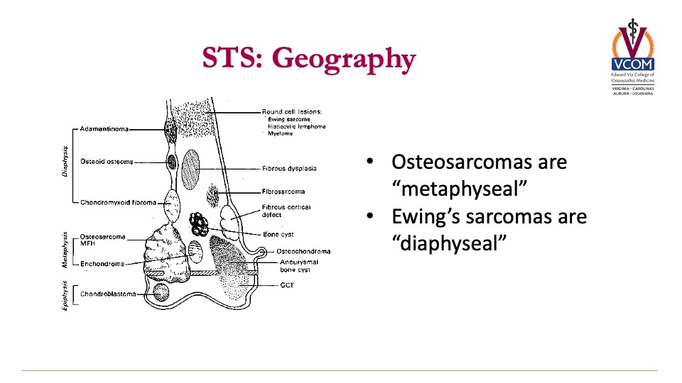
What is the geography of osteosarcomas are _____________ while Ewing's sarcomas are _________________.
metaphyseal
diaphyseal
diaphyseal
36
New cards
How do you diagnose STS (imaging)?
1. plain radiographs (AP and orthogonal views)
2. CT (small cortical lesions & lung windows for metastases)
3. angiogram (vascular structures & resectability)
4. Magnetic Resonance Imaging (MRI)
-Gold Standard!!!!!
-The most helpful test is an MRI!
-with contrast tells you more
-Collect x-ray first and if the x-ray is suspicious collect MRI
2. CT (small cortical lesions & lung windows for metastases)
3. angiogram (vascular structures & resectability)
4. Magnetic Resonance Imaging (MRI)
-Gold Standard!!!!!
-The most helpful test is an MRI!
-with contrast tells you more
-Collect x-ray first and if the x-ray is suspicious collect MRI
37
New cards
What is the preferred method for diagnosis and grading of STS?
pretreatment biopsy
38
New cards
Why is it important to have someone experienced performing and reading biopsies for STS?
track of needle matters in & out
"biopsy should be placed along the future resection axis with minimal dissection and careful attention to hemostasis"
grade can be underestimated
"biopsy should be placed along the future resection axis with minimal dissection and careful attention to hemostasis"
grade can be underestimated
39
New cards
What type of needle should you use in STS biopsy?
core needle biopsy preferred (because you want more tissue)
do not perform fine needle aspiration
open excision may be required
do not perform fine needle aspiration
open excision may be required
40
New cards
What 8 things should the pathology report include in STS?
1. organ/site
2. depth
3. size
4. grade
5. margin status
6. lymph nodes
7. +/- necrosis
8. mitotic rate, vascular invasion, inflammatory infiltration
2. depth
3. size
4. grade
5. margin status
6. lymph nodes
7. +/- necrosis
8. mitotic rate, vascular invasion, inflammatory infiltration
41
New cards
How do you stage STS?
T=size
N=# lymph nodes
M=metastasis
G=Grade (which is specific for STS and prostate cancer)
In sarcoma once the nodes are involved it's stage 4
Stage drives prognosis
N=# lymph nodes
M=metastasis
G=Grade (which is specific for STS and prostate cancer)
In sarcoma once the nodes are involved it's stage 4
Stage drives prognosis
42
New cards
How do you treat STS generally?
multidisciplinary and tertiary/university based team with surgeon, rad onc, med onc, & PT/OT
limb sparing is preferred, but need good margins
neoadjuvant chemo +/ XRT if primary resection difficult
limb sparing is preferred, but need good margins
neoadjuvant chemo +/ XRT if primary resection difficult
43
New cards
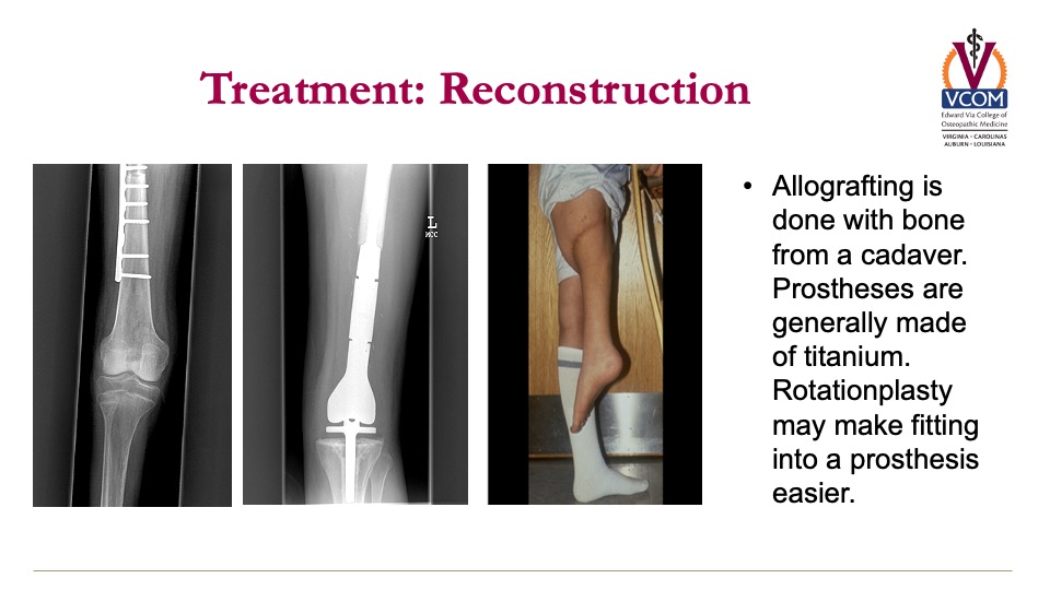
What is the difference between allografting, prostheses, and rotationplasty?
allografting: bone from cadaver
prostheses: made of titanium
rotationplasty: makes fitting into prosthesis easier
The ankle is now the knee joint
Nerves can be re-taught
prostheses: made of titanium
rotationplasty: makes fitting into prosthesis easier
The ankle is now the knee joint
Nerves can be re-taught
44
New cards
What are the options for radiation in the treatment of STS?
external beam radiation
axial lesions problematic to resect with good margins, so may use intraoperative RT or proton beam therapy
axial lesions problematic to resect with good margins, so may use intraoperative RT or proton beam therapy
45
New cards
Can you treat osteosarcoma with chemo and radiation?
no --> radio-resistant & chemo-sensitive
**neoadjuvant chemotherapy is common
**neoadjuvant chemotherapy is common
46
New cards
Can you treat Ewing's sarcoma with chemo and radiation?
yes --> radio- and chemo-sensitive
47
New cards
Can you treat chondrosarcoma with chemo and radiation?
no --> radio- and chemo-resistant typically
48
New cards
How does osteosarcoma typically present?
painful swelling around knee or humerus
night pain & limping
firm/soft mass fixed to underlying bone
serum alkaline phoshate elevated
night pain & limping
firm/soft mass fixed to underlying bone
serum alkaline phoshate elevated
49
New cards
What is the prognosis of osteosarcoma?
~20% survived 5 years before chemotherapy era
-60-70% 5-year survival currently
limb sparing in 80% currently
long term morbidity from chemo (heart disease, secondary cancers, infertility)
-60-70% 5-year survival currently
limb sparing in 80% currently
long term morbidity from chemo (heart disease, secondary cancers, infertility)
50
New cards
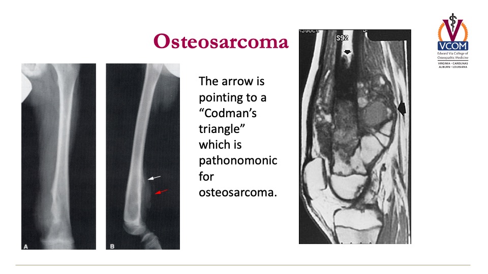
What is the Pathognomonic "appearance" of osteosarcoma?
Codman's Triangle
51
New cards
How does osteosarcoma appear on pathology?
derived from osteoblasts and will secrete osteoid (cotton candy appearance)

52
New cards
How do you treat osteosarcoma specifially?
chemo-sensitive with neoadjuvant chemotherapy --> crucial for limb preservation and tretament of "micro-metastatic" disease
surgery s/p chemo
"no viable cancer at resection is a good prognostic sign" --> but recurrence usually occurs in the lungs
surgery s/p chemo
"no viable cancer at resection is a good prognostic sign" --> but recurrence usually occurs in the lungs
53
New cards
How does Ewing's Sarcoma present?
similar to osteosarcoma, but is AGGRESSIVE (grows & spreads rapidly)
pain & constitutional symptoms present (mimics osteomyelitis)
metastatic disease in 25% at presentation (lungs, bone, bone marrow)
pain & constitutional symptoms present (mimics osteomyelitis)
metastatic disease in 25% at presentation (lungs, bone, bone marrow)
54
New cards
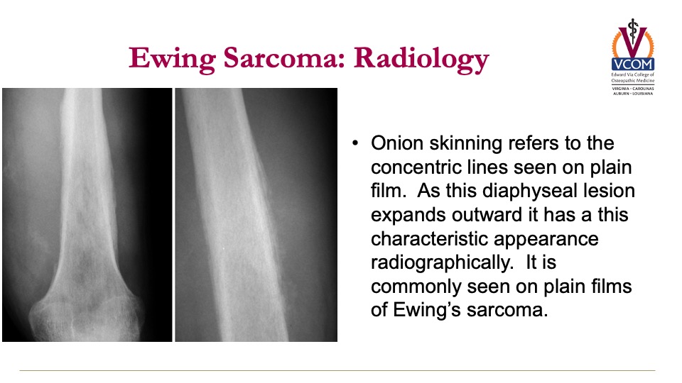
How does Ewing sarcoma appear on x-ray?
Onion skinning refers to the
concentric lines seen on plain
film. As this diaphyseal lesion
expands outward it has a this
characteristic appearance
radiographically.
concentric lines seen on plain
film. As this diaphyseal lesion
expands outward it has a this
characteristic appearance
radiographically.
55
New cards
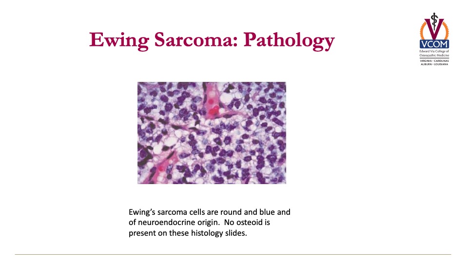
How does Ewing Sarcoma appear on pathology slides?
cells are small, round, and blue of neuroendocrine origin without osteoid
56
New cards
What is the presentation of chondrosarcomas?
- pain related to location of mass lesion, typically in shoulder
- previous site of enchondroma
- suspect if tumor involves shoulder/sternum
- rapid expansion suggests higher grade or de-differentiation
- previous site of enchondroma
- suspect if tumor involves shoulder/sternum
- rapid expansion suggests higher grade or de-differentiation
57
New cards
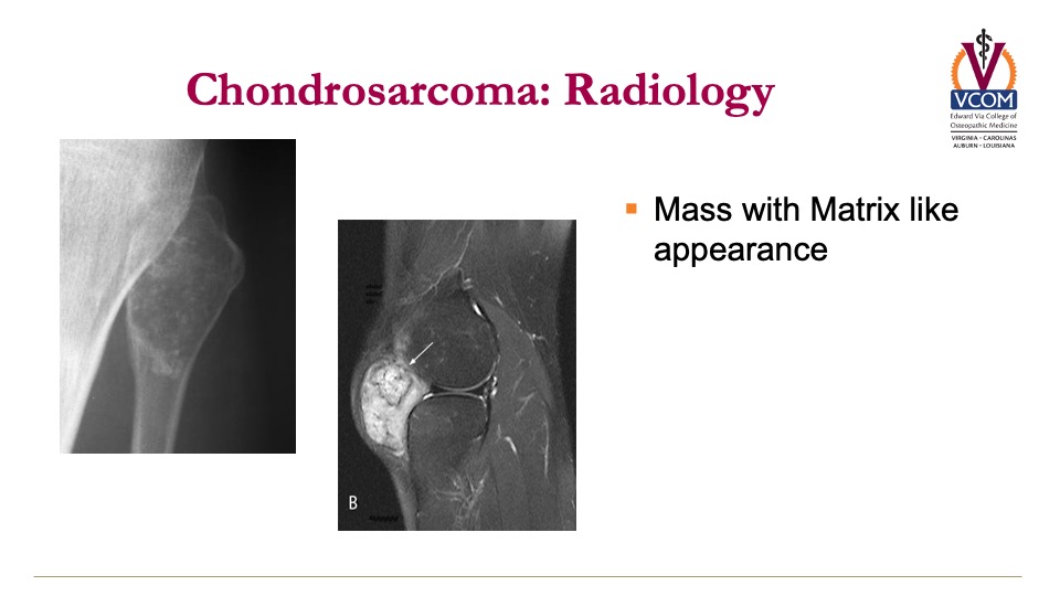
How does chondrosarcoma appear on x-ray?
mass with matrix-like appearance
x-ray left; mri on right
x-ray left; mri on right
58
New cards
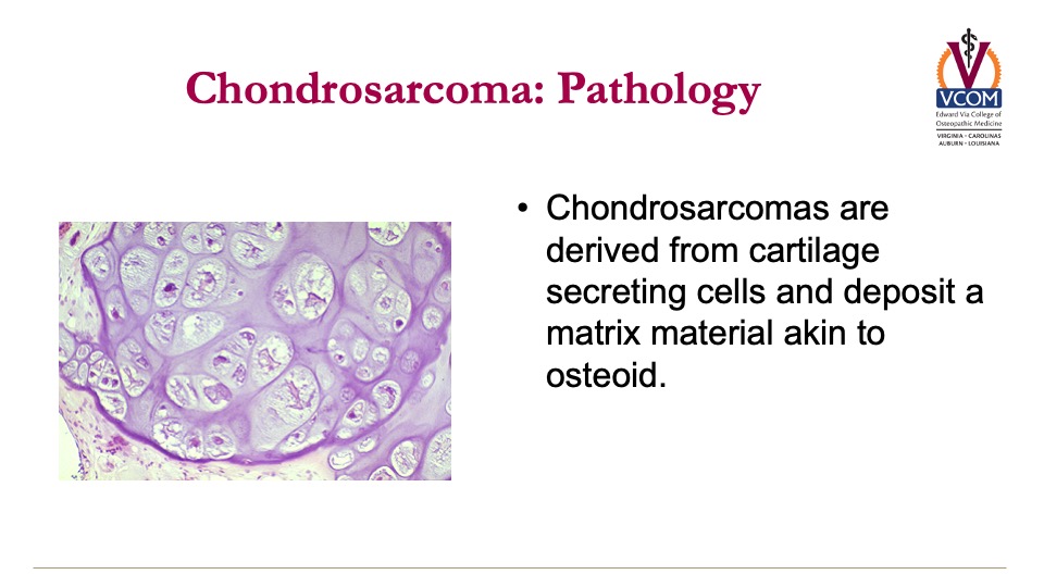
How does chondrosarcoma appear on pathology?
derived from cartilage secreting cells and deposit a matrix material akin to osteoid
59
New cards
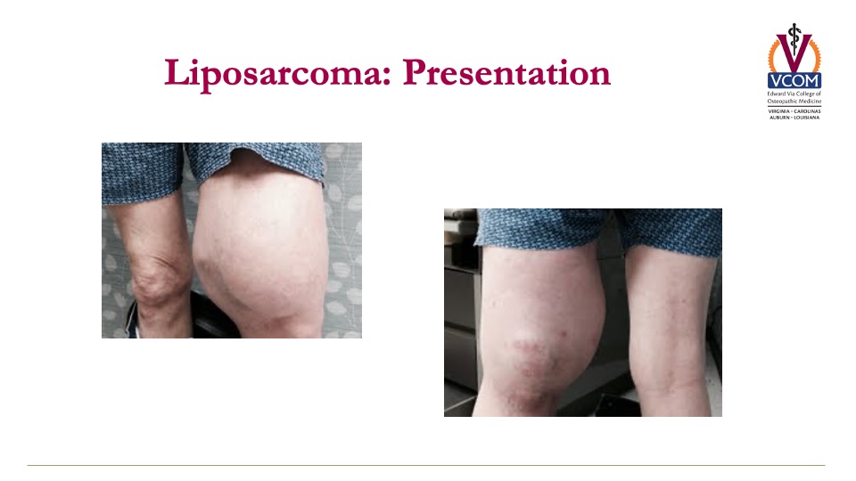
How does liposarcoma typically present?
- develop from well-differentiated tumors
- rapid growth suggests de-differentiated pathology
- develops in retroperitoneum preferentially and limbs next (rare)
- non-painful
- rapid growth suggests de-differentiated pathology
- develops in retroperitoneum preferentially and limbs next (rare)
- non-painful
60
New cards
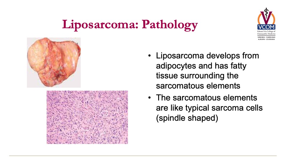
How does liposarcoma appear on pathology?
develops from adipocytes and has fatty tissue surrounding the sarcomatous elements, which are spindle shaped
61
New cards
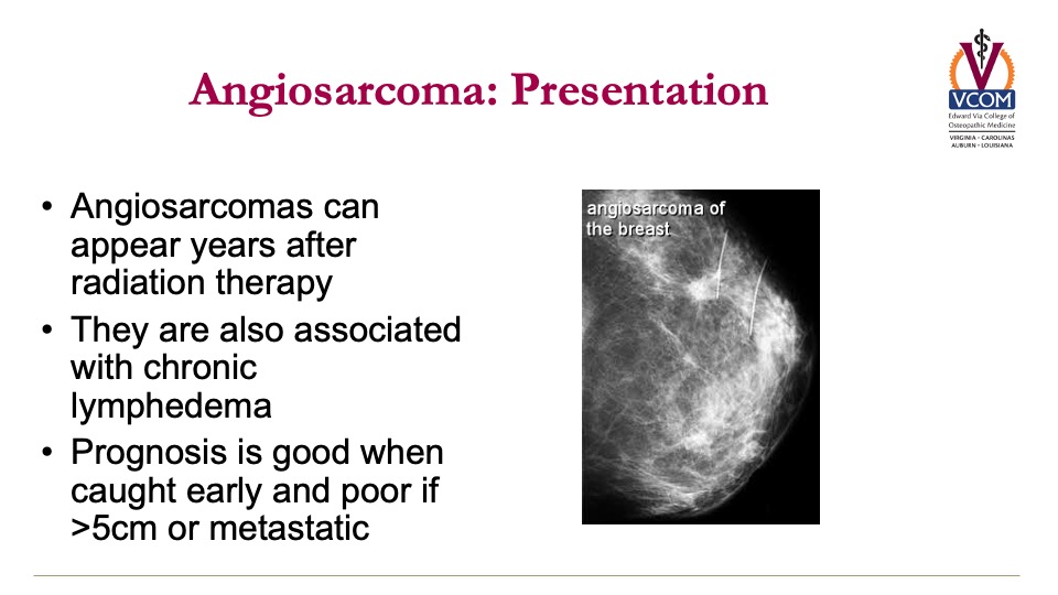
What is the typical presentation of angiosarcoma?
can appear years after
radiation therapy (anywhere)
are also associated with chronic lymphedema
radiation therapy (anywhere)
are also associated with chronic lymphedema
62
New cards
What is the prognosis of angiosarcoma?
prognosis is good when caught early and poor if >5cm or metastatic
63
New cards
How do gastrointestinal stromal tumors present (GIST)?
can occur anywhere in the GI tract & typically metastasize to the liver
64
New cards
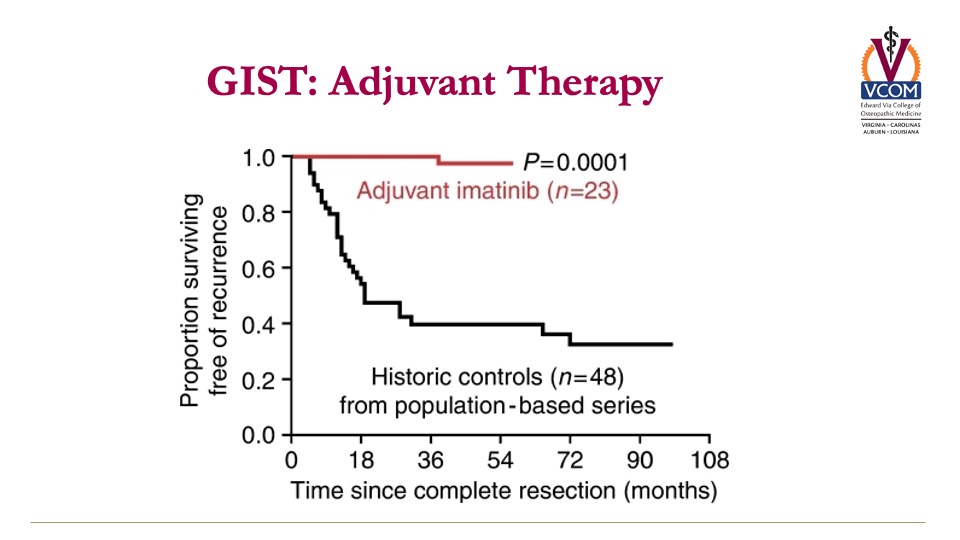
How can you treat GIST?
***Somatic tumors
***Somatic tumors
c-KIT, PDGF and VEGF which can be inhibited by small molecule tyrosine kinases (Imatinib, Sunitinib, Regorafenib) --> aka tyrosine kinase inhibitors
Adjuvant therapy is generally employed with Imatinib for 3
years.
Adjuvant therapy is generally employed with Imatinib for 3
years.
65
New cards
If you see a bone lesion in an adult, what is it most likely?
metastatic carcinoma
-in adults MC is the most common cancer to see in the bone and started somewhere else
-in adults MC is the most common cancer to see in the bone and started somewhere else
66
New cards
How does metastatic carcinoma typically present? What do labs look like?
- Pain and pathologic fracture
- Lytic (myeloma/lung) or -blastic lesions (breast/prostate)
- Occasional mild hypercalcemia and elevated alkaline phosphatase
- Rare cytopenias
- Lytic (myeloma/lung) or -blastic lesions (breast/prostate)
- Occasional mild hypercalcemia and elevated alkaline phosphatase
- Rare cytopenias
67
New cards
What cancers typically spread to bone?
breast, prostate, lung, thyroid, renal
68
New cards
What are the most common cases of metastatic carcinoma?
prostate & breast (the most common cancers)
69
New cards
How does metastatic carcinoma develop?
hematogenous spread to bone marrow
70
New cards
What is the prognosis of metastatic carcinoma?
prognosis better for bone only mets than visceral plus bone
***If you only have bone metasis they live a lot longer than breast cancer that has gone to the lungs and liver
***If you only have bone metasis they live a lot longer than breast cancer that has gone to the lungs and liver
71
New cards
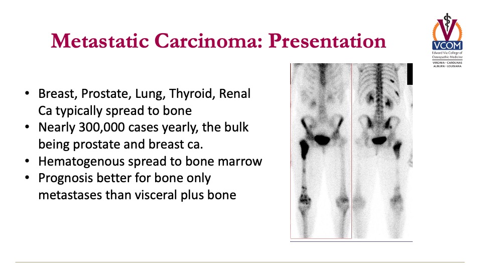
What does a nuclear medicine bone scan look like in metastatic carcinoma?
look for asymmetry
72
New cards
Summary Slide