organisation topic 2 biology
1/60
Earn XP
Description and Tags
Name | Mastery | Learn | Test | Matching | Spaced |
|---|
No study sessions yet.
61 Terms
What are cells? Specialised cells..? Whats differentiation? Specialised cells form.. & so on? Large multicellular organisms have…?
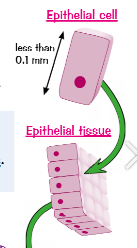
cells → basic building blocks that make up all living organisms
Specialised cells carry out a particular function
differentiation → process by which cells become specialised for particular job, occurs during development of multicellular organism
Specialised cells form tissues, which form organs, which form organ systems
Large multicellular organisms have different systems inside them for exchanging & transporting materials
Whats tissue? 3 examples in mammals?
tissue → group of similar cells that work together to carry out a particular function (includes more than 1 type of cell)
In mammals (humans) examples of tissues include:
Muscular tissues → contract (shortens) to move whatever its attached to
Glandular tissue → makes & secretes chemicals like enzymes & hormones
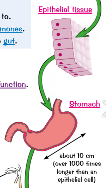
Epithelial tissue →covers some parts of the body (inside the gut)
What are organs? what is the stomach organ made up of?
organ → group of different tissues that work together to perform a certain function
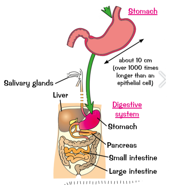
The stomach organ is made up of these tissues:
muscular tissue → moves the stomach wall to churn up food
Glandular tissues → makes digestive juices to digest food
Epithelial tissue → covers the outside & inside of stomach
Whats an organ system? What does the digestive system do? What organs is it made out of?
organ system → group of organs working together to perform a particular function
Digestive system (humans & other mammals) breaks down & absorbs food, made up of these organs:
Glands (pancreas & salivary glands) → produces digestive juices
Stomach & small intestine → digest food
Liver → produces bile
Small intestine → absorbs soluble food molecules
Large intestine → absorbs water from undigested food leaving faeces
(organ systems work together to make entire organisms)
Whats needs to be controlled in living things? How do you make a reaction happen quicker? Whats a catalyst? Whats an enzyme?
living things have thousands of different chemical reactions going on inside them → need to be carefully controlled (get right amounts of substances)
make a reaction happen quicker → raising temperature (speed up useful reactions & unwanted ones), there’s a limit to how far you can raise temperature before cells start getting damaged
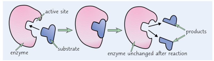
Catalyst (enzymes) → substance which increases the speed of reaction without being changed or used up in the reaction
Enzymes → are large proteins made up of chains of amino acids (chains are folded into unique shapes, enzymes need this to do their jobs)
chemical reactions usually invlove? enzymes usually.? for enzymes to work? What does the diagram of ‘lock & key’ model show & in reality?
chemical reactions usually involve things being split apart or joined together
Enzymes usually only catalyse one specific reaction
For enzymes to work substrate has to fit into its active site (unique shape), substrate doesn’t fit = reaction won’t be catalysed
Diagram of ‘lock & key’ model shows enzyme action, simpler than how enzymes actually work. In reality active site changes shape a little as substrate binds to get tighter fit (‘induced fit’ model of enzyme action)
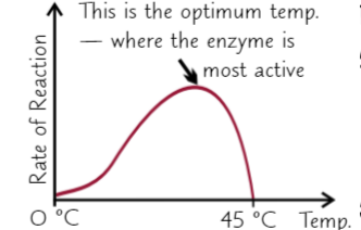
changing temp changes? What happens w higher temps? All enzymes have?
changing temperature changes rate of enzyme-catalysed reaction
higher temperature increases rate at first but if it gets too hot, some of bonds holding enzyme together break, this changes shape of enymes active site so substrate won’t fit anymore (enzyme is denatured)
All enzymes have an optimum temperature they work best at
How does pH affect enzymes? All enzymes have?
pH affects enzymes, if its too high/low pH interferes with bonds holding enzyme together = changes shape of active site & denatures enzyme
All enzymes have optimum pH they work best at, often neutral pH 7 (pepsin is an enzyme used to break down proteins in stomach, works best at pH 2, well suited for acidic conditions)
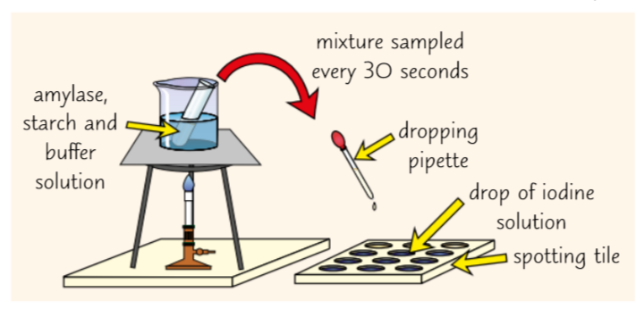
Investigate the effect of pH on enzyme activity (practical)
amylase catalyses (enzyme) breakdown starch to maltose, easy to detect starch using iodine solution (if starch present iodine solution change =brown-orange to blue-black. investigate how Ph affects amylase activity:
Put drop of iodine solution into every well of spotting tile
place Bunsen burner on heat-proof mat, & tripod & gauze over the Bunsen burner. Put beaker of water on top tripod & heat water until its 35C (use thermometer to measure temperature), try keep water temperature constant
using syringe add 1cm3 of amylase solution & 1cm3 of buffer solution with pH of 5 to boiling tube, using test tube holders put tube into beaker of water & wait 5 mins
use different syringe add 5cm3 starch solution to boiling tube
Immediately mix contents of boiling tube & start stopwatch
use continuous sampling to record how long it takes for amylase to break down all of starch, use dropping pipette to take fresh sample from boiling tube every 30s & put drop into well. when iodine solution remains brown-orange, starch is no longer present
Repeat whole experiment with buffer solutions of different pH values to see how pH affects time taken for starch to be broken down
Remember to control any variables each time (concentration & volume of amylase solution) to make it a fair test

Calculate rate of reaction (effect of pH on enzyme activity practical)
rate is a measure of how much something changes over time
for this experiment use: Rate=1000/time
(units given s-1 )
if experiment measures how much something changes over time, calculate rate of reaction by dividing the amount it has changed by the time taken
What molecules are too big? What do digestive enzymes do? Smaller soluble molecules can? What does the body make good use of?
starch, proteins & fats = big molecules (too big to pass through walls),
digestive enzymes break big molecules down into smaller ones = sugars (glucose, maltose), amino acids, glycerol & fatty acids.
Smaller soluble molecules pass easily through walls of digestive system, allowing them to be absorbed into bloodstream
Body makes good use of products of digestion, can be used to make new carbohydrates, proteins & lipids. some of glucose (carbohydrate) that’s made in respiration
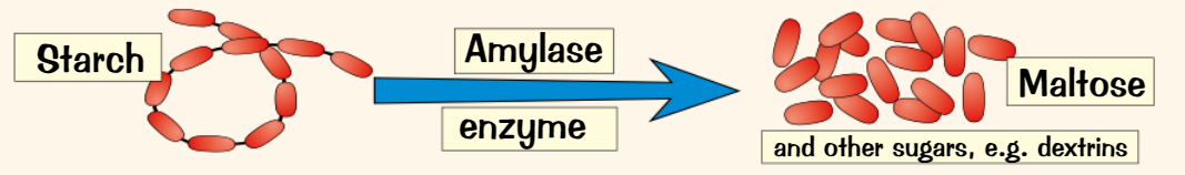
What does amylase convert? where is it produced?
Starch (carbohydrate)→ amylase enzyme (carbohydrase) → maltose & other sugars (simple sugars)
Amylase is made in 3 places:
The salivary glands
the pancreas
The small intestine
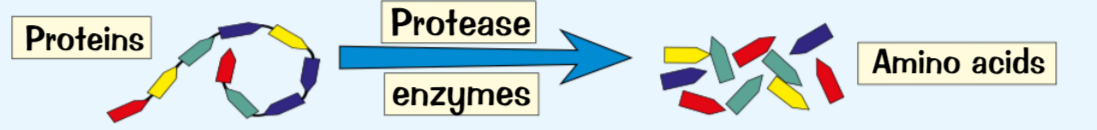
What does protease enzyme convert? Where is it produced?
Proteins → broken down by protease enzymes → amino acids
Proteases are made in 3 places:
The stomach (pepsin)
The pancreas
The small intestine
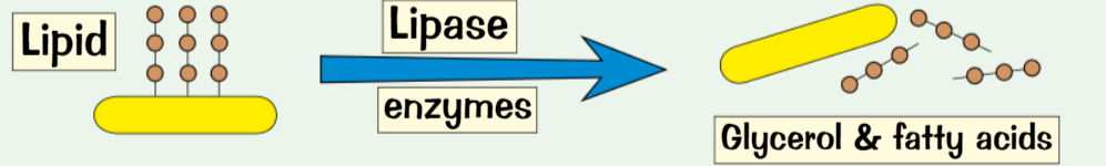
What does lipase enzymes convert? Where is it produced?
Lipids (oils & fats)→ broken down by lipase enzymes → glycerol & fatty acids
Lipases are made in 2 places:
Pancreas
small intestine
What produces & stores bile? Why is bile alkaline? How does it emulsifies fats?
Bile is produced in liver, stored in gall bladder before it’s released into small intestine
Hydrochloric acid in stomach = pH too acidic for enzymes in small intestine to work properly, bile is alkaline (neutralises acid & makes conditions alkaline) → enzymes in small intestine work best in alkaline conditions
Emulsifies fats (breaks down fat into tiny droplets) → gives much bigger surface area of fat for enzyme lipase to work on (makes digestion faster)
Enzymes in digestive system are produced by? Different enzymes catalyse…? Describe the enzymes/food pathway (9 points)
enzymes in digestive system are produced by specialised cells in glands & gut lining
different enzymes catalyse breakdown of different food molecules
Salivary glands → produce amylase enzyme in the saliva
Gullet (oesophagus)
Liver → where bile is produced, bile neutralises stomach acid & emulsifies fats
stomach → pummels food with its muscular walls, produces protease enzyme (pepsin), produces hydrochloric acid (kill bacteria, give right pH for protease enzyme to work)
Gall bladder → bile is stored before released into small intestine
Pancreas → produces protease, amylase, lipase enzymes & releases these into small intestine
Small intestine → produces protease, amylase & lipase enzymes to complete digestion, where digested food is absorbed out of digestive system into blood
Large intestine → excess water is absorbed from food
Rectum → faeces (mainly indigestible food) are stored before leave through anus
Preparing food sample (Food tests practical) (break, transfer, dissolve, filter)
To identify what type of food molecules a sample contains you need to prepare the food sample first:
Get a piece of food & break it up using a pestle & mortar
Transfer ground up food to a beaker & add some distilled water
Giver the mixture a good stir with glass rod to dissolve some of food
Filter solution using funnel lined with filter paper to get rid of solid bits of food
Use benedict’s test to test for sugars (food tests practical) (found & which, prepare, water, add, place, contains)
Sugars found in biscuits, cereal & bread (more), 2 types of sugars → non-reducing & reducing. Can test for reducing sugars in foods using benedict’s test:
Prepare food sample & transfer 5cm3 to test tube
prepare water bath so its set to 75C
add some benedict solution to test tube (~10 drops) using a pipette
Place test tube in water bath using test tube holder & leave for 5 mins, make sure tube is pointing away from you
If food sample contains reducing sugar = solution changes from normal blue to green, yellow or brick red (depends on sugar concentration)
Use iodine solution to test starch (food tests practical)
Foods like pasta, rice & potatoes contains lots of starch, method:
Make food smaple & transfer 5cm3 of sample to test tube
Add few drops of iodine solution & gently shake tube to mix contents, if sample contains starch = colour changes from browny-orange to black or blue-black
Use biuret test to test for proteins (food tests practical)
Meat & cheese are protein rich & good foods for test, method:
Prepare food sample & transfer 2cm3 of sample to test tube
add 2cm3 of biuret solution to sample & mix contents of tube by gently shaking
If food sample contains protein = changes from blue to purple
Use sudan III test to test for lipids (food tests practical)
Lipids found in olive oil, margarine & milk, method:
Prepare food sample (don’t need to filter it), transfer ~5cm3 into test tube
use pipette to add 3 drops of Sudan III stain solution to test tube & gently shake tube
Sudan III stain solution stains lipids, if sample contains lipids = mixture separates out into 2 layers, top layer will be bright red
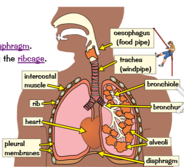
Whats the thorax? What are lungs? Describe airs pathway & how you ends up at gas exchange place?
Thorax is top part of body, separated from lower part of body by diaphragm
lungs = big pink sponges & protected by ribcage, surrounded by permeable membranes
Air you breathe in goes through trachea → splits into 2 tubes → bronchi (each is bronchus), one going to each lung
Bronchi split into progressively smaller tubes → bronchioles
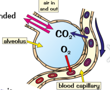
Bronchioles finally end at small bags called alveoli where gas exchange take place
Lungs contain? The blood…? When blood reaches body cells? At the same time…?
Lungs contain millions of little air sacs → alveoli, surrounded by network of blood capillaries (where gas exchange happens)
blood passing next to alveoli just returned to lungs from rest of body, contains lots of CO2 & little oxygen. Oxygen diffuses out of alveolus (high concentration) into blood (low concentration) to be breathed out
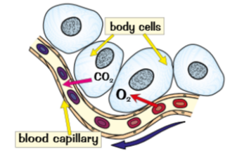
When blood reaches body cells oxygen is released from red blood cells (high concentration) & diffuses into body cells (low concentration)
at same time CO2 diffuses out of body cells (high concentration) into blood (low concentration), then carried back to lungs
Calculate the breathing rate in breaths per minute
Breaths per minute = number of breaths/ number of minutes
Whats the circulatory system? Describe the proccess
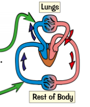
Circulatory system made up of heart, blood vessels & blood, humans have double circulatory system (two circuits joined together):
In first one right ventricle pumps deoxygenated blood to lungs to take in oxygen, blood returns to heart
In second one, left ventricle pumps oxygenated blood around all other organs of body, blood give up its oxygen at body cells & deoxygenated blood returns to heart to be pumped out of lungs again
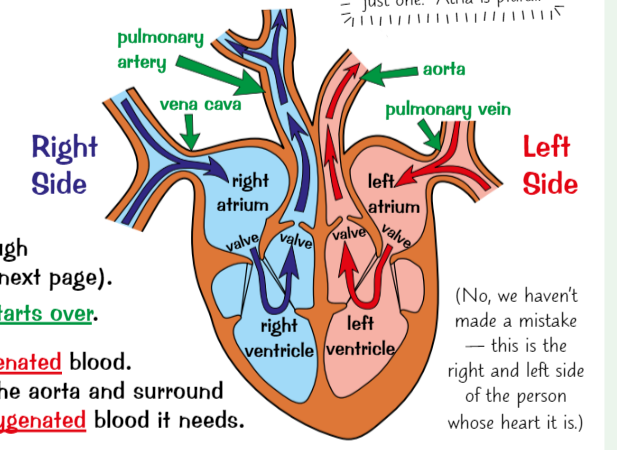
Whats the heart? What are valves? How does the heart use its 4 chambers (5 points)
heart = pumping organ that keeps blood flowing around body, walls of heart are mainly made of muscle tissue
Heart has valves to make sure blood flows in right direction (prevents it flowing backwards)
This is how the heart uses its 4 chambers (right atrium, right ventricle, left atrium & left ventricle) to pump blood around:
Blood flows into 2 atria from vena cava & pulmonary vein
Atria contract, pushing blood into ventricles
Ventricles contract, forcing blood into pulmonary artery & aorta, & out of heart
blood flows to organs through arteries & return through veins
atria fill again & whole cycle starts over
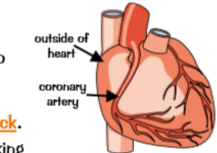
(heart also needs its own supple of oxygenated blood, Arteries called coronary arteries branch off aorta & surround heart, making sure it gets all oxygenated blood it needs)
whats a pacemaker? they produce? whens artificial pacemaker used?
resting heart rate is controlled by group of cells in right atrium wall that act as pacemaker.
These cells produce small electric impulse which spreads to surrounding muscle cells causing them to contract
artificial pacemaker often used to control heartbeat if natural pacemaker cells don’t work properly (patient has irregular heartbeat), little device that’s implanted under skin & has wire going to heart →produces electric current to keep heart beating regularly
3 different types of blood vessels?
Arteries → carry blood Away from heart
Capillaries → involved in exchange of materials at tissues
Veins → carry blood To the heart
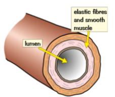
Why and how do arteries carry blood under pressure?
Heart pumps blood out at high pressure so artery walls are strong & elastic
Walls are thick compares to size of hole in middle → lumen
Contain thick layers of muscle → make them strong & elastic fibres to stretch & spring back
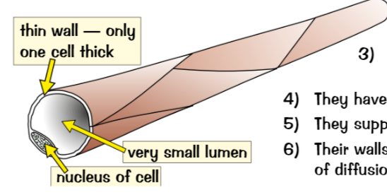
why & how Capillaries are really small
arteries branch into capillaries
Really tiny (too small to see)
Carry blood really close to every cell in body to exchange substances with them
Have permeable walls → substances can diffuse in & out
Supply food & oxygen, take away waste like CO2
Walls usually one cell thick → increases rate of diffusion by decreasing distance
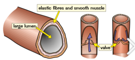
How & why do veins take blood back to the heart?
Capillaries join up to form veins, blood is at lower pressure in veins so walls don’t need to be as thick as artery walls
Bigger lumen than arteries to help blood flow despite lower pressure
Valves to help keep blood flowing in right direction
Calculate the rate of blood flow
Rate of blood flow = volume of blood divided by number of minutes
units = ml/min
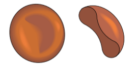
How & why do red blood cells carry oxygen?
function → carry oxygen from lungs to all of cells in body
Shape = biconcave disc (donught) → gives larger SA for absorbing oxygen
Dont have a nucleus → more room to carry oxygen
In lungs, haemoglobin binds to oxygen = oxyhaemoglobin, in body tissues they split up into haemoglobin & oxygen → release oxygen to cells
how do white blood cells defend against infection? (3 points)
Some change shape to gobble up unwelcome microorganisms → phagocytosis
Others produce antibodies to fight microorganisms & antitoxins to neutralise any toxins produced by microorganisms
Contain a nucleus
How do platelets help blood clot?
small fragments of cells that have no nucleus
help blood clot at wound → stop blood pouring out & stop microorganisms getting in (float around waiting for accidents)
Lack of platelets → cause excessive bleeding & bruising
What does plasma the liquid that carries everything in blood carry? (7 points)
Red & white blood cells & platelets
Nutrients → glucose & amino acids (soluble products of digestion, absorbed from gut & taken to cells of body)
Carbon dioxide from organs to lungs
Urea from liver to kidneys
Hormones
Proteins
Antibodies & antitoxins produced by white blood cells
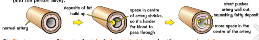
What is coronary heart disease? What are stents?
Coronary heart disease → coronary arteries that supply blood to muscle of heart get blocked by layers of fatty material building up → arteries become narrower so blood flow is restricted & there’s lack of oxygen to heart muscle → result in heart attack
Stents → tubes that are inserted inside arteries, keep them open making sure blood passes through to heart muscles → keeps persons heart (beating & person alive)
Risks & benefits of stents
Stents are a way of lowering risk of heart attack in people with coronary heart disease → effective for long time & recovery time from surgery is relatively quick
On down side → risk of complications during operations (heart attack) & risk of infection from surgery. Also risk of developing blood clot near stent → thrombosis
Whats cholesterol? Too much of LDL cholesterol? What are statins?
Cholesterol → essential lipid body produce & needs to function, too much of a certain type of cholesterol (‘bad’/ LDL cholesterol) can cause health problems
Too much of LDL cholesterol in bloodstream can cause fatty deposits to form inside arteries → leads to coronary heart disease
Statins → drugs that reduce amount of LDL cholesterol present in bloodstream, slows down rate of fatty deposits forming
3 Advantages of statins
reduce amount of LDL cholesterol in blood → reduce risk of strokes, coronary heart disease & heart attacks
Reduce amount of LDL cholesterol, statins increase amount of beneficial cholesterol (HDL cholesterol) in bloodstream → removes LDL cholesterol from blood
studies suggest Statins may also help prevent other diseases
3 disadvantages of statins
statins → long-term drug that must be taken regularly, risk someone could forget to take them
Statins can cause negative side effects → headaches, serious side effects can be kidney failures, liver damage & memory loss
Effect of statins isn’t instant, takes time for their effect to kick in
Whats a artificial heart?(advantage & 3 risks)
artificial hearts (mechanical devices) → pump blood for person whose own heart has failed, usually used as temporary fix → keep person alive until donor heart is found/ help person recover by allowing heart to heal & rest. Some cases → used as permanent fix (reduces need for donor heart)
Main advantage → less likely to be rejected by immune system than donor heart → made from metals/ plastics so body don’t recognise them as foreign
Risk → needs surgery → lead bleeding & infection. Don’t always work as well → parts of heart could wear out/ electrical motor fails. Blood doesn’t flow as smoothly → cause blood clots & lead to strokes (has to take drugs → thin blood & make sure no problems with bleeding if hurt in accident)
What happens to valves? What may damage cause? What is severe valve damage treated w? Replacing valves less drastic procedure than…?
valves in heart can be damaged or weakened by heart attacks, infection or old age
Damage may cause valve tissue to stiffen → wont open properly. Or valve becomes leaky → allowing blood flow in both directions = blood doesn’t circulate as effective
Severe valve damage treated by replacing valves → taken from humans/ other mammals (biological) or man-made (mechanical)
Replacing valve less drastic procedure than whole heart transplant → fitting artificial valves still major surgery & problems with blood clots
If someones loses lots of blood…? Whats artificial blood? Arficial blood product would replace..?
someone loses lots of blood → heart still pumps but remaining blood cells around (get oxygen to organs) long as volume of blood can be topped up
Artificial blood → blood substitute → salt solution (saline) used replace lost volume of blood → safe (if no air bubbles) & keeps people alive even if lose 2/3 of red blood cells → give time to produce new blood cells, if not patient needs blood transfusion
Artificial blood product would replace function of lost red blood cells → no need for blood transfusion, scientist currently working on products like that
What are communicable diseases? Caused by? sometimes described? Examples? What are non-communicable diseases? description? Examples?
communicable disease → can spread from person to person/ between animals & people.
Caused by bacteria, viruses, parasites & fungi
Sometimes described → contagious/ infectious diseases
Examples → measles & malaria
Non-communicable diseases → cannot spread between people/ between animals & people
Generally last long time & get worse slowly
Examples → asthma, cancer & coronary heart disease
Problems w immune system? Types of cancer? Immune system reactions? Mental health issues?
problems with immune system → increased chance suffering from communicable diseases (influenza - flu) → body less likely able to defend itself against pathogen (cause disease)
Types of cancer → triggered infection by certain virus → infection with types of hepatitis virus cause long-term infections in liver (virus lives in cells) → lead to increased chance developing liver cancer. Another example → infection with HPV → cause cervical cancer in women
Immune system reactions → caused infection by pathogen → trigger allergic reactions (skin rashes/ worsen symptoms of asthma - asthma sufferers)
mental health issues → depression → triggered by suffering from severe physical health problems → particularly if impacts persons ability to carry out everyday activities/ affect life expectancy
Other factors affecting health (diet, stress, situation)
Good, balanced diet that provides body with everything it needs & right amounts, poor diet → affect physical & mental health
Stress → constantly under lots stress can lead to health issues
Life situation → easy access to medicines to treat illness/ access to things that prevent you getting ill in first place (healthy food, condoms - prevent transmission of STD)
Whats a risk factor? Examples? Many non-communicable diseases r caused by? give examples of lifestyle factors nationally & globally
risk factors → linked to increase in likelihood a person develops certain disease during lifetime (not guaranteed)
Risk factors → often aspects of persons lifestyle (exercise)/ presence of certain substances in environment (air pollution)/ substances in body (asbestos fibres - block airways & cause diseases → cancer)
Many non-communicable diseases caused by several different risk factors interacting with each other
lifestyle factors have different impacts locally, nationally & globally
(developed countries non-communicable diseases more common → higher income & buy high-fat food. Nationally, deprived areas → more likely smoke, poor diet & not exercise. cardiovascular disease, obesity, type 2 diabetes higher in those areas. individual choices affect local incidence of disease)
5 risk factors that have been proven to cause what diseases?
smoking → proven directly cause cardiovascular disease, lung disease, lung cancer ( damage walls of arteries & cells in lining of lungs)
Obesity → thought directly cause type 2 diabetes (make body less sensitive/ resistant to insulin → struggles control concentration of glucose in blood)
Too much alcohol → proven directly cause liver disease, can affect brain function (damage nerve cells in brain causing it to lose volume)
Smoking when pregnant → lots of health problems for unborn baby (drinking alcohol similar)
Cancer → directly caused by exposure to certain substances/ radiation. Things that cause cancer → carcinogens → Ionising radiation (X-rays) example
How can non-communicable diseases be costly? (human cost, financial cost)
Tens of millions die from non-communicable diseases per year, people with these may have lower quality of life/ shorter lifespan → affects sufferers themselves & loved ones
Financial cost → families may have to move/ adapt their home to help family member with disease (costly) → if family member with disease has to give up work/ dies, families income reduced (can affect country’s economy)
Whats is uncontrollable growth & division a result of? What are the 2 types of cancers?
Uncontrolled growth & division → result of changes occur to cells & result in formation of tumour (mass of cells), not all tumours cancerous:
Benign → where tumour grows until no more room, stays in 1 place (usually within membrane) rather than invading other tissues in body (type isn’t normally dangerous & tumour isn’t cancerous)
Malignant → where tumour grows & spreads to neighbouring healthy tissues, cells break off & spread to other parts of body by travelling in bloodstream. Malignant cells invade healthy tissues in body & form secondary tumours (are dangerous & can be fatal - cancers)
risk factors associated with lifestyle (cancers) (4 factors)
Smoking → lung cancer & mouth, bowel, stomach & cervical cancer
Obesity → bowel, liver & kidney cancer (2nd biggest preventable cause of cancer after smoking)
UV exposure → exposed to UV radiation from sun have increased change developing skin cancer, sunny climates & people who spend lots time outside & frequent use sun beds → higher risk of skin cancer
Viral infection → increase changes certain type cancer, example: infection with hepatitis B & hepatitis C virus increase risk liver cancer. Likelihood becoming infected depends on lifestyle (unprotected sex/ sharing needles)
Risk factors associated with genetics (cancers) (example)
sometimes inherit faulty genes → more susceptible to cancer
for example → mutations in BRCA genes linked to increase developing breast & ovarian cancer
How are plant cells organised into tissues & organs (made, covers, leaf, spaces, transport, growing)
made of organs (stems, roots & leaves), work together to make organ systems (Perform various tasks plants need to carry out to survive & grow → transporting substances). Plant organs made of tissues:
Epidermal tissue → covers whole plant
Palisade mesophyll tissue → part of leaf where photosynthesis happens
Spongy mesophyll tissue → part of leaf & contains big air spaces to allow gases to diffuse in & out of cells
Xylem & phloem → transport things (water, mineral ions & food) around plant (through roots, stems & leaves)
Meristem tissue → found at growing tips of shoots & roots, able to differentiate into lots different types plant cells = allows growth
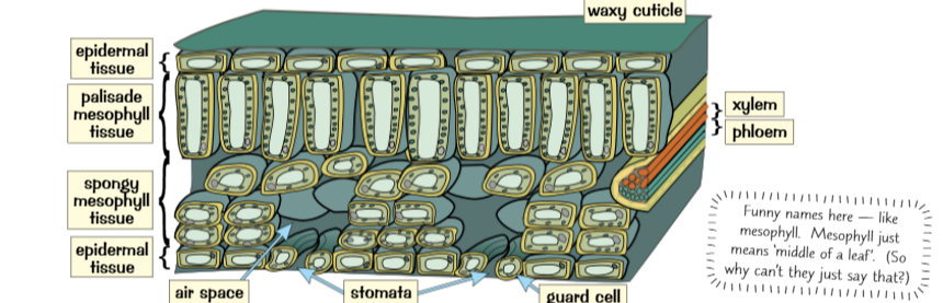
structure and function: epidermal tissue? Upper epidermis? Palisade layer? Xylem & phloem? Tissues of leaves? Opening and closing stomata?
Epidermal tissue → covered with waxy cuticle, helps reduce water loss by evaporation
Upper epidermis → transparent so light can pass through it to palisade layer
Palisade layer → lots chloroplasts (photosynthesis takes place) near top leaf where can get most light
Xylem & phloem → form network of vascular bundles, deliver water & other nutrients to entire leaf & take away glucose produced by photosynthesis , help support structure
Tissue of leaves adapted → efficient gas exchange → lower epidermis full of little holes (stomata) let CO2 diffuse directly into leaf.
Opening & closing of stomata → controlled by guard cells (response to environmental conditions). Air spaces in spongy mesophyll tissue → increase rate of diffusion in gases
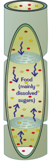
How do phloem tubes transport food (made, transport, direction, process)
Made of columns of elongated living cells with small pores in end walls to allow cell sap flow through
Transport food substances (mainly dissolved sugars) made in leaves to rest of plant for immediate use (in growing regions)/ for storage
Transport goes in both directions
This process is called translocation
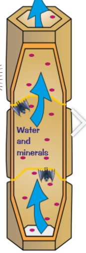
How do xylem tubes take water up (process)
made dead cells joined end to end with no end walls between & hole down middle, strengthened with material → lignin
Carry water & mineral ions from roots to stem & leaves
Movement of water from roots, through xylem & out leaves→ transpiration stream
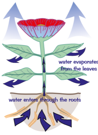
what is transpiration caused by, what does it create, what does it mean? (what’s it a side-effect of)
transpirations → caused by evaporation & diffusion of water from plants surface, most transpiration happens at leaves
Evaporation creates slight shortage of water in leaf & more water is drawn up from rest of plant through xylem vessels to replace it
Means more water is drawn up from roots & there’s constant transpiration stream of water through plant
(side-effect of way leaves adapted for photosynthesis, have stomata so gases exchange easily because more water inside than air outside, escapes from leaves through stomata by diffusion
4 main things affecting transpiration rate ( light, temperature, air, humidity)
Light intensity → brighter the light = greater transpiration rate. Stomata begin close when dark (photosynthesis isn’t happening) don’t need to let CO2 in, very little water escapes
Temperature → warmer = faster transpiration happens ( more energy evaporate & diffuse out stomata)
Air flow → better air flow around leaf = greater transpiration rate. Air flow poor → water vapour surrounds leaf & inside it (diffusion doesn’t happen quickly). Good air flow → water vapour swept away maintaining higher concentration to area lower concentration (air), diffusion happens quickly
Humidity → drier air around leaf = faster transpiration happens. Air humid → lots of water in already ( not much difference between inside & outside), diffusion happens fastest if really high concentration to really low concentration
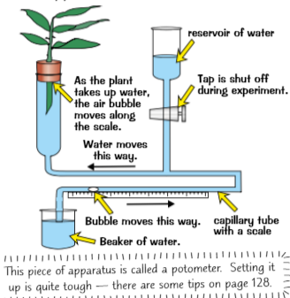
Estimate rate of transpiration
measure uptake of water by plant, can assume water uptake by plant is directly related to water loss by leaves (transpiration)
Set up apparatus in diagram & record starting position of air bubble, start stopwatch & record distance moved by bubble per unit time ( oer hours)
keep conditions constant throughout (Temperature & air humidity)
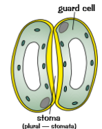
How are guard cells adapted to open & close stomata (7 points)
kidney shape → opens & closes stomata
plant has lots water guard cells fit with it & go plump & turgid → stomata open so gases can be exchanged for photosynthesis
Plant is short of water, guard cells lose water & become flaccid → making stomata close (helps stop water vapour escaping)
Thin outer cells & thickened inner walls make opening & closing worksensitive to light & close at night to save water without losing out on photosynthesis
Find more stomata on underside of leaves→ lower surface shaded & cooler (Less water lost through stomata than if were on upper surface)
Guard cells adapted → for gas exchange & controlling water loss within leaf