UFCVM 1st Year Parasitology Final Exam
1/294
Earn XP
Name | Mastery | Learn | Test | Matching | Spaced | Call with Kai |
|---|
No study sessions yet.
295 Terms
What is the basic morphology of trematodes?
Dorsoventrally flattened
Solid body
Variable shape and size
Tegument is either smooth or covered with tiny spines
Anterior oral sucker and ventral acetabulum
Digestive system (oral sucker → pharynx → esophagus → 2 blind ceca (either branched or unbranched)
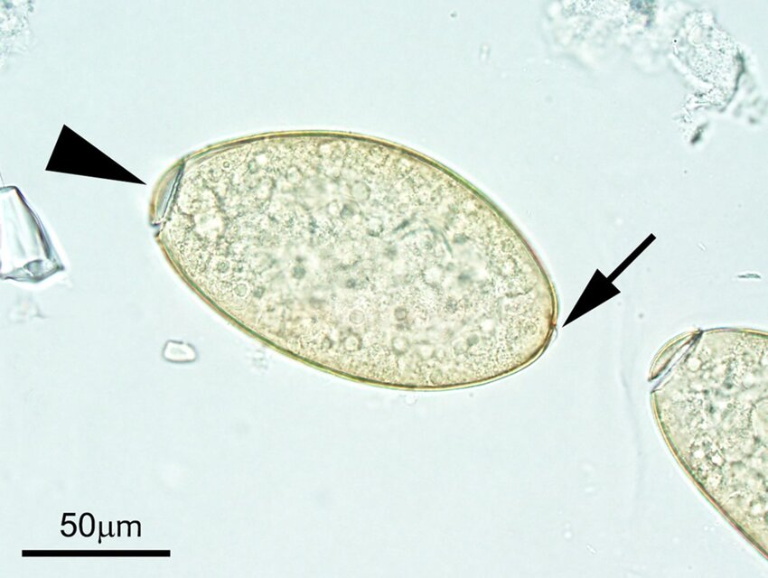
Trematode Egg
Operculated
Contains embryo and yolk cells
Colorless to dark brown
What type of ceca does this trematode have?
A simple ceca

What type of ceca does this trematode have?
A branched or dendritic ceca
How do trematodes reproduce?
Trematodes are hermaphroditic, meaning they possess both male and female reproductive organs, allowing them to produce eggs and cross-fertilize.
What characteristics differentiate schistosomes from other trematodes?
Dioecious (separate males and females)
Females lie in the male’s gynecophoral canal
Live in blood vessels, not small intestines
Eggs lack operculum and may lack spines
Infects host by skin penetration
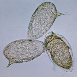
Schistosome Egg
Lacks operculum
May have spines
What is the general life cycle of trematodes?
Adults produce operculated eggs → Ciliated miracidia larve develops within and emerges → miracidium infects a snail host → sporocyst forms → rediae develop → cercariae encysts, enters water and is either ingested or attaches to vegetation, loses tail→ metacercariae infects definitive host and excyst in small intestine and migrates→ adult trematodes mature in host
What is the general life cycle of a schistosome?
Adults produce eggs that are expelled in urine or feces → eggs hatch into miracidia → miracidia infect snails → sporocysts form → cercariae released into water → cercariae penetrate skin of definitive host → adults reside in blood vessels.
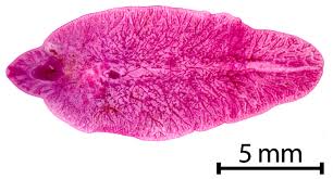
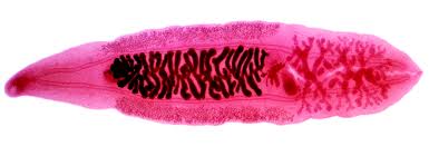
Identify this parasite
Fasciola hepatica
Broad “shoulders”
Cephalic cone
Tegument covered in spines
Clouded morphology
What are the characteristics of Fasciola hepatica?
Mainly distributed in SE and W. US (need IH aquatic snail)
Definitive hosts (DH): Cattle, sheep, goats, and other ruminants
Zoonotic
Lives in bile ducts
Operculated egg
How do F. hepatica larvae migrate?
Juvenile flukes penetrate the small intestinal wall, enter the abdominal cavity, penetrate and migrate through the liver, and enter the bile ducts and mature
Prepatent period: ~5 months
What is “Acute Fascioliasis?”
A syndrome caused by large amounts of F. hepatica metacercariae infecting a host in a short period.
Causes: traumatic hepatitis and Black Disease (2ry bacterial disease that proliferates and damages tissues)
What is chronic fascioliasis?
A syndrome caused by the presence of 200-500 F. hepatica metacercaria that extends over a certain period of time. Causes hepatic fibrosis, hyperplastic cholangitis, and pipestem liver (cattle).
How do you diagnose F. hepatica?
Clinical signs and presence of snail IH
Eggs can be identified in fecal sedimentation
Postmortem lesions and presences of flukes in liver bile ducts
How do you prevent and control F. hepatica"?
Improve drainage and fence off flood-prone areas
Anthelmintic treatment to reduce environmental contamination
Vaccinate against black disease
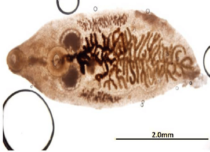
Identify this parasite
Platynosomum fastosum
Brown, operculated eggs
What are the characteristics of Platynosomum fastosum?
Distributed in Florida, other SE states, and Hawaii
DH: cats
They inhabit bile ducts
What is the lifestyle of P. fastosum?
Adults live in bile ducts→ eggs containing miracidia shed in feces→ eggs ingested by terrestrial snail (1st IH)→ develops into sporocysts→ leaves snail and develops into cercariae→ ingested by pill bugs (2nd IH) and encysts→ Pill bugs are ingested by Anolis spp. lizards (paratenic host)→ Lizards are ingested by cats→ Metacerciae excyst and enter bill duct/gall bladder→ Matures
How does disease manifest with DH are infected with P. fastosum?
Normally disease is not severe, temporary inappetence
Chronic infection can lead to biliary hyperplasia (fibrosis, cholestasis, hepatic failure)
“Lizard poisoning” → anorexia, lethargy, depression, v/d, fever, jaundice, death
How do you diagnose P. fastosum?
Eggs can be found in fecal sedimentation (only if the bile ducts are not occluded)
Ultrasound can be used to identify blocked bile ducts
Necropsy can show flukes in the bile ducts and hepatic lesions
How do you prevent P. fastosum infection?
Prevent predation of lizards by DH
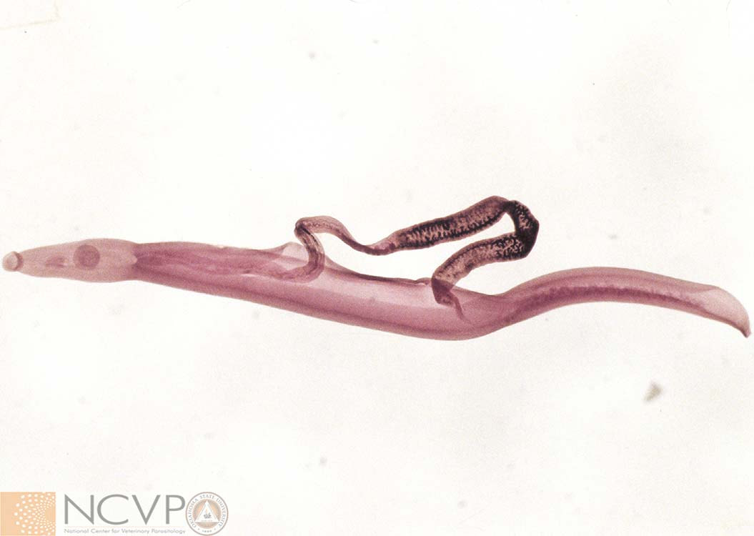
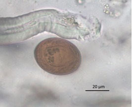
Identify this parasite
Heterobilharzia americana
Schistosome
Non-operculated egg with miracidium
What are the characteristics of Heterobilharzia americana?
Distributed mostly along the Gulf Coast, southern Atlantic coast (starting to migrate west)
DH: Dogs, racoons (natural host), bobcats, horses
Females reside in males’ gynecophoral canal
What is the life cycle of H. americana?
Adults reside in mesenteric and hepatic veins→ eggs are laid in veins then pass through SI wall and are shed into feces→ Miracidium emerges in water and penetrates freshwater snail IH→ Develops into sporocysts→ Develops into cercariae→ Cercariae leaves the snail and penetrates the DH and migrates to veins→ Adults pair up and mate
Prepatent period: ~10 weeks
How does disease present with a H. americana infection?
Is often asymptomatic with adults
Cercarial penetration can lead to a rash (“Swimmer’s itch” in humans)
Eggs in tissue can cause granulomatous inflammation and calcification
Possible clinical signs
Lethargy
Weight loss
Inappetence
V/D
Hematochezia (fresh blood from anus)
How do you diagnose a H. americana infection?
Eggs can be found in a fecal sedimentation using 0.9% saline
PCR on fecal or tissue biopsy
Histopathology
How do you prevent a H. americana infection?
Avoid fresh bodies of water in endemic areas
What is a cestode?
Flat, solid-bodied parasite
Hermaphroditic
Segmented - muscular systems enables movements
Endoparasitic
Adults live in GI tract
What are characteristics of adult cestodes?
No body cavity
No digestive tract - absorbs nutrients through tegument
At least one set of male and female reproductive organs per segment
Can be tiny or large
What are characteristics of cyclophyllidean tapeworms?
Scolex to attach them to the intestines
Have 4 suckers (acetabula)
Armed scolex v. unarmed
Neck has a germinal region where body segments are generated
Strobila - chain of proglottids
Proglottids
Contains reproductive organs
Genital pores (where sexual reproduction occurs)
Nervous system
Excretory system
What are the different types of proglottids and what differentiates them?
Immature: Most proximal to neck, not yet functional
Mature: Middle of body, fully functional
Gravid: Most distal; degenerated; only the uterus remains
What is the general life cycle for cyclophyllideans?
Adults reside in GI tract→ Gravid proglottids shed in feces, disintegrates, and releases eggs→ Eggs containing hexacanth embryo is ingested by IH→ Larval cestodes (metacestodes) develops→ IH is ingested by DH and metacestodes develop to adults and reproduce by self- or cross-fertilization
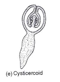
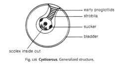
What characterizes this metacestode?
Single scolex and in invertebrate IH

What metacestode type is this?
Single scolex in vertebrate IH
What metacestode type is this?
Single scolex in vertebrate IH
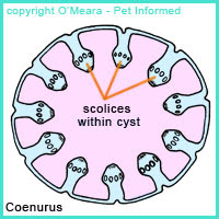
What metacestode type is this?
Many scolices vertebrate IH
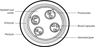
What metacestode type is this?
Many many protoscolices with vertebrate IH
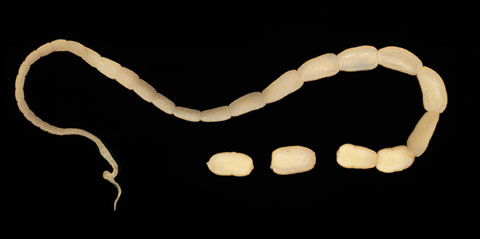
Identify this parasite
Dipylidium caninum
Egg packets contain multiple eggs with hexacanth (6 hooks) embryo
What are the general characteristics of Dipylidium caninum?
Distributed worldwide
DH: Canids, felids, humans (zoonotic)
They inhabit the small intestines
Adults have 4 suckers, a retractable rostellum with hooks, and bilateral genital pores (2 sets of M/F organs)
What is the lifestyle of D. caninum?
Gravid proglottids are released in feces→ Proglottids rupture and release egg packets→ Egg packets are ingested by larval fleas or chewing louse (IH)→ Embryo develops into cysticercoid→ IH is ingested by DH→ Scolex attaches to SI and develops into an adult
Prepatent period: ~2-3 weeks
How does disease presents with a D. caninum infection?
Usually the adults are non-pathogenic
Scooting due to crawling proglottids (itchiness)
Heavy infections can lead to constipation or diarrhea, unthriftiness, or pot belly
Impaction is sometimes reported in small puppies with heavier infections
How do you diagnose D. caninum?
Proglottids are identified by their 2 lateral genital
Can be broken open, mixed with saline, and examined for egg packets
Looks like dried rice granules
A fecal flotation with Sheather’s solution can be used to identify eggs (usually only seen when proglottid is present)
Coproantigen ELISA
How do you prevent/control D. caninum infection?
Control the flea and louse population
Do regular fecal monitoring and treat as needed
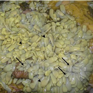
Identify this parasite
Anoplocephala perfoliata
Most commom and most pathogenic in N. America
Live in SI and LI (ileocecal valve)
Identify this parasite
Anoplocephala magna
Resides in the SI
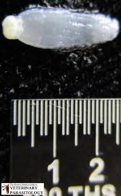
Identify this parasite
Anoplocephaloides mamillana
Resides in the small intestine
What are the general characteristics of Anoplocephalid tapeworms?
They are distributed worldwide
DH: Equids
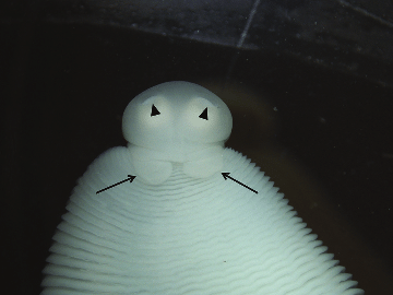
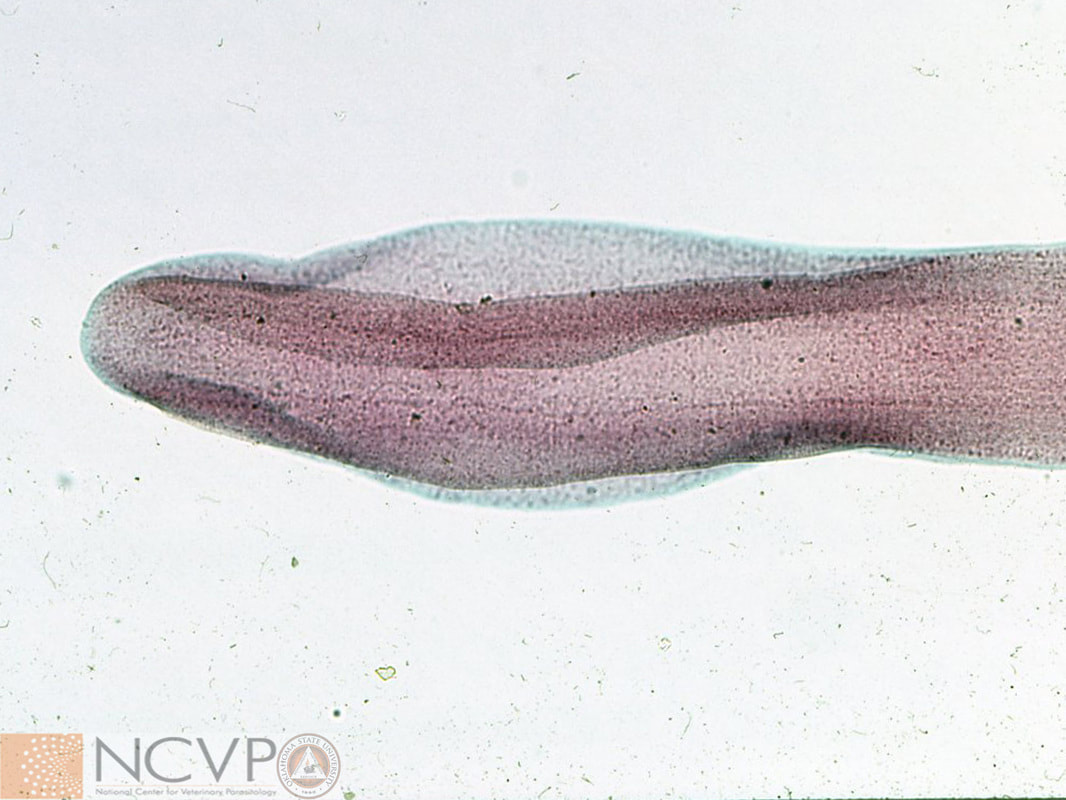
Identify this structure on A. perfoliata
Lappets “bunny ears” - located on the anterior end of the parasite
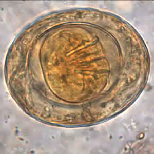
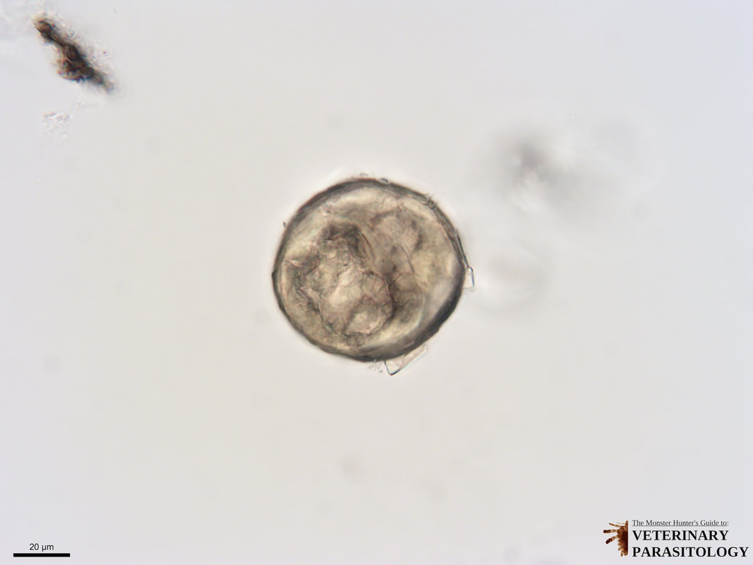
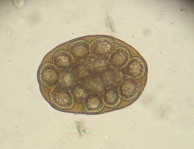
What type of egg is this?
Anoplocephalid spp.
Hexacanth embryo
Pyriform apparatus (pear-shaped structure surrounding the embryo)
What is the life cycle of Anoplocephalid spp.?
Adults live in the GI tract→ Gravid proglottids pass through feces and disintegrates→ Eggs are ingested by oribatide mite (IH) and develops into a cysticercoid→ Horses ingests the mite on grasses/grains→ Scolex attaches to intestines and matures into an adult
Prepatent period: ~2 months
How is disease presented in an Anoplocephalid spp. infestation?
Light infections usually are asymptomatic but heavy infections can be deadly
A. perfoliata: Ulcers at attachment sites increase the likelihood of intussusception
A. magna: Hemorrhagic enteritis
A. mamillana: Minimal pathology
How do you diagnose an Anoplocephalid spp. infection?
Perform a fecal examination - can sometimes find proglottids and perform sedimentation and flotations with Sheather’s solution
ELISA detects antibodies
PCR on feces
Necropsy
How do you control Anoplocephalid spp. infection?
If infection is detected, treat all horses on property
What are the general characteristics of pseudophyllidian tapeworms?
Has an aquatic part of the life cycle
Scolex has 2 deep, weakly muscular grooves (bothria) used for attachment
Proglottids have 1 set of M/F organs that are centrally located
Medial genital pore
Medial uterine pore where eggs can exit and mix with feces
What is the general life cycle of pseudocyclophyllidean tapeworms?
Adults live in the SI→ Operculated eggs are released in feces→ Develops into ciliated coracidium→ Coracidium is ingested by 1st IH (aquatic crustacean) and develops into a procercoid→ 1st IH is ingested by a 2nd IH (vertebrate) and develops into a plerocercoid→ 2nd IH is ingested by DH→ Develops into adults and reproduce by self- or cross-fertilization
Identify this parasite
Spirometra mansonoides
Psuedocyclophyllidean tapeworm
Operculated egg
What are the characteristics of Spirometra mansonoides?
Distributed in the S. US, Hawaii, and a few NE states
DH: Cats, dogs, racoons
They inhabit the small intesting
Zoonotic (humans are IH)
What is the life cycle of S. mansonoides?
Adults reside in the SI→ Eggs are shed into feces→ In water, coracidium develops and emerges→ Coracidium is ingested by microscopic copepod (1st IH) and develops into procercoid→ Copepod is ingested by 2nd IH (frog, snake, rat, bird) and develops into plerocercoid/sparganum→ DH ingests 2nd IH→ Plerocercoid attaches to SI and matures into adults
Prepatent period: 10-30 days

How does disease present with a S. mansonoides infection?
Infections with adult tapeworms is mostly asymptomatic, but can see v/d and weight loss with severe infections
Sparganosis (larval tapeworms) infect normal DH.
IH can get an infection from ingesting 1st or 2nd IH or using IH as a poultice (cloth used for wounds)
Migrating spargana can lead to painful subcutaneous nodules
How is a S. mansonoides infection diagnosed?
Perform a fecal floatation in Sheather’s solutions
Proglottids can be seen as chains in vomit or feces
Identify proglottids based off of their medial uterine pore
How do you prevent a S. mansonoides infection?
Prevent DH predation on IH
Avoid drinking water that may contain copepods
Regularly monitor fecal and treat as needed
What are general characteristics of Oxyroidea spp?
Double-bulbed esophagus
Direct life cycle (no migration)
Can use “scotch tape” to identify eggs on perineum
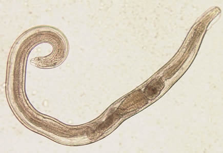
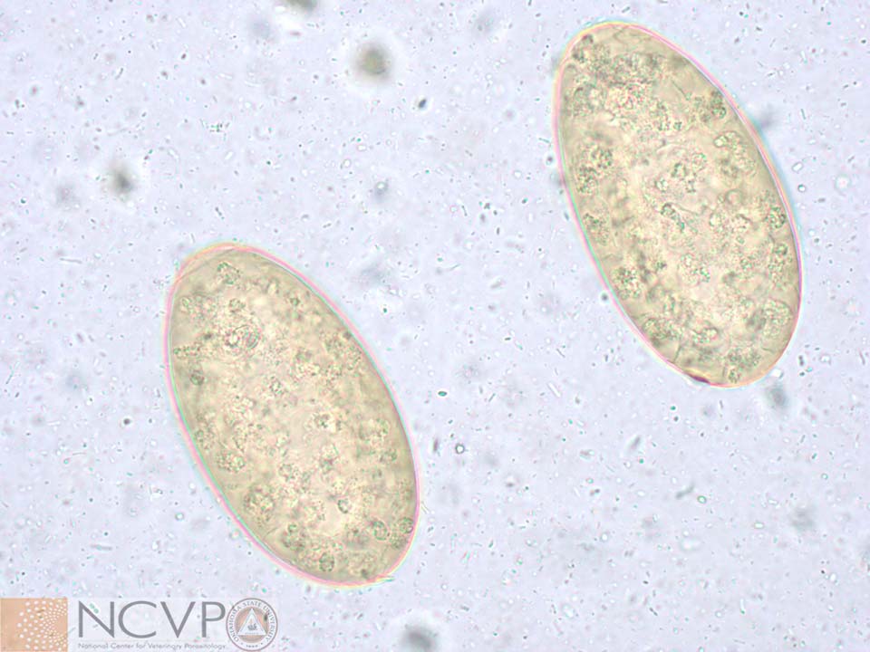
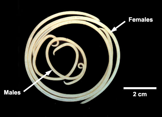
Identify this parasite
Oxyuroidea equi
Nematode
Asymmetrical, operculated/plugged, non-larvated eggs
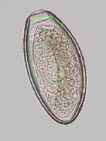
What are the characteristics of Oxyuris equis?
Resides in the LI of horses
Common distribution pretty much everywhere
Adults - whitish, thick-bodied, club shapes esophagus
Females are much larger than males
Not zoonotic
What is the life cycle of O. equi?
Transmission is from horse→ horse
Adult female deposits eggs in perianal region in a sticky matrix then returns into the horse→ eggs fall and larvate in the environment→ Larvated eggs ingested by horse→ Eggs hatch and larvae develops in crypts of cecum and ventral colon→ Larvae leaves crypt, enters lumen, and move to the dorsal colon where they develop into adults
How does disease present with an O. equi infection?
No major pathology - pinworms aren’t too bad!
“Pruritis Ani” or “Seat itch” can lead to hair loss or secondary bacterial infections
May cause colitis, but this is not the primary source of pathology
Summer sores - thickened, scaly, “rat tailed” appearance
How do you diagnose O. equi?
Eggs can be collected by “scotch tape” method or by scraping them with a tongue depressor and applying mineral oil
Diagnose infection based on clinical signs and history
How do you prevent an O. equi infection?
Use proper sanitary practices
Use routine anthelmintic use
What are general characteristics of Ascoridea spp?
Very large (>6” in length)
3 anterior lips
Direct life cycle (sometimes will infect paratinic hosts) with often larval migration
Non-larvated eggs in feces
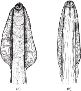
Identify this parasite
Toxocara canis
Round, non-larvated eggs
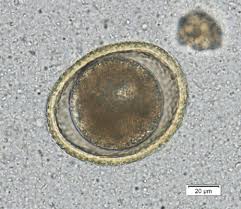
What are the characteristics of Toxocara canis?
Live in the small intestine of dogs
Females are larger than males
One of the largest nematodes
Have muscular groves called alae (pictured)
What is the life cycle of T. canis?
Adults are fond in the SI of the DH→ Non-larvated eggs leave DH in feces→ Eggs larvate in the environment→ Larvae infects new DH through 1) Ingestion, 2) Prenatal/transuterine transmission, 3) Ingestion of paratenic host→ Larvae develops into an adult
Direct transmission of T. canis in dogs < 3 months old
Direct ingestion of infective stage (L3)
Larvae undergo “tracheal migration”
Larvae hatch from the egg in the duodenum, penetrate the intestines, and migrate to the lymph nodes.
Larvae will then migrate to the liver, heart, or pulmonary arteries
L3 larvae will infect the alveoli, bronchioles, trachea, and stomach. L3 can be coughed up and swallowed
Larvae molt to L4/L5 in SI where they mature into adults
Prepatent period: 3-4 weeks
Direct transmission of T. canis in dogs > 3 months old
Ingests the infective stage (L3)
“Somatic migration” where larvae hatches from egg in the duodenum
Larvae penetrates the intestine and enters systemic circulation
L3 larvae encysts, becoming hypobiotic, in various tissues until conditions are favorable and the larvae is prompted by the dog’s immune system to become active again
No maturation occurs
Prenatal transmission to DH
Larvae become hypobiotic (mobilize day 42 or later of pregnancy)
The larvae migrates to the fetus and lives in the liver of the fetus
L3 migrates to the lungs at birth, infecting the alveoli, bronchioles, trachea - they are coughed up, swallowed, then enters the stomach
L4/L5 in SI and mature into adults in about 2 weeks
Eggs are found in puppy feces by23-40 days old
Transmission of T. canis in paratenic hosts
DH ingests a paratenic host with encysted L3s (rodents usually)
L3s enter directly into the stomach, no migration needed
How does disease present with a T. canis infection?
Infection is more problematic in young puppies with undeveloped immunity
Heavy infection - death is rare, but may occur due to tracheal migration
Pneumonia
V/D
Pot belly
Focal lesions on CNS (migrations) leading to neurologic disorders (rare)
How do you diagnose T. canis?
Eggs can be found in a fecal float
Adults can be seen in feces or vomitus
How do you control a T. canis infection?
Eggs are persistent in the environment due to their sticky, durable outer coating
Remove feces daily (good sanitation)
Control paratenic hosts (rodents)
Routine antihelminth treatment
Zoonosis of T. canis
Visceral Larval Migrans (VLM)
Chronic granulomatous lesions due to larval migrations when humans ingest L3
Often infects liver, lings, brain, eye
Enlarged liver
Leads to weight loss, appetite, persistent cough
Humans are paratenic hosts
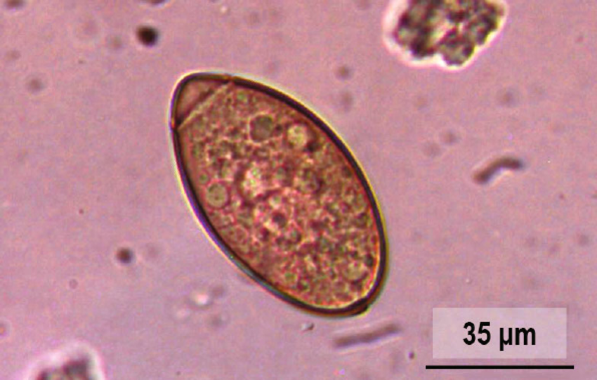
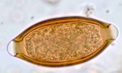
What are general characteristics of Trichuroidea spp.?
Short, fat posterior ends; long whip-like anterior end
Beaded esophagus (stichocyte)
Usually have a direct life cycle
Example of Trichuris egg pictured
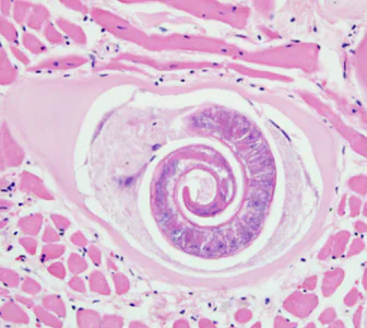
Identify this parasite
Trichinella spiralis
What are the general characteristics of Trichinella spiralis?
Adults live in SI and larvae encysts in striated muscles of many mammals
Female worms are much bigger
Female produce eggs that hatch in her uterus and “gives birth” (larviparous)
Nurse cells use host tissues to protect larvae from the immune system of the host and drugs in muscle tissues
What is the epidemiology of T. spiralis?
Development in both sylvatic (from wild carnivores and prey animals) and urban (between pets/domesticated animals and humans) cycles.
Zoonotic infections occur through the ingestion of raw meat
How does T. spiralis develop in hosts and what are their pathology?
Limited pathology in swine
Intestinal infection with adults - Direct infection; clinical signs develop 1-2 days post-infection
Nausea
v/d
Fatigue
Fever
Abdominal discomfort
Skeletal muscle invasion by L1 - L1 encysted in muscles are ingested; clinical signs develop 2 weeks post-infection
Eosinophilia
Fevers
Chills
Cough
Periorbita edema
Joint/muscle pain
L1 larvae enter lymphatics, migrate through the heart/lungs and enter circulation (goes to striated muscles)
How do you diagnose T. spiralis?
Muscle biopsy
Serum samples (IgG, IgM, IgE is detectable for up to two years, but is after the fact information). There’s nothing you can do once the larvae encysts in muscle
How do you control a T. spiralis infection?
Prevent cannibalism
Cook meat thoroughly
Freezing for some species
What are general characteristics of Trichostrongyloidea spp.?
Hairlike worms with tiny mouths
Males have a copulatory bursa with similar spicules
Direct life cycles
Usually found in the GI tract
Non-larvated eggs
Fairly small
What are the characteristics of Trichostongyles?
Small, bursate, mostly in ruminants
Inhabit abomasum (4th chamber of the stomach) and SI
What is the general lifestyle of Trichostrongyle?
Eggs are shed in feces→ L1 develop into L3 (infective stage) and feed on fecal bacteria→ L3 ingested while DH grazes→ L3 enters the wall of the abomasum or SI and develop into L4→ L4 develop into lumen-dwelling adults in mucosa of predilection site→ No migration
Prepatent period is about 3 weeks, but L4 can undergo hypobiosis
What is Parasitic Gastroenteritis (PGE)?
A disease caused by a complex of mainly Trichostrongyloid nematodes that affects cattle and small ruminants (predominately)
Signs:
Rough hair coat
Diarrhea (more pronounced in cattle)
Anemia (more pronounced in sheep and goats)
Submandibular edema (bottle jaw)
Normal adults become immune >18 months of age (usually in cattle)
What animals are most susceptible to PGE?
Young animals
New animals in a herd
Pregnant or immunocompromised animals
Older animals
How do you diagnose PGE?
Clinical signs
High fecal egg count
FAMACHA - Faffa Malan Chart (ocular mucosa membrane color)
Adult worms can be seen at necropsy - some species produce gross lesions
How do you prevent PGE?
Management of parasite - labor intensive and unlikely to eradicate due to the number of eggs and survival of infective stage (L3)
Move animals to a worm-free pasture, pen, or barn to reduce rate of reinfection
Use preventative deworming
Most treatments no longer work due to resistance
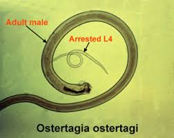
Identify this parasite
Ostertagia ostetagi
What are the characteristics of Ostertagia ostetagi?
Larvae resides in the abomasum and adults reside in the lumen
Larvae develop in gastric glands
Highly pathogenic due to protein losing gastropathy
Adults are bloodsucking and can cause anemia
Why is O. ostertagi so pathogenic?
Larvae destroys the gastric glands by rupturing tight junctions between cells and damaging HCl-producing parietal cells.
Protein leaks into the abomasum
Abomasal pH becomes more neutral, leading to the loss of acid bacteriostasis that leads to bacterial overgrowth
What is the epidemiology of O. ostertagi?
Distributed in Northern US and Canada
L3 larvae persistant over winter and up to 1 year in pasture
Distributed in Southern US
Cool, wet winters are favorable for L3 larvae
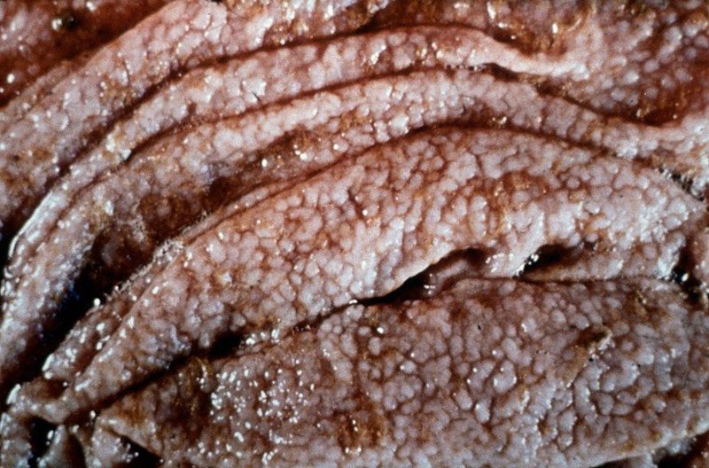
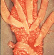
What is Type I Ostertagiasis?
Occurs mid to late favorable season (cool and wet climate)
Large numbers of larvae are acquired from pasture, heavily contaminating the environment
This leads to substantial damage during their departure from the gastric glands, causing “Moroccan leather” appearance in the abomasum (pictured)
Young cattle is most susceptible