Chapter 6: The Muscular System
1/163
Earn XP
Name | Mastery | Learn | Test | Matching | Spaced | Call with Kai |
|---|
No analytics yet
Send a link to your students to track their progress
164 Terms
What are Muscles?
Responsible for body movements, stabilizing joints, and generating heat
How do Muscles Work?
Generate the force required by contracting, a process in which proteins inside the muscle fibers overlap more than when they are at rest.
Why do we need Muscles?
Needed to move substances in our body like air, food and blood.
What unique characteristic sets muscles apart from other body tissues? (Essential Function)
Contract, or Shorten.
3 Types of Muscle Tissue
Skeletal, Smooth and Cardiac
Skeletal Muscle Location
Attached to bones, or for some facial muscles, to skin.
Cardiac Muscle Location
Walls of the heart
Smooth Muscle Location
Walls of hollow organs (other than the heart)
What Muscle Types are only considered as “Muscle Fibers”?
Skeletal & Smooth
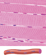
Skeletal Cell Shape
Single, very long, cylindrical, multinucleate cells with very obvious striations
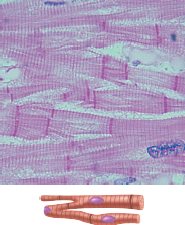
Cardiac Cell Shape
Branching chains of cells; uninucleate, striations; intercalated discs
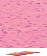
Smooth Cell Shape
Single, fusiform, uninucleate; no striations
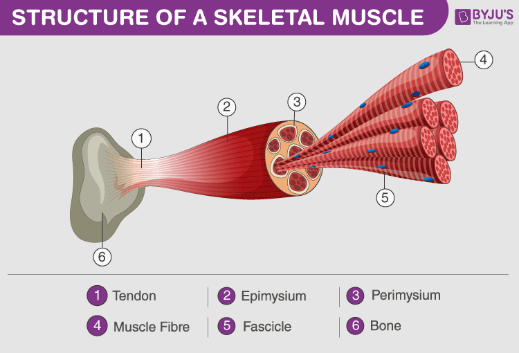
Skeletal Tissue components
Epimysium, perimysium, and endomysium

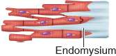
Endomysium (Responsiveness)
Thin Connective Tissue surrounding each muscle cell
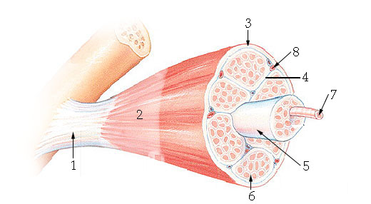
Epimysium (‘Overcoat” of Connective Tissue)
Sheath of fibrous connective tissue surrounding a muscle

Fascicle
Bundle of nerve or muscle fibers
What unique characteristic sets muscles apart from other body tissues? (Essential Function)
Contract, or Shorten.
Cardiac Tissue components
Endomysium attached to the fibrous skeleton of the heart

Smooth Tissue components
Attached to bones, or for some facial muscles, to skin.
Skeletal Regulation of Contraction
Voluntary; via nervous system controls
Cardiac Regulation of Contraction
Involuntary; internal heart pacemaker; nervous system controls; hormones
Smooth Regulation of Contraction
Involuntary; nervous system controls; hormones, chemicals, stretch
Skeletal, Cardiac, and Smooth Speed of Contraction
Slow to Fast
Slow
Very Slow
Skeletal, Cardiac, and Smooth Rhythmic Contraction
No
Yes
Yes, in some
T or F: Skeletal and Smooth Muscle Cells are ELONGATED
T
Skeletal Muscle Fibers
Packaged into organs

Skeletal Cell Shape
Single, Very long, Cylindrical, Multinucleate cells with very obvious striations
Skeletal muscle Location
They are:
Large (of the Muscle Fibers)
Cigar-Shaped
Multinucleate Cells
Cardiac muscle Location
Walls of the heart
Tendon (“cord-like”)
A cord of dense fibrous tissue attaching a muscle to a bone.
Aponeurosis (“sheet-like”)
Indirectly attaches the muscle to bone, cartilage, or another connective tissue covering.
What are tendons made out of?
Collagen Fibers
How are cardiac muscles joined together?
Special Gap Junctions (Intercalated Discs)
3 Important Roles in the Body
Maintain posture and body position
Stabilizes joints
Generates Heat
Visceral Organs
Soft Internal organs of the body
Microscopic Anatomy of Skeletal Muscle
Muscle - Bundle of muscle fiber - Muscle Fiber - Myofibrils - Sacromere
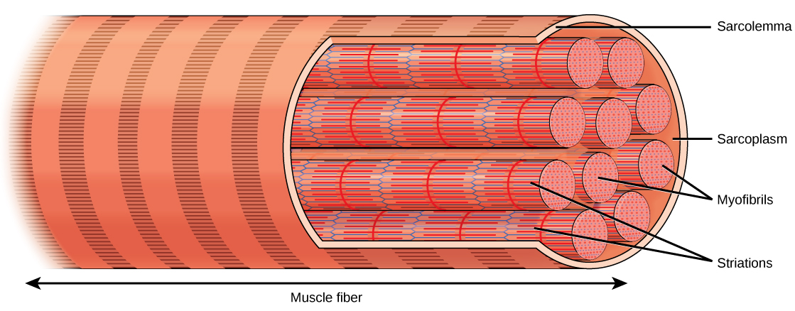
Sarcolemma (“Muscle Hunk”)
Many oval nuclei can be seen just beneath the plasma membrane
Sarcomere | Muscle Cell
Contractile unit of muscle fiber
Structural and functional unit of skeletal muscle
Myofibrils
A complex organelle or a Muscle Fiber
Myofilaments
Produce banding (striped pattern)
What do Banding Patterns reveal?
The working structure of MYOFIBRILS

I Band
Has a Darker Area (Z-Disc)
Has a Midline Interruption
Contains only thin filaments
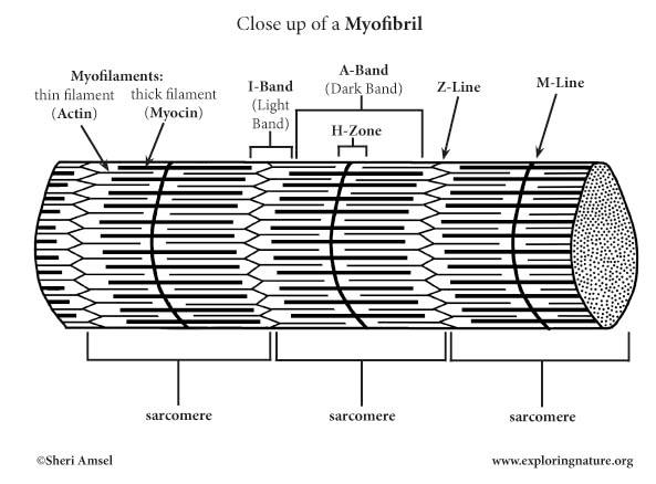
A Band
Has a lighter area (H zone)
Entire length of thick filaments

What line is in the middle of the H-Zone?
M Line
Contains tiny protein rods that hold adjacent thick filaments with together
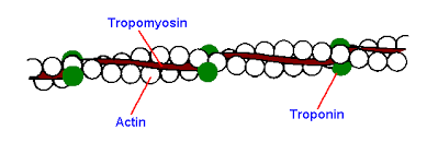
Thin Filament (Actin)
Actin
Actin-containing Thin Filaments
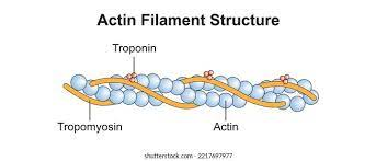
Actin
Contractile Protein
Regulatory proteins prevent binding of myosin heads to actin Anchored to the Z discs
Actin-containing Thin Filaments
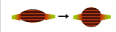
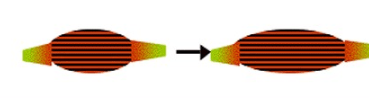
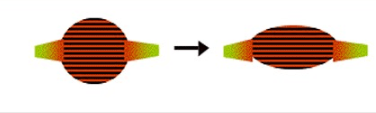
Slide toward each other during contraction

Sarcoplasmic Reticulum
Interconnecting tubules and sacs surround every myofibril
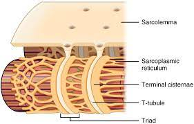
What does the Sarcoplasmic Reticulum (SR) do?
Stores CALCIUM and RELEASE it on demand when a muscle fiber is stimulated to contract
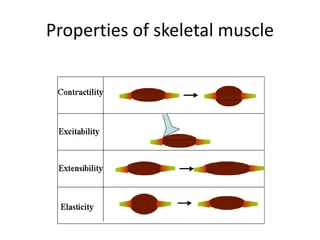
4 Special Function Properties of Skeletal Muscle
Irritability / Responsiveness
Contractibility
Extensibility
Elasticity
Irritability / Responsiveness
Ability to receive and respond to stimulus
Contractibility
Ability to forcibly shorten when an adequate stimulus is received
Extensibility
Ability to be stretched
Elasticity
Ability to recoil and resume resting length after stretching
What do Nerve Impulses do?
Stimulate muscle fibers to contract
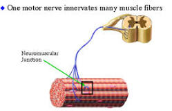
Motor Unit
Consist of one neuron and all the skeletal muscle fibers it stimulates
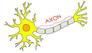
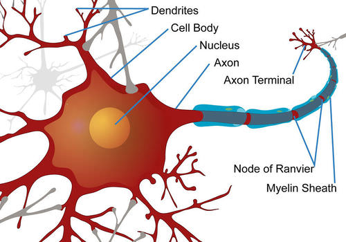
Axon
Long, threadlike extension of a neuron
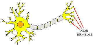
Axon Terminals
Branches that form junctions with the sarcolemma of a different muscle fiber
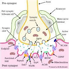
Neuromuscular Junctions
Contain synaptic vesicles filled with neurotransmitter
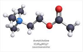
Acetylcholin (ACh)
Specific neurotransmitters that stimulates skeletal muscle fibers
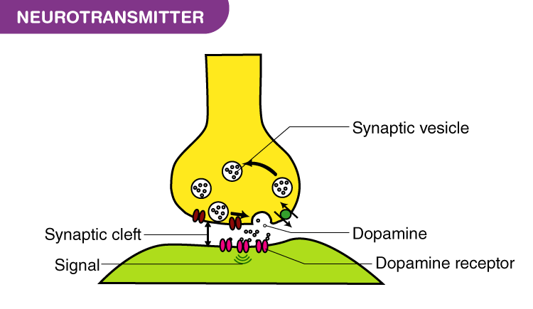
Synaptic Cleft
Nerve endings and muscle fiber never touch; the gap is filled with interstitial fluids
Thick Filament (Myosin)
Bundled molecules of myosin; extend the entire length of the dark A band
Midpart
- Smooth ends are studded with small projections
- Containing ATPase enzyme activity and split ATP to release the “energy” used for muscle contraction
1st event at the neuromuscular junction
Nerve impulse reaches axon terminal of motor neuron
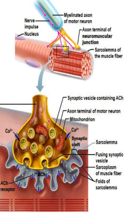
2nd event at the neuromuscular junction
In response to a nerve impulse, calcium (Ca2+) channels open and calcium enters the axon terminal

3rd event at the neuromuscular junction
Calcium entry causes some synaptic vesicles to release their contents (the neurotransmitter acetylcholine) by exocytosis
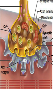
4th event at the neuromuscular junction
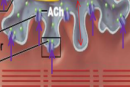
Acetylcholine diffuses across the synaptic cleft and binds to the receptors in the sarcolemma
5th event at the neuromuscular junction
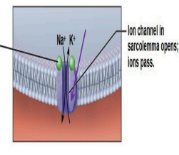
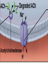
ACh binds and opens channels that allow simultaneous passage of Na+ into the muscle fiber and K+ out of the muscle fiber.
More Na+ ions leave, producing a local change in the electrical conditions of the membrane (depolarization) which leads to an Action Potential
6th event at the neuromuscular junction
The enzyme acetylcholinesterase breaks down ACh in the synaptic cleft, ending stimulation of the muscle fiber
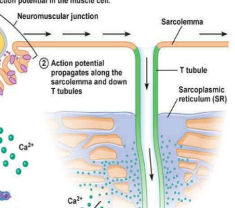
7th event at the neuromuscular junction
The action potential triggers the sarcoplasmic reticulum to release Calcium
2 Events that return the cell to its resting state
Diffusion of potassium ions out of the cell
Sodium-potassium pump
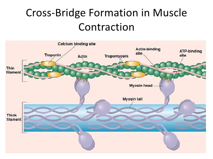
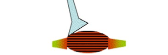
Formation of Cross Bridges
Myosin heads attach to actin which requires calcium ions (Ca2+) and ATP (to “energize” the myosin heads)
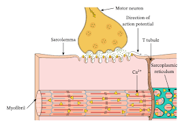
Action Potential
Pass deep into the muscle fiber along membranous tubules that fold inward from the sarcolemma.
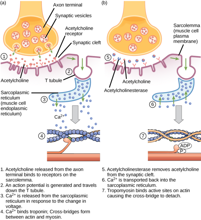
Excitation-contraction coupling
Action potential stimulates the sarcoplasmic reticulum to release calcium ions into the cytoplasm
Calcium ions trigger the binding of myosin to actin = filament sliding
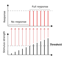
“All-or-none” Law
a muscle fiber will contract to its fullest extent when it is stimulated adequately; it never partially contracts
Graded Responses
Whole muscle reacts to stimuli
Different degrees of shortening = different amount of force
Graded muscle contractions production
Changing the frequency of muscle stimulation
Changing the number of muscle fibers being stimulated at one time

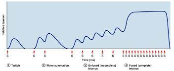
Muscle Twitches
Result from certain nervous system problems; not the normal way of operation
Unfused (incomplete) Tetanus
Nerve impulses are delivered to the muscle at a rapid rate
The effects of the successive contractions are “summed” (added) together
The muscle contractions get stronger and smoother.
Fused (complete) Tetanus
Muscle is stimulated so rapidly that no evidence of relaxation is seen, and contractions are completely smooth and sustained
Tetanus
The primary role is to produce smooth and prolonged muscle contractions.
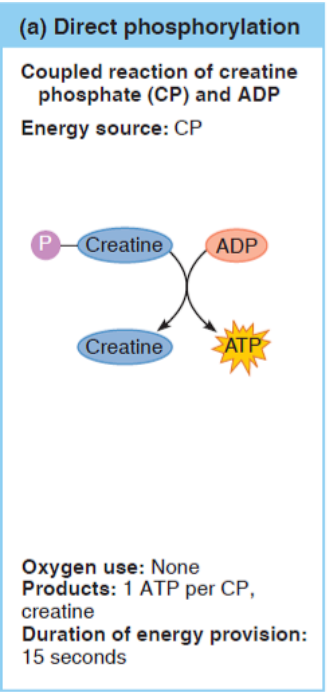
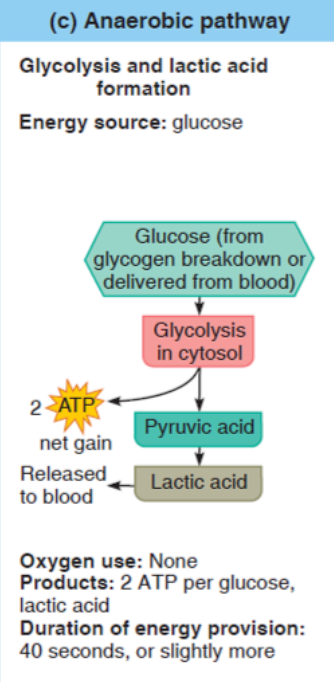
3 Metabolic Pathways that regenerate ATP
Direct Phosphorylation of ADP by Creatine Phosphate
Anaerobic Glycolysis
Aerobic Respiration
Direct Phosphorylation of ADP by Creatine Phosphate
Fastest
Anaerobic Pathway (Glycolysis)
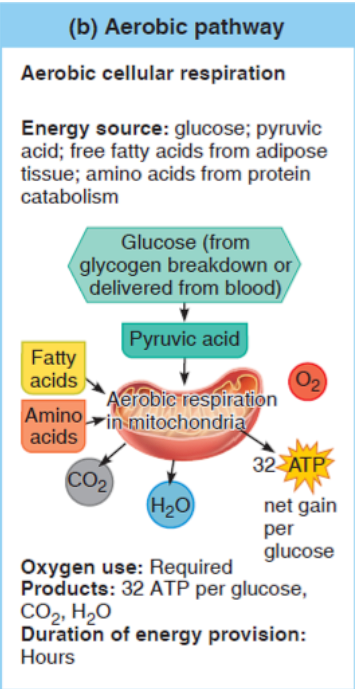
Aerobic Pathway
Slowest
2 Types of Head Muscles
Facial
Chewing
Facial Muscles
Insert into soft tissues like other muscles or skin
Chewing Muscles
Break down food for the body
What are the 8 Facial Muscles?
Frontalis
Obicularis Oculi
Zygomaticus
Buccinator
Orbicularis Oris
Temporalis
Occipitalis
Masseter

Frontalis
Raises eyebrows

Orbicularis Oculi
Blinks and closes eye

Zygomaticus “Smiling Muscle”
Raises corner of mouth

Buccinator
Compress cheek, hold food between teeth during chewing

Orbicularis Oris “Kissing Muscle”
Closes and protrudes the lips

Temporalis “Time Muscle”
Closes Jaw
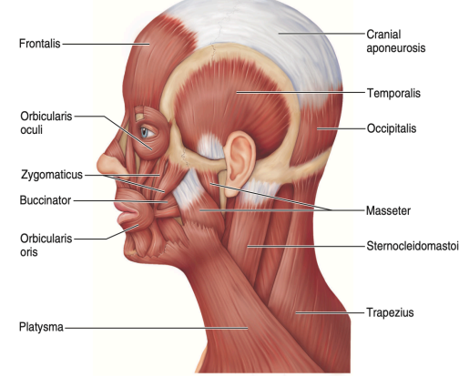
Occipitalis
Pulls the scalp posteriorly

Masseter
Closes Jaw

What are the 2 Neck Muscles?
Platysma
Sternocleidomastoid
What do Neck Muscles do?
They move the head and shoulder girdle and are small and strap-like

Platysma
Tenses skin of the neck (as in shaving)