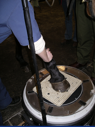
Equine diagnostic imaging
1/31
There's no tags or description
Looks like no tags are added yet.
Name | Mastery | Learn | Test | Matching | Spaced | Call with Kai |
|---|
No analytics yet
Send a link to your students to track their progress
32 Terms
What are the main imaging modalities used in equine?
Radiography
Nuclear scintigraphy
Ultrasonography
MRI
Computed tomography (CT)
What is radiography used to image?
Mainly bone
Some articular and periarticular structures
Can see some swelling and mineralisation of soft tissues
(contrast techniques)
What are the differences between portable and gantry machines?
Portable
Stationary anode —> minimal heat absoprtion
Limit to kVp and mAs
Fewer parts = longer life
Gantry mounted
Rotating anode —> maximum heat absorption
High kVp and mAs possible
More complex = more risk of malfunction
Three phase power supply
What machine settings (exposure factors) can be set on a radiography machine?
kVP = speed of electrons
mA = number of electrons released
S = time
What factors are taken into consideration for optimum film:focal distance?
By body part
By size of animal
By cassette or film type
By processing type
What are the different image aquisition systems?
Computed radiography
Direct radiography
Conventional
(Everything mainly digital and instant imaging now)
How is film stored as a permanent record?
As DICOM image —> unique number
As hard copy
When reporting on images what process should you follow?
State area that has been imaged
Critique quality of film
Make a radiography report
1. Recognition phase (search)
2. Descriptive phase (report)
3. Interpretation phase (Ddx)
What features of film quality should be assessed?
Positioning
Collimation
Contrast
Exposure
Labeling
Artefacts
(Pink Camels Collect Extra Large Apples)
What should be done in the recognition phase of the report?
Lesion localisation
Systematic search pattern
Identify visible structures
Normal/abnormal findings
Normal variations
What should be done in the descrriptive phase of the report?
Categorise according to Rontgen signs
Size
Shape
Position
Number
Margination —> well/poorly defined, sharp/blunt margins
Opacity
What should be done in the interpretive phase of the report?
Take into consideration:
History
Signalment
Clinical signs
Diagnostic tests
What are ultrasounds used to view and how do they work?
Tendons and ligaments
Sound wave produced by piezoelectric crystal
Reflected from tissue interface
Degree of reflection determined by tissue density and whether US wave is reflected, absorbed or scattered
What is the advantage of ultrasonography
Safety:
No ionising radiation!!
No radiation protection issues!
What are the standard frequencies used in equine ultrasounding?
2.5 - 14 mHz
What are the different configuration of ultrasound probes?
Linear
Sector
Convex/microconvex
What should you choose a probe based off?
Area dependent —> adequate for depth and resolution
Type of head and frequency
How is a patient prepared for ultrasound?
Clip and clean skin
Ultrasound gel
Standoff
Alcohol —> not clipped
What features are used to describe a lesion on an ultrasound?
Size – actual size or CSA %
Shape –
Position
Number
Margination - Fibre alignment
Echogenicity
Assess in transverse and longitudinal images
How does nuclear medicine/gamma scintigraphy work?
Use of radio-isotope —> technetium 90
Gamma ray emission
Polyphosphonate binds HAP crystals
Gamma rays detected
How does the isotope used for nuclear medicine work?
Binds actively to remodelling bone
IV injection
Excreted in urine, half life of 6 hours
What is used to acquire an image in nuclear scintigraphy?
Gamma camera —> a giant Geiger counter

What are the different types of image acquisition using a gamma camera?
Static —> horse must remain mobile for 60-120 seconds
Dynamic —> same length but corrects for movement by realigning images
What are the phases of scanning in nuclear scintigraphy?
Phase I – blood pool
Rarely used in equine
immediate
Phase II – soft tissue
2-15 minutes
Diffusion to extracellular fluid
Phase III – bone
2-3 hours
Bound to HAP
In what cases is nuclear scintigraphy used?
Mainly bone abnormalities
Commonly used to diagnose lameness, but can also be used to evaluate other conditions
more intense colour = more gamma rays detected, compare intensity of L vs R, compare regions of interest & ratios
Where is there increase binding of HAP to crystals in nuclear scintigraphy?
Inflammation
Bone repair/remodelling
Osteomyelitis
Neoplasia
What safety precautions need to be taken in cases of nuclear scintigraphy?
Isolation of horse for 48 hours
Minimise contact with radioactive horse
Wear gloves when handling
Contain urine and faeces
Monitor exposure
EPD and personal dose meters
Radiation badges
What are the advantages and disadvantages of nuclear scintigraphy?
Useful addition to the diagnostic work up
Not to be used as a replacement for nerve blocks or thorough clinical examination.
Conditions that have primarily soft tissue pathology will not be identified
Sensitive but not very specific
Use in combination with other imaging modalities and diagnostic analgesia
What is MRI (magnetic resonance imaging) used for?
Use after basic imaging modalities to help advance diagnoses further or obtain a diagnosis if basic imaging modalities have failed to do so
Very limited to lower limb and extremities if not under GA
What are the advantages of MRI?
Increase detail
Specific and sensitive
Directed treatment towards lesions
What are the limitations of MRI?
Takes a very long time
Lesions can't really be aged
Sometimes poorly worked up case
What areas can be examined using equine CT?
Standing CT —> limited to heads and some parts of the cervical spine
GA —> distal limbs, head and cervical spine