Cell and Tissue Unit
 Call Kai
 Learn
Learn Practice Test
 Spaced Repetition
 Match
Match Flashcards
 Knowt Play
1/74

Earn XP
Description and Tags
Cells, organelles, tissues, specialized cells
Name | Mastery | Learn | Test | Matching | Spaced | Call with Kai | Chat |
|---|
No analytics yet
Send a link to your students to track their progress
75 Terms
Interstitial fluid
extracellular fluid between cells in a tissue
Extracellular fluid
watery substance that surrounds all cells in our body
Cytosol
fluid component of the cell (intracellular fluid=inside)
Nonmembranous organelles
cytoskeleton, cilia, microvilli, flagella, centrioles, ribosomes
Membranous organelles
mitochondria, nucleus, E.R, Golgi apparatus, Lysosomes, peroxisomes
Function of the cytoskeleton
strengthens and supports the cell

Function of centrioles
Each cell has 2 centrioles that aid in division during mitosis
What is the difference between microvilli and cillia?
Microvilli increase the surface area of the cell for absorption and are shorter, while Cillia can usually move and are longer, hair-like projections that help move things from the surface of the cell.
What is the function of a ribosome? Where can you find them?
Makes proteins. Can either be attached to the rough endoplasmic reticulum or free-floating

What is the function of mitochondria?
Produces energy for the cell via cellular respiration and krebs cycle
What does the nucleus of a cell contain?
DNA/RNA, proteins, and enzymes
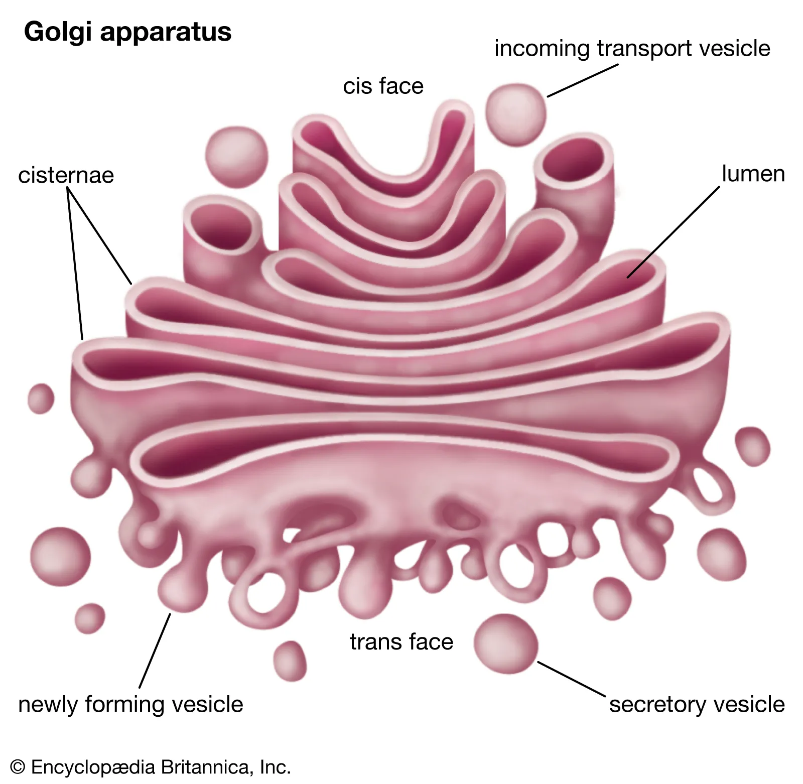
What is the function of the Golgi apparatus?
Stores and packages proteins “UPS of the cell”

Endoplasmic reticulum
Function: makes secretory products, intracellular storage and transport, removal of drugs/toxins. Can be rough or smooth
Lysosomes
Contain digestive enzymes. Breakdown organic compounds, damaged organelles, and pathogens
Peroxisomes
Vesicles that contain degrading enzymes. Breakdown organic compounds and neutralize toxins.
Describe the phospholipid bilayer
Hydrophillic heads, and hydrophobic tails. Makes the plasma membrane selectively permeable.
Integral vs. Peripheral proteins
integral means part of the membrane structure, peripheral means bound to the surface but can be removed.
What is the difference between the cytoplasm and cytosol?
cytosol is just the fluid part of the cell, while the cytoplasm is the fluid plus the organelles
What is the function of vesicles inside of the cell?
They are small containers that are used to communicate between the membranous organelles. Can transport proteins/glycoproteins and contain digestive enzymes. “Shipping boxes”
What is an example of a cell that would have a lot of mitochondria and why?
A skeletal muscle cell would have a lot of mitochondria because it’s energy demands are higher than that of other cells.
What role does the nucleus play in cell differentiation?
The nucleus contains all the information for what specific proteins the cell needs to make and what functions the cell will perform
Nuclear pores
small passages in the nuclear envelope
Nucleolus
Inside of the nucleus. Where rRNA synthesis occurs and ribosomes assembled. Non-membranous
Osmosis
The diffusion of water across a selectively permeable membrane. Water will always follow solute.
Tonicity
describes how the solution will effect a cell. Isotonic, hypertonic, hypotonic.
What are the four main types of tissues?
Muscle, connective, epithelial, nervous
What is the function of epithelial tissue?
Provides protection from external environment, controls what gets in and out, and secretes product. Can be absorptive.
What type of tissue would you find in the lining of the blood vessels and digestive tract?
Epithelial
Is epithelial tissue avascular or vascular?
Avascular. Epithelial tissue obtains nutrients from diffusion usually from underlying connective tissues.
Endocrine glands
Epithelial. Secretes hormones.
Exocrine glands
Lacrimal (tears), salivary, sweat, and sebaceous glands.
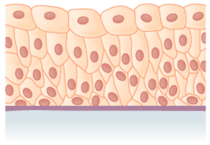
What are the three shapes of epithelial cells?
Squamous (flat), cuboidal, and columnar

What type of tissue is depicted in the image?
Simple squamous epithelium
Simple squamous epithelium
Single layer of cells, highly permeable. Located in regions of the body where absorption happens. Alveoli, kidneys, etc.
Stratified squamous epithelium
Consists of multiple layers and occurs in places where friction and damage are more common. Mouth, skin, esophagus, vagina. Can be keratinized or nonkeratinized.
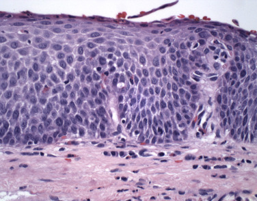
What type of tissue is depicted in the image?
Stratified squamous epithelium

What type of tissue is being depicted in the image?
Keratinized stratified squamous epithelium
Cuboidal epithelium
Round, or cube-like shape. Simple cuboidal is found where secretion and absorption take place. Stratified is rare.

What tissue is depicted in this image?
Simple cuboidal. Could look stratified but it surrounds each individual lumen.
Where would you find transitional epithelium?
In the bladder because it needs to expand to hold urine.

What type of tissue is depicted in the image?
Transitional epithelium. “Balloon-like” apical surface.
Where in the body would you find simple columnar epithelium?
Small intestine, stomach, gallbladder. Anywhere where absorption or secretion occurs.
True or false: Columnar epithelium can either be ciliated or nonciliated
True
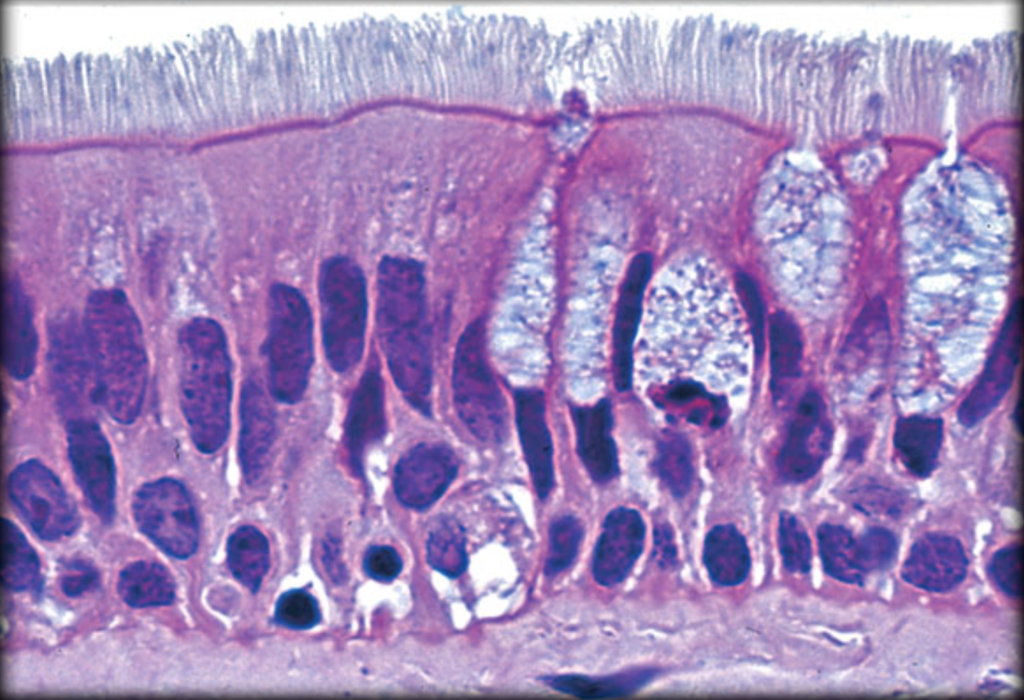
What type of tissue is depicted in the image?
Pseudostratified columnar epithelium. It is pseudostratified because the nuclei sit at different heights.
Goblet cells
Specialized, glandular epithelial cells that secrete mucous to trap particles in the respiratory and digestive tract.

What are the 3 methods exocrine glands use to secrete into ducts?
Merocrine, 2. Apocrine, 3. Holocrine
Merocrine secretion
Secretion released by vesicles that dump their contents outside of the cell membrane. Exocytosis.
Apocrine excretion
Cytoplasm breaks off and the secretory vesicles go with it. Example: milk production in the mammary glands.
Holocrine secretion
The cell becomes packed with secretory products and bursts.

What type of epithelial tissue is depicted in the image?
Simple squamous epithelium
What two parts make up the matrix in connective tissue?
Ground substance and extracellular protein fibers
What are the functions of connective tissue?
Structure, connect tissues, transport fluids, store energy
What two types of connective tissue fall under the category of connective tissue proper?
Loose and dense connective tissue
Which is the most common type of connective tissue?
Areolar connective tissue. “Packing material” of the body.

What type of tissue is depicted in the image?
Areolar connective tissue. Fibers, fixed cells, wandering cells.

What type of connective tissue is depicted in the image?
Adipose (fat) tissue. Made up of adipocytes.
What are the types of loose connective tissue?
Areolar, adipose, reticular,

What tissue is being depicted in this image?
Blood
What is the name for the ground substance in blood?
Plasma
Erythrocytes
Red blood cells. Responsible for carrying oxygen to the tissues by hemoglobin.
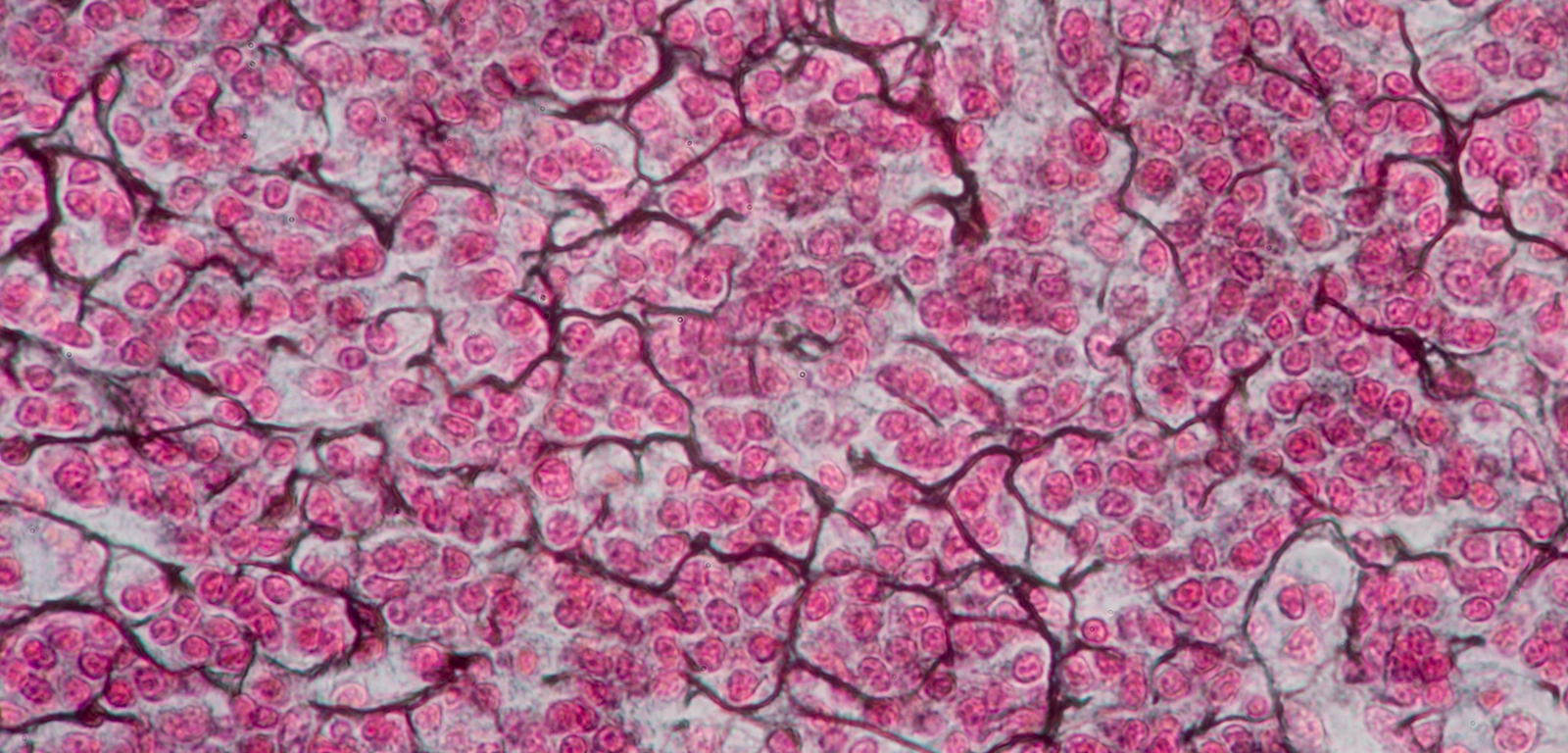
What type of tissue is being depicted in the image?
Reticular tissue.
What are the functions of reticular tissue?
Provides support, filtration, new blood vessel development, and facilitates movement.
What are the 3 types of dense connective tissue?
Dense regular, dense irregular, and elastic
Where in the body would you find dense regular tissue?
Tendons and ligaments. Collagen fibers prevent ripping and stretching.

What type of tissue is being depicted in the image?
Dense regular connective tissue

What type of tissue is being depicted in this image?
Dense irregular connective tissue

What type of connective tissue is being depicted in this image?
Elastic dense connective tissue
What are the 3 types of cartilage?
Fibrocartilage, elastic, and hyaline
What type of cells are present in cartilage?
Chondrocytes
What is the function of hyaline cartilage?
Reduces friction between bony surfaces
What is the function of elastic cartilage?
More flexible than hyaline. Provides support and returns to its original shape.
Where in the body would you find elastic cartilage?
External ear
Fibrocartilage
Most durable and tough, resists compression, limited movement. Made of dense bundles of collagen fibers.
What are the three types of muscle tissue?
Smooth, cardiac, and skeletal

What types of muscle tissues are striated?
Cardiac and skeletal muscle. This is because they both have contractile sarcomeres.