fill in the blank - ch 19 heart
1/49
There's no tags or description
Looks like no tags are added yet.
Name | Mastery | Learn | Test | Matching | Spaced | Call with Kai |
|---|
No analytics yet
Send a link to your students to track their progress
50 Terms

The dark lines connecting cardiac muscle cells are called __________
Intercalated discs
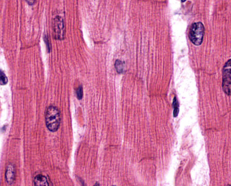
The visible banding pattern in cardiac muscle is called __________
Striations
Cardiac muscle cells typically have __________ (one/multiple) centrally located nucleus/nuclei
One (sometimes two) centrally located nuclei
Intercalated discs contain __________ junctions that allow electrical signals to spread quickly.
Gap junctions
Compared to skeletal muscle, cardiac muscle cells are more __________ (branched/unbranched)
Branched
The tip of the heart is called the __________
Apex
The superior portion of the heart is called the __________
Base
The ear-like flaps on the heart are called __________
Auricles
The __________ ventricle is thicker because it pumps blood to the entire body
Left
The heart is shaped like an upside-down __________
Triangle
The white string-like structures are called __________
Chordae tendineae
Chordae tendineae attach to __________ muscles
Papillary
The function of chordae tendineae is to prevent valves from __________
Prolapsing / flipping backward
Ridges found in the atria are called __________ muscles
Pectinate
Ridges found in the ventricles are called __________
Trabeculae carneae
The wall separating the ventricles is the __________ septum
Interventricular
The fossa ovalis is a remnant of the fetal __________
Foramen ovale
The valve between the right atrium and right ventricle is the __________ valve
Tricuspid
The valve between the left atrium and left ventricle is the __________ valve
Bicuspid / Mitral
The valve leading into the pulmonary trunk is the __________ semilunar valve
Pulmonary
The valve leading into the aorta is the __________ semilunar valve
Aortic
Heart sounds “lub-dub” are caused by valve __________
Closing
Blood returning from the body enters the right atrium through the __________ and __________
Superior vena cava and inferior vena cava
Blood flows from the right ventricle into the __________ trunk
Pulmonary
Blood traveling to the lungs is part of the __________ circuit
Pulmonary
Blood traveling to the body is part of the __________ circuit
Systemic
Blood in pulmonary arteries is __________ (high/low) in oxygen
Low
Blood in pulmonary veins is __________ (high/low) in oxygen
High
Coronary arteries supply oxygen to the __________
Myocardium
A blockage in a coronary artery can cause a __________
Myocardial infarction (heart attack)
Blood from the heart muscle drains into the __________ sinus
Coronary
The __________ cardiac vein collects blood from multiple veins
Great
The heart’s natural pacemaker is the __________ node
SA (sinoatrial)
If the SA node fails, the __________ node takes over
AV (atrioventricular)
Electrical signals travel from the AV node to the __________ of His
Bundle
The final conduction fibers are called __________ fiber
Purkinje
Nodal cells are never at __________
Rest
The P wave represents __________ depolarization
Atrial
The QRS complex represents __________ depolarization
Ventricular
The T wave represents ventricular __________
Repolarization
The PR interval represents the time from atria to __________
Ventricles
There is no atrial repolarization wave because it is hidden by the __________ complex
QRS
S1 (“lub”) is caused by closure of __________ valves
AV (tricuspid and mitral)
S2 (“dub”) is caused by closure of __________ valves
Semilunar
Normal heart rate is __________ to __________ bpm
60 to 100
A heart rate above 100 bpm is called __________
Tachycardia
A heart rate below 60 bpm is called __________
Bradycardia
Pulse pressure = __________ − __________
Systolic − Diastolic
MAP = __________ + 1/3 (pulse pressure)
Diastolic pressure
If MAP falls below __________ mmHg, organs may not receive enough blood
60