Final Exam Questions
1/73
There's no tags or description
Looks like no tags are added yet.
Name | Mastery | Learn | Test | Matching | Spaced |
|---|
No study sessions yet.
74 Terms
Describe the four phases of the action potential and the order in which they occur?
Rising Phase (depolarisation): Sodium channels open and sodium enters the cell, depolarising it.
Overshoot Phase: The peak of the action potential where membrane potential is positive after potassium channels start to open and sodium channels start to deactivate.
Repolarisation: Membrane begins to repolarise as potassium channels are activated and potassium leaves the cell to restore membrane potential. Sodium channels are inactivated by a flap.
Undershoot Phase (Hyperpolarisation): Too much potassium is let out the cell and it hyperpolarises (more negative than original potential) before potassium gates close, and it slowly returns to the resting membrane potential (-70 mV). Potassium and sodium gates eventually close, and the membrane potential slowly returns to the resting level of -70 mV. During this phase, it is more difficult to initiate another action potential, as the membrane potential is more negative, but it can be done because sodium channels are closed not inactivated.
Rising Phase (depolarisation):
Sodium channels open and sodium enters the cell, depolarising it.
Overshoot Phase:
The peak of the action potential where membrane potential is positive after potassium channels start to open and sodium channels start to deactivate.
Repolarisation:
Membrane begins to repolarise as potassium channels are activated and potassium leaves the cell to restore membrane potential. Sodium channels are inactivated by a flap.
Undershoot Phase (Hyperpolarisation):
Too much potassium is let out the cell and it hyperpolarises (more negative than original potential) before potassium gates close, and it slowly returns to the resting membrane potential (-70 mV). Potassium and sodium gates eventually close, and the membrane potential slowly returns to the resting level of -70 mV. During this phase, it is more difficult to initiate another action potential, as the membrane potential is more negative, but it can be done because sodium channels are closed not inactivated.
how the selectivity filter in K+ channels work
Because sodium is smaller than Potassium the ions in cushion of water around sodium must be removed to pass through the filter. K has 8 water molecules which are removed when it passes through but because Na is smaller it is too small to interact with oxygen lining the pore wall ions that remove the water and the water is not able to be removed and it is therefore unable to pass through because Na more stable water shell outside channel than inside.
Because chlorine & calcium are larger than potassium, they cannot fit through the 4 subunits are assembled to form a k channel and 4 poor loops form a poor that serves as a narrow tunnel allowing k to move across but blocking larger ions.
Larger molecules selectivity
Because chlorine & calcium are larger than potassium, they cannot fit through the 4 subunits are assembled to form a k channel and 4 poor loops form a poor that serves as a narrow tunnel allowing k to move across but blocking larger ions.
Smaller molecules selectivity
Because sodium is smaller than Potassium the ions in cushion of water around sodium must be removed to pass through the filter. K has 8 water molecules which are removed when it passes through but because Na is smaller it is too small to interact with oxygen lining the pore wall ions that remove the water and the water is not able to be removed and it is therefore unable to pass through because Na more stable water shell outside channel than inside.
Ion channels and active transporters
Ion channels: Allow the passive flow of ions across their electrochemical gradient and does not require energy.
Active transport: Use energy in the form of ATP to transport ions against their electrochemical gradient.
The LGN in the thalamus is the primary relay center for visual information it processes and organizes visual information before transmitting it to the primary visual cortex (V1) for further processing. The LGN helps in forming a coherent visual representation by maintaining the segregation of various types of visual information, such as colour, brightness, and motion.
Alpha motor neurons
Gamma motor neurons
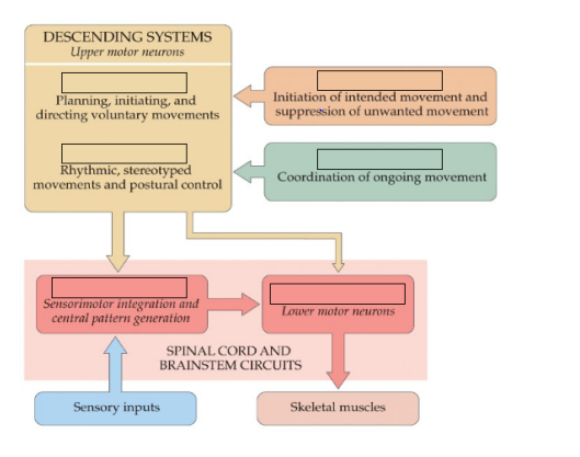
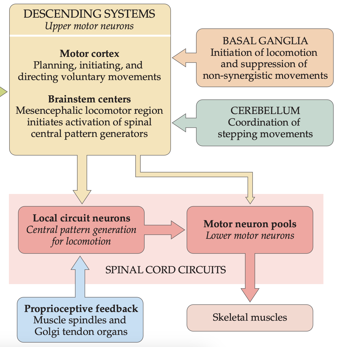
Basal ganglia also modulate activity of the motor cortex and other motor areas, to select appropriate movements and suppress unwanted or competing movements for smooth voluntary movements.
Therefore, both the cerebellum and basial ganglia have a role in ensuring movements are accurate, coordinated and smooth by modulating other systems, which is why they are said to be modulatory.
When the patellar tendon is tapped sensory information from muscle spindles travels up afferents to the spine. Sensory neurons make a direct monosynaptic connection with motor neurons that innervate the muscle. The motor neurons are excited, and the muscle contracts and the leg extends. It is a monosynaptic stretch reflex that causes the lower leg to extend (kick out) when the patellar tendon is tapped or stretched. - For tripping
Foveal vision and attention: The fovea is the region of the retina with the highest concentration of photoreceptors (cones) and provides the sharpest, most detailed vision. Eye movements allow us to focus moving objects or areas of interest onto the fovea, where we can perceive them with the highest visual acuity and colour sensitivity. Eye movements, such as fixations and micro saccades, allow us to direct our visual attention and focus on specific areas or objects of interest. This selective attention is important for tasks that require detailed visual processing, like reading, object recognition, or scanning the environment.
Tracking moving objects: Eye movements, particularly smooth pursuit movements, enable us to track and maintain focus on moving objects in our visual field.
Image stabilization: Vestibulo-ocular and optokinetic reflexes help stabilize the image on the retina during head and body movements, preventing blurred vision. This image stabilization is crucial for maintaining a clear and stable visual perception of the world around us, even when we are in motion.
Aligned visual and motor maps in the superior colliculus facilitate the direct translation of sensory information into motor commands, supporting the integration of visual perception and eye movement control for effective interaction with the visual environment.
Retinotopic visual representation: Contains a topographic map of the visual field representing the spatial arrangement of the visual field, with nearby regions of the visual space being represented by nearby neurons in the superior colliculus.
Saccadic motor representation: Contains a motor map that represents the generation of saccadic eye movements which is aligned with the visual map.
Sensorimotor integration: The alignment of the visual and motor maps in the superior colliculus allows for direct and efficient coupling between sensory information (visual input) and motor commands (saccadic eye movements). When a visual stimulus is detected in the visual field, the corresponding region of the visual map in the superior colliculus is activated. This, in turn, activates the aligned region of the motor map, resulting in the generation of a saccadic eye movement that shifts the gaze to the location of the visual stimulus.
Describe the 4 main similarities in structure across association areas.
Similarities in structure across association areas:
• Highly interconnected with other brain regions
• Receive inputs from multiple sensory modalities.
• Lack clear topographical organization (unlike primary sensory cortices)
• Involved in higher-order cognitive functions.
Brain areas not typically characterized as association cortices include the primary sensory and motor cortices.
Distinctive features of association areas:
• Involved in complex cognitive functions like attention, memory, language, decision-making.
• Receive inputs from multiple sensory modalities.
• Lack clear topographical organization.
Wernicke's aphasia is characterized by fluent but nonsensical speech, with difficulties in comprehension. Broca's aphasia is characterized by non-fluent, effortful speech, with relatively preserved comprehension.
Striate cortex layers
LGN is in Layer 4 C and A
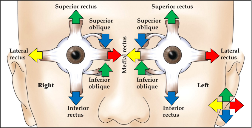
Eye muscles
pupillary light response
LGN relays information (in thalamus) EW constricts ciliary muscle does pupillary light response
Layers of LGN
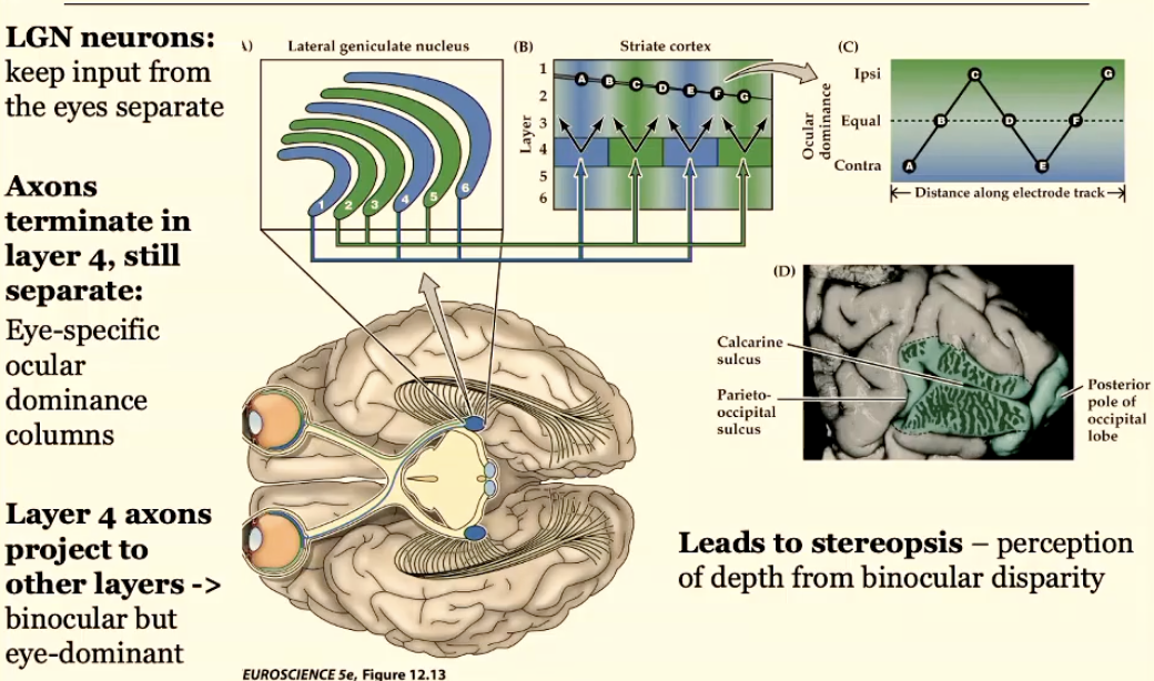
LGN neurons: keep input from eyes separate
Layer 4 neurons (axon terminals) are eye specific and layer 4 neurons that project to other layers which accept all information are binocular but still eye dominate but you still have some equal
Stereopsis: depth vision from binocular
Layers of striate cortex
Primary Visual (striate) cortex 2mm thick 6 layers
Layer 4 of the visual cortex has serval layers
Layer 4 is important for input and output due to the pattern of connection which allows the to support vision
4C has spiny stallate neurons which get LGN info and passes it onto other layers
4B sends axons to extrastriate areas
Neurons in outside layers go to other brain areas (extrastriate areas)
Neurons in deeper layers send axons to subcortical targets (to thalamus then to the LGN and the superior colliculus) 6