Week 5 - Cell Biology
1/64
There's no tags or description
Looks like no tags are added yet.
Name | Mastery | Learn | Test | Matching | Spaced |
|---|
No study sessions yet.
65 Terms
What is cell biology?
Cell biology is the study of:
How cells grow, divide, move, and maintain themselves.
How these cellular processes are controlled metabolically.
How different cell forms and functions are generated.
How cellular processes are regulated and respond to internal and external signals.
What are the 3 main methods to study cell biology?
Microscopy
Biochemistry
Genetics
What are the 4 ways of studying cell biology with microscopes?
Conventional light microscope
With green fluorescence protein (GFP)
Flourescence microscopy
Electron microscopy
How do different microscopy techniques differ in resolution and use?
The naked eye has very low resolution; light microscopes resolve to ~250 nm, electron microscopes far finer (<250 nm), and X-ray crystallography to atomic detail.
Light microscopy needs contrast (pigments, dyes, fluorescent probes) and can see organelles but not fine structure.
Fluorescence microscopy uses fluorophores like GFP to visualise proteins, organelles, and cell dynamics with high contrast; confocal systems can improve resolution and generate 3D images.
Electron microscopy allows ultrastructural detail: TEM shows internal structure via transmitted electrons, while SEM reveals surface topography.
Describe how biochemistry can be used to understand cell biology.
Biochemistry helps us understand cell biology by studying molecules directly.
By purifying molecules and testing their activity, we can determine what they do and how they contribute to cellular processes. To do this, scientists may:
Use whole-cell extracts or separate the cell into different compartments to study specific molecules.
Perform in vitro (cell-free) assays, where purified molecules are tested outside the cell to observe their function in a controlled environment.
Describe how genetics can be used to understand cell biology.
Genetics helps study cell biology by linking changes in genes (genotype) to changes in cell structure or behavior (phenotype).
Scientists can use mutants—organisms with altered genes—to see what happens when a specific component is disrupted. This reveals the normal function of the gene in the cell.
There are two main approaches:
Forward genetics: Start with a phenotype (e.g., a defective cellular process) and work backwards to identify the gene responsible.
Reverse genetics: Start with a known gene and deliberately alter or remove it to observe how the phenotype changes, revealing the gene’s role.
What are the 3 main components of the cytoskeleton?
Actin
Microtubules
Intermediate filaments
What is the cytoskeleton involved in and what is it important for?
Involved in many processes including cell growth, division, shape, movement (whole cells and inside cells), communication between cells.
Critical for cellular organisation and polarity.
What is the structure and assembly process of actin filaments?
Actin filaments (microfilaments) are helical polymers made of actin monomers with plus and minus ends. Actin monomers bind ATP, which allows them to assemble into the polymer. After incorporation, ATP is hydrolysed to ADP, and actin-ADP subunits eventually dissociate from the filament, especially at the minus end.
What are the subunits of actin?
Actin monomers (G-actin)
What is the general structure of actin?
A helical polymer.
What are the main functions of actin?
Functions in cell shape (mechanical support), intracellular movement (moves things around the cell), cell movement (moves the cell).
Describe the structure of microtubules
Composed of α/β-tubulin heterodimers
Polar polymer: has both plus (+) and minus (−) ends
Tubulin-GTP adds to plus end; tubulin-GDP dissociates
GTP hydrolysis → causes rapid growth/shrinkage (showing dynamic instability)
What are the main function of microtubules in the exoskeleton?
They act for intracellular transport (act as tracks), mitosis (form the mitotic spindle during cell division), cell shape (provide structural support), cell motility (form cilia and flagella).
What are the subunits of microtubules?
Tubulin dimers (alpha and beta tubulin).
What is the general structure of microtubules?
They are hollow tubes.
What is the general structure of intermediate filaments?
Rope-like fibres.
What are the subunits of intermediate filaments?
Various proteins.
What are the main functions of the intermediate filaments?
They provide mechanical strength, with clear nuclear positioning.
Which components of the exoskeleton show polarity
Actin filaments (microfilaments) and microtubules.
Compare actin filaments, microtubules and intermediate filaments (the key components of the exoskeleton).

What is compartmentalisation?
Compartmentalisation is the segregation of cellular components into separate regions (organelles) by membranes.
These compartments are separated by protein and lipids, and their composition has determined organelle ‘boundary’.
What are the 3 reasons to show why compartmentalisation is important?
Different processes in the cell need different environments
This enables the concentration of the reactants to be increased
And ensures incompatible reactions are separated.
What is protein targeting determined by?
By signal sequences, which are cellular “postcodes”.
These are amino acid sequences that determine where proteins go.
How do signal sequences determine where proteins go?
- Signal sequences bind receptors which direct them to their location.
- There are different signal sequences for different locations.
- These can be at the N or C terminal of a protein.
Why is protein targeting needed to and from the nucleus?
Because transcription and translation is spatially separated.
Some proteins e.g., histones and transcription factors need to go from cytoplasm back into the nucleus
Other proteins such as RNA associated proteins, need to move out the nucleus.
Small molecules need to diffuse across the pore, larger molecules need targeting sequences for this (through protein targeting).
How is protein targeting controlled to and from the nucleus?
Nuclear localisation signal (NLS) and nuclear export signal (NES) control this.
These can be found at either the N or C terminals of proteins.
What is exocytosis?
Moving material out of the cell.
What is endocytosis?
Moving material into the cell.
What role do coat proteins play in vesicles?
Coat proteins help form and define vesicles.
Different coat proteins are used for vesicles going to different cellular compartments.
They act like labels and scaffolds, ensuring the vesicle carries the right cargo to the right place.
How does vesicle trafficking work?
A vesicle is a small, phospholipid-enclosed sac that carries fluid, proteins, and macromolecules.
It forms when the donor membrane curves and buds off.
The vesicle then moves to the target membrane and fuses, delivering its cargo.
Why is protein targeting to the mitochondria and the chloroplast hard?
They have to cross multiple membranes.
This makes import complex.
How do organelles move within the cell? Why do organelles move around the cell?
Organelles move around to perform their functions.
Movement is controlled by two main mechanisms:
Cytoskeleton and motor proteins – act as tracks and motors for transport.
Tethers (‘piggybacking’) – hold organelles together or allow one organelle to move with another.
What are the motor proteins of actin filaments?
Myosin
What are the motor proteins of microtubules?
Kinesin and dynein
Are there motor proteins in intermediate filaments?
No
What are motor proteins?
Motor proteins are specialized proteins in cells that generate force and movement along cytoskeletal filaments, helping transport organelles, vesicles, and other cargo within the cell.
How does the cytoskeleton control intracellular movement?
Movement is controlled by: cytoskeleton + motor proteins.
Motor proteins: convert chemical energy (ATP) → mechanical energy for movement.
ATP/ADP binding changes motor shape, allowing it to “walk” along filaments.
Motors pull cargo (e.g., organelles) along cytoskeletal tracks.
What are the motor types of the cytoskeleton? What is their role in movement?
Motor types:
Myosin: moves along actin filaments (usually toward plus end of F-actin)
Kinesin: moves along microtubules (usually toward plus end)
Dynein: moves along microtubules (usually toward minus end)
What is the actin-myosin system important for?
Moving organelles (e.g., lysosomes, mitochondria) and vesicles around the cell.
Generating muscle contraction.
How do myosin and kinesin motors move organelles?
Both use ATP hydrolysis to power movement.
Myosin: moves along actin filaments, releases from actin after ATP binding.
Kinesin: moves along microtubules, releases from microtubules after ATP hydrolysis.
Give an example of organelle movement mediated by actin.
Chloroplasts move in response to light.
Movement is actin-dependent, but not mediated by myosin.
Specialized proteins, e.g., CHUP1, help attach chloroplasts to actin for movement.
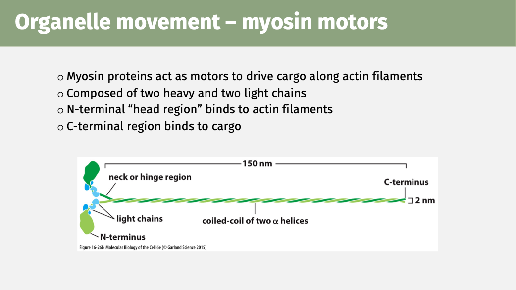
Describe the structure of myosin motors and how this relates to their function?
Myosin proteins act as motors to drive cargo along actin filaments.
They are composed of two heavy, and two light chains.
The N terminal “head region” binds to the actin filament.
The C terminal region binds to the cargo.
Why do unicellular organisms need to move/migrate?
To respond to environmental signals (E.g., In chemotaxis (which is a response to chemicals) and phototaxis (which is a response to light))
Why do multicellular organisms need to move/migrate?
Like unicellular organisms, to respond to environmental signals
For development (e.g., migration of sperm cells for embryo development)
For repair (e.g., skin cell migration to close the wound)
For defence (e.g., white blood cell movement)
What drives movement in unicellular organisms? How do they act?
Flagella/cilia drive movement
These are membrane protrusions from the cell containing microtubules
Flagella: move with an undulating, wave-like motion.
Cilia: move with a whip-like motion, similar to a breaststroke, often in coordinated waves.e
What is axoneme, how does it power movement?
Flagella and cilia are built around a core called the axoneme.
The axoneme is a bundle of microtubule doublets.
Dynein arms connect adjacent microtubules.
Dynein “walks” along one microtubule using ATP, trying to slide the microtubules past each other.
This sliding is converted into bending, producing the flagella or cilia motion.
What are the roles/different types of cilia?
Motile cilia:
Move cells or fluid over tissues.
Examples:
Respiratory tract – move mucus & trapped bacteria to mouth
Oviduct – move eggs toward uterus
Non-motile cilia (primary cilium):
Found on many cell types.
Sense and respond to the environment.
Important for sight and smell.
What are bacterial flagella made of?
Bacterial flagella are composed of the protein flagellin, not microtubules.
They are a special case compared to eukaryotic flagella/cilia.
How do multicellular organisms achieve cell migration?
Cilia/flagella: swim (e.g., sperm).
Crawling: cells move over surfaces (majority of animal cell movement).
Controlled by: growth factor signalling.
Mechanism: cytoskeletal dynamics + receptors guide movement.
How do cells migrate during tissue repair?
Fibroblasts move to the wound site.
Signalling factors detect the wound and guide migration.
Actin polymerisation pushes out the cell edges.
Lamellipodia (actin mesh) extend the front of the cell to drive movement forward.
What are the steps of amoeboid cell migration?
Protrusion: actin filaments push the plasma membrane outward.
Attachment: the extended edge of the cell sticks to the surface.
Traction: the rest of the cell (trailing cytoplasm) is pulled forward.
Which 2 things does cell migration involve?
- Cell movement / cell migration involves:
‘swimming’: flagella and cilia
‘crawling’: protrusion, attachment, traction
What is cell migration dependant on?
Dependant on the cytoskeleton.
What is import and export into the nucleus controlled by?
Nuclear import and export are controlled by receptor proteins called importins and exportins.
These receptors rely on the small GTPase Ran, which provides directionality by switching between Ran-GTP and Ran-GDP states.
Where is Ran-GDP found, and what converts Ran-GTP to Ran-GDP?
Ran-GDP is found in the cytosol. A GAP (GTPase-activating protein) stimulates Ran to hydrolyze GTP → GDP.
Where is Ran-GTP found, and what converts Ran-GDP to Ran-GTP?
Ran-GTP is found in the nucleus. A GEF (guanine nucleotide exchange factor) replaces GDP with GTP.
Describe how proteins are imported into the nucleus (protein targeting to the nucleus).
A cargo protein with a nuclear localization signal binds to its receptor (importin) in the cytoplasm.
The cargo–importin complex enters the nucleus through a nuclear pore.
Inside the nucleus, Ran-GTP binds to importin, causing the cargo to be released.
Ran-GTP displaces the cargo by binding importin more strongly.
The importin–Ran-GTP complex is exported back to the cytoplasm.
In the cytoplasm, Ran-GTP is hydrolyzed to Ran-GDP, releasing importin, which is now free to begin another import cycle.
How are proteins exported from the nucleus? Describe the steps of nuclear export involving Ran-GTP.
Cargo binds to export receptor (exportin).
Ran-GTP binds the receptor, which promotes tight cargo binding.
Cargo–receptor–Ran-GTP complex exits through the nuclear pore into the cytosol.
Ran-GTP is hydrolyzed to Ran-GDP, causing a conformational change.
Complex dissociates—cargo is released into the cytosol, and the receptor returns to the nucleus for reuse.
Why doesn’t Ran-GDP accumulate in the cytoplasm, and how is it returned to the nucleus for nuclear import?
Ran-GDP does NOT build up in the cytoplasm because it is quickly bound by Nuclear Transport Factor 2 (NTF2).
Ran-GDP–NTF2 complex enters the nucleus through the nuclear pore.
Inside the nucleus, Ran-GEF converts Ran-GDP → Ran-GTP, which causes Ran-GDP to dissociate from NTF2.
This maintains the gradient: Ran-GTP high in nucleus, Ran-GDP high in cytoplasm, enabling directionality for nuclear import/export.
How are proteins directed to the ER for synthesis?
They contain an ER signal sequence at their N-terminus, which ensures they are targeted to the ER during translation.
What recognizes the ER signal sequence, and what happens next?
The Signal Recognition Peptide (SRP) binds the N-terminal ER signal sequence and pauses translation.
How does the ribosome-SRP complex get to the ER?
The SRP–ribosome complex binds to the SRP receptor on the ER membrane.
What triggers the release of SRP and the SRP receptor?
The translocator binds the ribosome, which pushes out the SRP–SRP receptor complex, allowing it to dissociate and be recycled.
What happens to the growing protein once SRP is released?
Translation resumes, and the protein is threaded (translocated) across the ER membrane through the protein translocator into the ER lumen.