Cranial Nerves
1/214
Name | Mastery | Learn | Test | Matching | Spaced |
|---|
No study sessions yet.
215 Terms
CN I: name, sensory or motor, function
olfactory nerve
sensory
smell
CN II: name, sensory or motor, function
optic nerve
sensory
vision
CN III: name, sensory or motor, function
oculomotor nerve
motor
eye movement, accommodation, pupil constriction
CN IV: name, sensory or motor, function
trochlear nerve
motor
superior oblique innervation
CN V: name, sensory or motor, function
trigeminal nerve
both
facial sensation, mastication
CN VI: name, sensory or motor, function
abducens nerve
motor
lateral rectus innervation
CN VII: name, sensory or motor, function
facial nerve
both
facial expressions, anterior 2/3 of taste, lacrimation, salivation (submaxillary and submandibular glands)
CN VIII: name, sensory or motor, function
vestibulocochlear nerve
sensory
hearing, balance
CN IX: name, sensory or motor, function
glossopharyngeal nerve
both
swallowing, salivation (parotid gland), posterior 1/3 of taste, monitoring of carotid sinus
CN X: name, sensory or motor, function
vagus nerve
both
taste, swallowing, palate elevation, talking, thoracoabdominal viscera
CN XI: name, sensory or motor, function
accessory nerve
motor
head turning, shoulder shrugging
CN XII: name, sensory or motor, function
hypoglossal nerve
motor
tongue movement
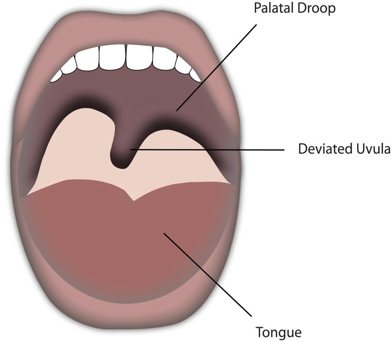
what are signs of a CN X palsy
uvula deviates away from side of lesion
palate does not elevate
patient reports hoarse voice
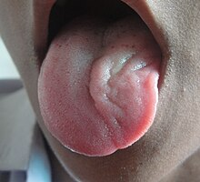
what is a sign of a CN XII palsy
tongue deviates towards the side with the lesion
the optic nerve carries ______ info only
sensory
what is the ON composed of
GC axons
path of GC axons that form ON
converge at optic disc, exit sclera through the lamina cribrosa, leave the orbit via optic foramen, travel through optic canal
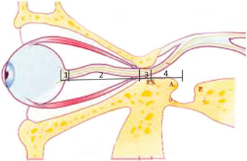
where does the ON leave the orbit
optic foramen
(nasal/temporal) fibers cross at the _________ to join the fibers in the other eye
nasal fibers cross at the optic chiasm to join the fibers in the other eye
the optic tract contains nasal fibers from the (contralateral/ipsilateral) eye
optic tract contains nasal fibers from the contralateral eye and temporal fibers from the ipsilateral eye
the fibers in the optic tract carry visual information from the (same/opposite) side of the visual field
the fibers in the optic tract carry information from the same side of the visual field
4 destinations of CN II fibers
LGN, pretectal nucleus, superior colliculus, hypothalamus
what do ON fibers that travel to the LGN do
relay visual info to the primary visual cortex (V1)
what do ON fibers that travel to the pretectal nucleus do
responsible for pupil innervation
what do ON fibers that travel to the superior colliculus do
involved in saccade control
what do ON fibers that travel to the hypothalamus do
involved in circadian rhythm
what are the nuclei of CN III and where are they located
right and left nucleus
located in the midbrain at the level of the superior colliculus
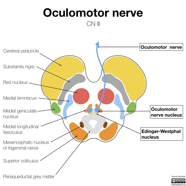
what does each nucleus (right and left) of CN III contain
each nucleus has a sub-nucleus and an Edinger-westphal nucleus
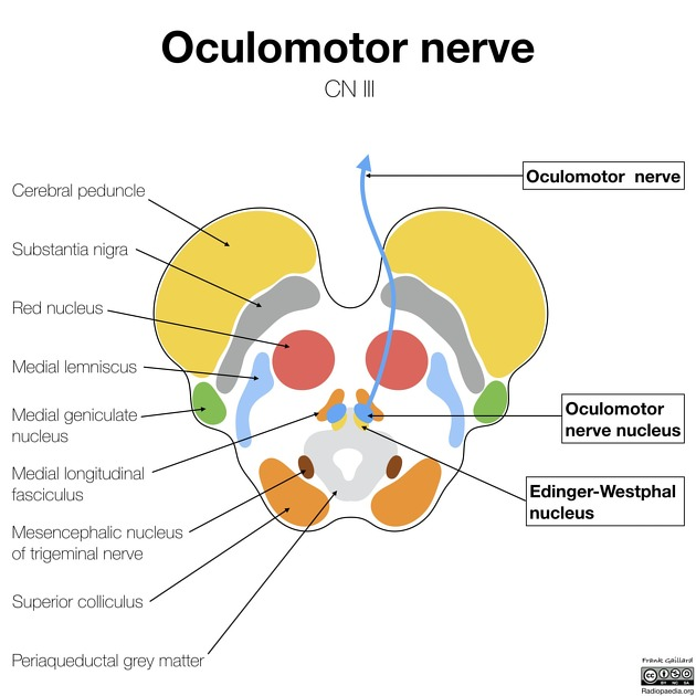
role of sub-nuclei of the CN III nuclei
provide voluntary motor control to SR, MR, IR, IO muscles
role of Edinger-Westphal nuclei of the CN III nuclei
provide parasympathetic innervation to the CB and iris sphincter muscles through the ciliary ganglion
the CN III nuclei are connected to which other nuclei, and how
connected to nuclei of CN IV, VI, and VIII via the MLF (medial longitudinal fasciculus)
what 2 structures do the CN III nuclei receive information from
superior colliculus, visual cortex
which CN III fibers project to ipsilateral muscles
fibers of the sub-nuclei that leave for the medial rectus, inferior oblique, and inferior rectus
which CN III fibers project to contralateral muscles
fibers of the sub-nuclei that leave for the superior rectus
what are the only fibers from CN III sub-nuclei that cross
SR fibers
a lesion in the right SR sub-nuclei will result in poor control of what and why
the left SR muscle
the SR CN III fibers are the only CN III fibers that cross the midline (contralateral innervation)
what controls the levator palpebrae muscle of each eye
one central CN III sub-nucleus
a lesion in the levator palpebrae sub-nucleus will result in what and why
sudden onset bilateral ptosis
one sub-nuclei of CN III control the levator palpebrae innervation of both eyes
when the sub-nuclei exit the brainstem, where do they go
cavernous sinus
what kind of innervation do CN III fibers receive in the cavernous sinus, and from where
CN III fibers get sympathetic fibers from the internal carotid artery plexusw
what does CN III divide into and where
superior and inferior divisions
divides just before it enters the orbit
where does CN III enter the orbit
superior orbital fissure
path of CN III sub-nuclei fibers from the brain to the eye
sub-nuclei fibers join together and enter brainstem > travel closely to posterior communicating artery (ICA branch) > enter cavernous sinus > get ICA plexus sympathetic fibers in cavernous sinus > divide into superior and inferior divisions > enter orbit through superior orbital fissure
what does the superior division of CN III innervate
superior rectus and superior levator palpebrae
what travels with the superior division of CN III, and what does it innervate
sympathetic fibers travel with the superior division and innervate Muller’s muscle of the UL
what does the inferior division of CN III innervate
medial rectus, inferior rectus, inferior oblique
what travels with the inferior division of CN III, and what does it innervate
parasympathetic fibers from the EW nuclei
innervate the iris sphincter and the ciliary muscle
what is the path of the EW nuclei fibers
PS EW nuclei fibers travel with the inferior division of CN III
go to the ciliary ganglion and leave the ganglion as SPCNs
innervate iris sphincter and ciliary muscler
role of EW nuclei fibers
innervate iris sphincter and ciliary muscle with parasympathetic innervation for pupillary constriction and accommodation
what does the CN III innervate
MR, SR, IR, IO
levator palpebrae
CB & iris sphincter (PS innervation)
Muller’s muscle (sympathetic innervation)
what does a complete CN III lesion look like
severe ptosis (levator not being innervated)
eye goes down and out (only LR and SO are getting innervation)
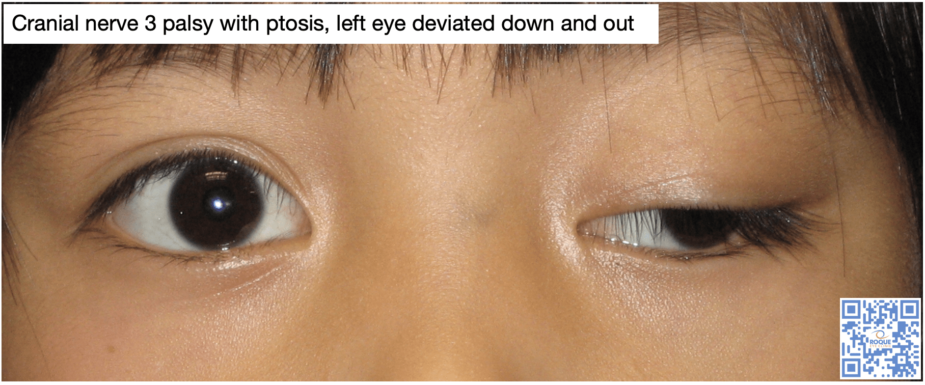
is ptosis more severe in horner’s or CN III palsy, and why
CN III palsy
horner’s is caused by lack of sympathetic innervation, causing muller’s muscle not to be innervated
muller’s is only responsible for 1-2 mm of lid elevation
CN III palsy causes lack of innervation to the levator (via superior division of CN III), which is the main elevator of the eye (causing large ptosis)
where are pupillary fibers of CN III located
on the outside of CN III
pupil-involving CN III palsy should raise suspicion for what and why
aneurysm of the posterior communicating artery (most likely at junction of ICA and PCA)
CN III fibers travel closely with PCA before reaching cavernous sinus
since pupillary fibers are on the outside of CN III, an aneurysm of the PCA will press on the pupil fibers, causing pupil involvement
what is the most likely cause of a pupil-sparing CN III palsy
ischemia of small BVs that nourish inner fibers of CN III (likely due to vascular disease like DM or HTN)
what type of CN III palsy is considered an emergency and why
pupil-involving CN III palsy
most likely caused by a compressive lesion (aneurysm or tumor) of the PCA pressing on the outer CN III fibers
CN rule of fours
CN 1-4 originate above the pons (CN 3 & 4 originate in the midbrain)
CN 5-8 originate in the pons
CN 9-12 originate in the medulla
remember midbrain, pons, medulla
what is the medulla responsible for
autonomic functions
breathing, heartrate, digestion
hearing and balance are the 2 main functions of what nerve
CN VIII: vestibulocochlear
what 2 nerves innervate taste in the tongue, and what part of the tongue
anterior 2/3 of tongue: CN VII (facial nerve)
posterior 1/3 of tongue: CN IX (glossopharyngeal nerve)
what nerve innervates tongue movement, and where will the tongue move if there is a lesion in that nerve
CN XII (hypoglossal nerve)
tongue will move toward direction of lesion
remember tongue toward
what nerve innervates the uvula, and where will the uvula move if there is a lesion in that nerve
CN X (vagus nerve)
uvula will move away from lesion
remember vagus = uVula
what 2 movements are associated with CN XI
CN XI is the accessory nerve
it is associated with head turning and shoulder shrugging
remember accessory nerve = student nerve, turning head and shrugging shoulders like you don’t know something
what are the only 2 nerves with contralateral innervation
nerves innervating SR and SO
CN IV (trochlear nerve)
only the superior division of CN III that innervates the SO
what is the main nerve responsible for mastication (chewing)
CN V (trigeminal nerve)
what is the main nerve responsible for lacrimation
CN VII (facial nerve)
what is the main nerve responsible for swallowing
CN IX (glossopharyngeal nerve)
what are 2 other names for V1 (visual cortex) in the brain
Brodmann’s area 17
striate cortex
(this is where the LGN projects visual info)
where does CN III start its course
midbrain
what is the sensory and motor innervation that occurs when you shine light into a patient’s eye to check their pupils
sensory: CN II (ON) brings the light signal to the brain by sending signals to the pretectal nucleus (light response, pupil control), which then signals to the EW nucleus
motor: CN III causes the EW nucleus to send PS innervation to the iris sphincter, causing the pupil to constrict
where do the preganglionic parasympathetic fibers start their course
EW nucleus
what happens when you have a lesion between the pretectal nucleus and EW nucleus and why
Argyll Robertson pupil: pupil does not constrict with light, but constricts with accommodation
pretectal nucleus is responsible for light response, is not able to tell the EW nucleus to signal to iris sphincter when light shines
EW nucleus still works, so can still respond to accommodation
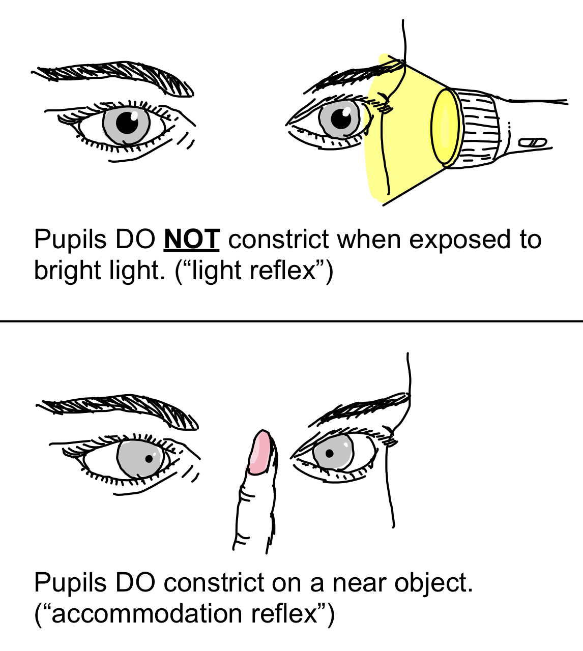
what happens when you have a lesion in the EW nucleus
Adie’s tonic pupil
pupils don’t respond to light and respond slowly to accommodation
where is the lesion most likely located if there is a pupil-involing CN III palsy
junction of ICA and posterior communicating artery
what is the longest cranial nerve
CN IV (trochlear nerve)
location and number of nuclei in CN IV
2 nuclei located in the midbrain at the level of the inferior colliculus
(slightly lower than CN III nuclei located in the midbrain at the level of superior colliculus)
what nuclei are the nuclei of CN IV connected to and how
nuclei of CN III, CN VI, CN VIII via the MLF
path of CN IV nuclei connection to visual cortex
CN IV nuclei use the tectobulbar tract to connect to the superior colliculus, which then connects to the visual cortex
what is the only CN that comes off of the dorsal (posterior) side of the brainstem
CN IV
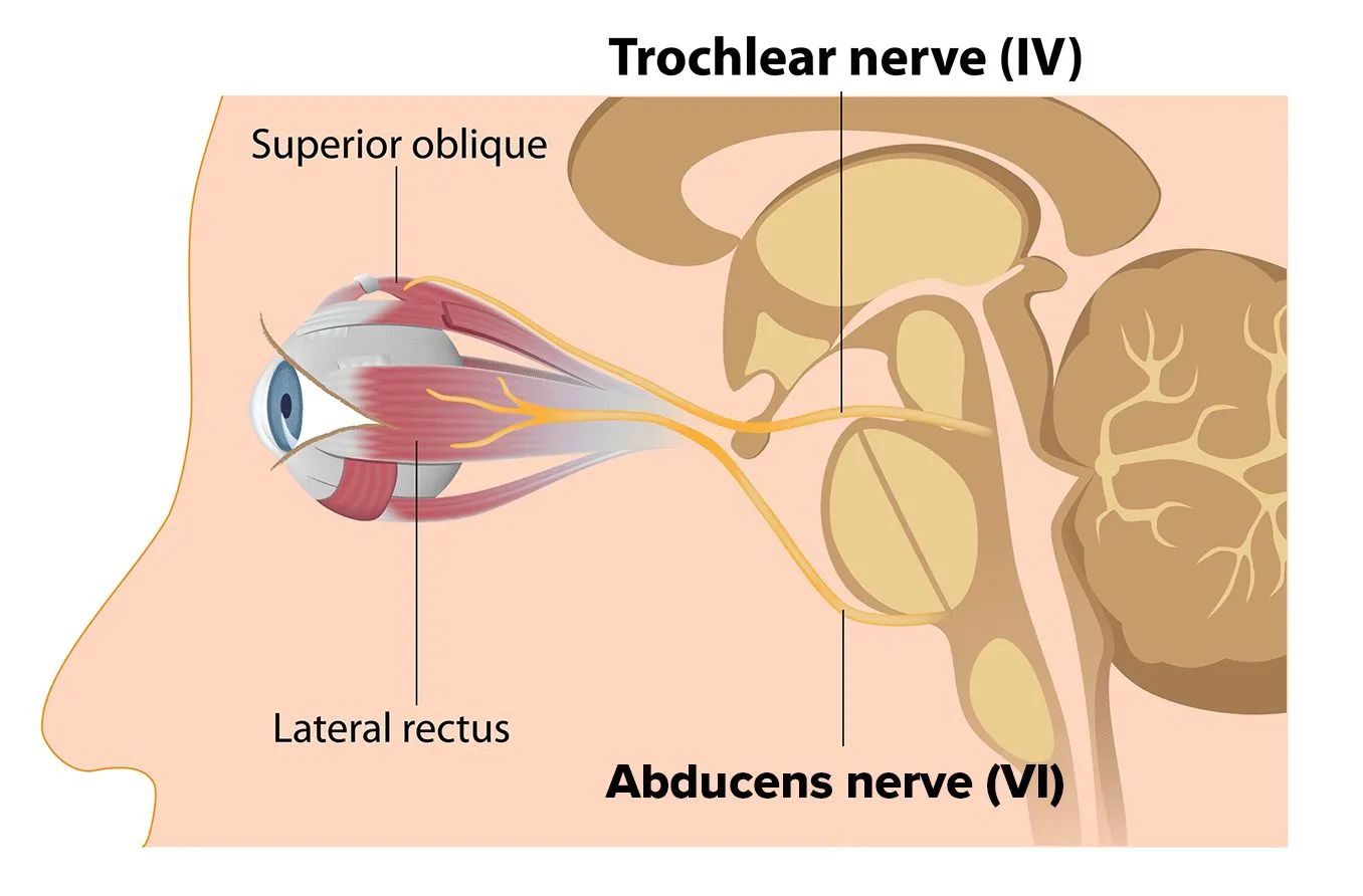
what is the only cranial nerve to fully decussate
CN IV
contralateral innervation of SO
a patient with a superior oblique palsy will tilt their head (towards/away) from the affected side
away from the affected side
actually tilting their head towards the location of the lesion since a left lesion would affect the right SO
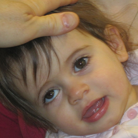
damage to the right trochlear nucleus will cause a _____ palsy, and the patient will tilt their head to the ______
left SO palsy
right
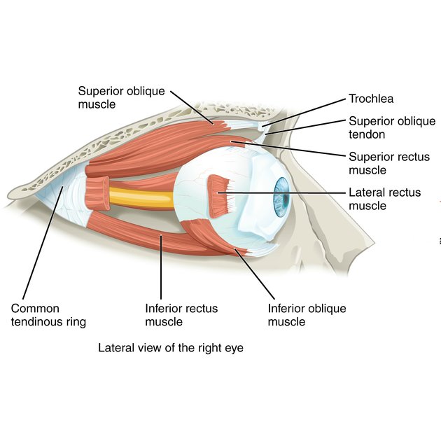
path of CN IV entering the orbit
enters the lateral wall of the cavernous sinus
travels under CN III
enters the orbit above the annulus of zinn through the superior orbital fissure
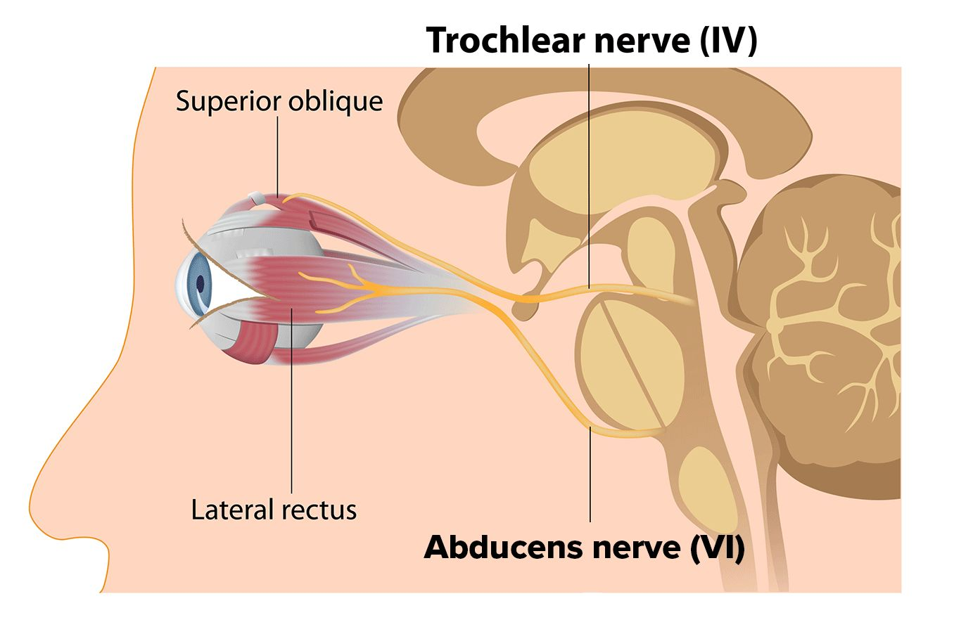
where does CN IV enter the orbit
superior orbital fissure
anatomical origin of the SO (where muscle attachment is)
lesser wing of the sphenoid
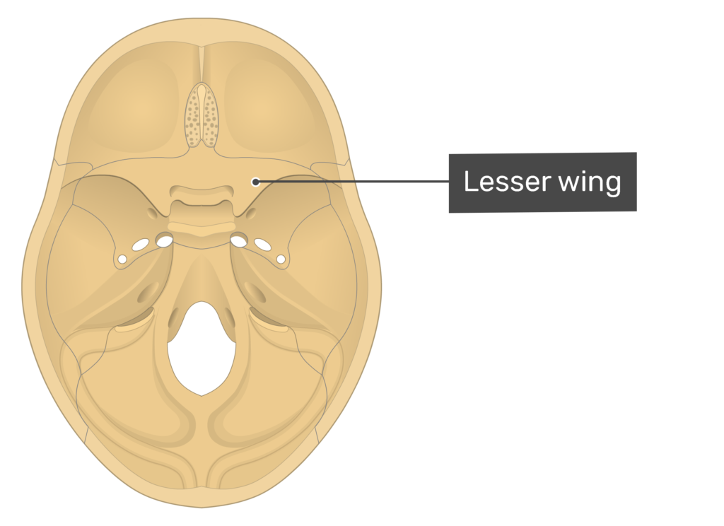
physiologic origin of the SO (functional origin)
trochlea
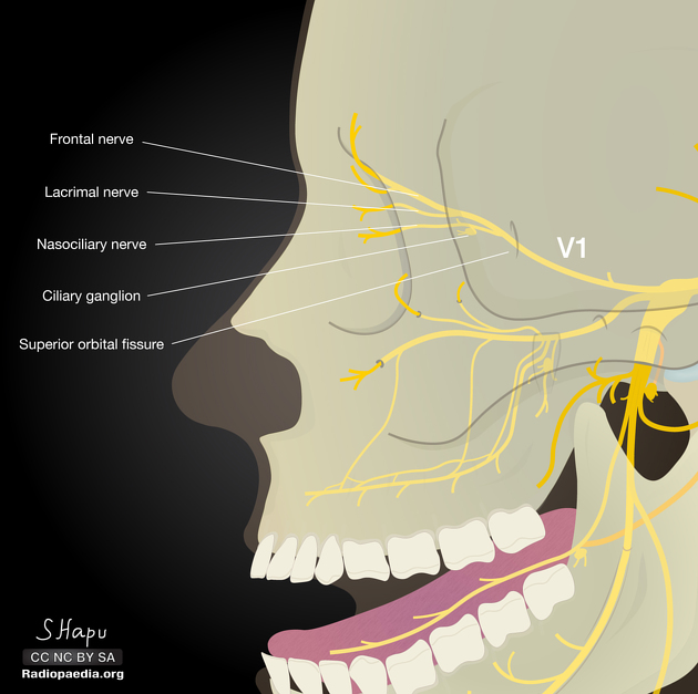
where are the roots of CN V located and what are they
1 motor root and 1 sensory root
located in the pons
sensory function of CN V
carries sensory info from the face and head, including all orbital structures
3 sensory divisions of CN V and where they provide sensory input
CN V1: sensory info from head to tip of nose
CN V2: sensory info from side of nose to mouth
CN V3: sensory info from head and ear to mandible
what branch of CN V provides sensory and motor input and what does it innervate
CN V3
sensory info from head and ear to mandible
motor info to muscles of mastication
the sensory fibers of CN V travel to the ____ nuclei in the _____ to relay sensory info to the ______
sensory fibers of CN V travel to the sensory nuclei in the pons to relay sensory info to the thalamus
path the sensory fibers of CN V take to relay sensory info
leave face and head structures
enter cavernous sinus
synapse in trigeminal ganglion in the posterior cavernous sinus
travel to the sensory nuclei in the pons
relay sensory info to the thalamus
where do sensory fibers of CN V synapse in the cavernous sinus
trigeminal ganglion in the posterior cavernous sinus
what are the 3 sensory nuclei where sensory fibers of CN V synapse
spinal nucleus
main sensory nucleus
mesencephalic nucleus
spinal nucleus
where CN V sensory fibers synapse in the medulla
gets touch, pain, and temperature info from the ipsilateral face
main sensory nucleus
where CN V sensory fibers synapse in the pons
gets light touch info and discriminates sensation
mesencephalic nucleus
where CN V sensory fibers synapse in the pons
proprioception
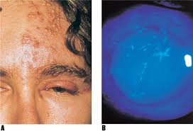
what part of the face is herpes simplex keratitis most likely to affect and why
upper eyelid and forehead
HSK can affect any or all branches of CN V1
branches of CN V1
nasociliary nerve
frontal nerve
lacrimal nerve
NFL is #1
function of nasociliary nerve (CN V1)
carries sensory info from the cornea, iris, and tip of nose
