Biopsych
1/121
Earn XP
Description and Tags
Name | Mastery | Learn | Test | Matching | Spaced | Call with Kai |
|---|
No analytics yet
Send a link to your students to track their progress
122 Terms
How does the peripheral nervous system divide
Automatic and somatic nervous
How does the automatic nervous system divide
Sympathetic and Parasympathetic nervous systems
Component of CNS
Brain and spinal cord
Function of the brain
Centre of conscious awareness
Cerebral cortex (outer layer) highly developed, separates humans and animals
Function of spinal cord
Extension of brain, responsible for reflex actions
Passes information to peripheral NS
Function of peripheral nervous system
Sends information to CNS from world
Transmits information from CNS to muscles and glands
Function of somatic nervous system
Associated with voluntary movement
Sends info to CNS from sensory receptors, send info from CNS to muscles
Function of automatic nervous system
Sends information to and from internal organs, operates involuntarily
Function of sympathetic nervous system
Responding to dangerous/ stressful systems, FIGHT OR FLIGHT
Function for parasympathetic nervous systems
Known as rest and digest, state body returns to after being in a stressed situation
What is the endocrine system
Information system of glands and hormones that works through the bloodstream
Key hormone glands
Hypothalamus, pituitary (brain)
Thyroid (neck)
Adrenals, pancreas (trunk)
Ovaries, testes (sex organs)
The master gland
Pituitary = Controls the release of hormones from all other endocrine glands in the body
Triggering of fight or flight
Hypothalamus triggers activity of sympathetic NS
Adrenaline released from adrenal medulla and triggers physiological changes
Leaving fight or flight
Parasympathetic NS returns body to resting state, PNS actions are antagonist of SNS
PNS reduces activities in body that were triggered by SNS and adrenaline
Sensory Neuron
Carry info from PNS to CNS
Long dendrites and short axons
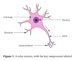
Relay Neuron
Connect sensory neurons to other relay or motor neurons
Short dendrites and short axons
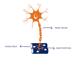
Motor Neurons
Connect CNS to effectors (muscles and glands)
Short dendrites and long axons
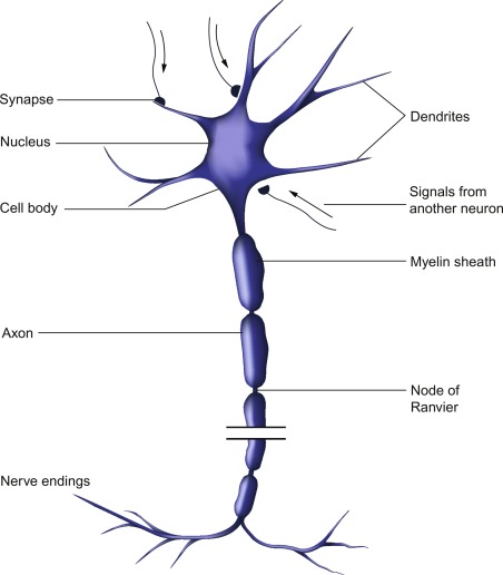
Cell body of a neuron
Includes a nuclear, genetic information of the neuron
Dendrites of neuron
Carry electrical impulse towards the cell body from neighbouring neurons
Axon of a neuron
Carry the impulse away from cell body to other neurons
Covered in myelin sheath
Myelin sheath of neuron
Fatty layer than protects the axon and helps speed up the electrical impulse
Segmented by nodes of ranvier
Nodes of ranvier in neuron
Gaps within the myelin sheath, force the impulse to jump across the gaps
Without gaps, the impulse is slowed
Electrical transmission between neurons
Resting neuron = Negative charged inside compared to outside
Stimulated neuron = Positive charge on inside, for split second, causes action potential
Neuron VS Synpase : Method of transmission
Signals within neurons are electrical
Signals between neurons, aka across a synapse, are chemical
Synaptic transmission
Electrical impulse reaches end of neuron (presynaptic terminal), triggers release of neurotransmitters from synaptic vesicles
Neurotransmitter crosses gap and taken up by post synaptic receptor sites
Message converted from chemical to electrical
Function of neurotransmitters
Relay message across synapse
Each neurotransmitters has specific molecular structure to fit to the post-synaptic receptor site
Each neurotransmitter has a specific function
Excitation
Excitation = Increasing positive charge of neighbouring neuron
Adrenaline causes excitation , increases positive charge of neighbouring neuron and increase chance the neighbouring neuron will fire and continue the impulse
Inhibition
Inhibition = Decrease the positive charge of neighbouring neuron
Serotonin causes inhibition when received by neuron, makes it negatively charged and less likely to fire
Summation
Excitatory and inhibitory influences from neurotransmitters are summed up
If net effect is inhibitory, post-synaptic neuron is less likely to fire
If net effect is excitatory, post-synaptic neuron more likely to fire
Action potential is only triggered is summation of signals reaches a threshold
Define localisation of function
Theory that different areas of the brain are responsible for different behaviours, processes or activities
Who were key names in developing localisation theories
Broca and Wernicke
How do hemispheres in the brain work
Left hemisphere controls right side of the body, right hemisphere controls left side of brain
Cerebral cortex
Outer layer of both hemispheres, about 3mm thick
Much more developed in humans than in animals
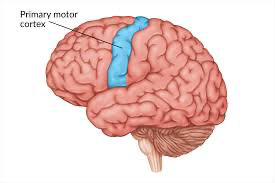
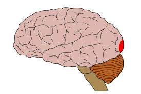
Motor cortex
Back of the frontal lobe, both hemispheres
Controls voluntary movement
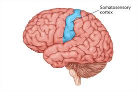
Somatosensory cortex
Front of both parietal lobes, separated from motor cortex by the central sulcus
Responsible for sensory information from skin, amount of area devoted = its sensitivity
Visual cortex
Occipital lobe, right visual field to left lobe, left visual field to right lobe
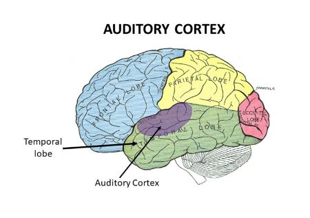
Auditory cortex
Both temporal lobes
Analyses speech based information
Broca’s Area
Small area in left frontal lobe, responsible for speech production
Damage = Broca’s Aphasia = laborious speech, lacks fluency
Wernicke’s area
In the left temporal lobe, responsible for language comprehension
Damage = Wernicke’s aphasia = Nonsense, but fluent, speech
EVAL : Evidence for localisation
Peterson et al : Brain scans to show how Wernicke’s was active in listening, and Broca’s active in reading
Tulving et al : Episodic memory in right prefrontal cortex, semantic memory in left prefrontal cortex
EVAL : Neurosurgical evidence
Dougherty et al: Cingulotomy on 44 OCD patients, 32 weeks after 1/3 ptp has successful response
EVAL: Phineas Gage
Tempering iron through his left frontal lobe, personality had a complete 180 turn
EVAL: Lashley’s Research on holistic
Removed 10-50% of the brains of rats running mazes
No area proven to be more important in the rats process to learning the mazes
Define brain plasticity
Tendency to change and adapt as a result of experiences and learning
Childhood brain growth
Gopnik et al : Ages 2/3, each neuron has around 15,000 synaptic connections
As we age, the connections we rarely use get deleted and the ones frequently used are strengthened = synaptic pruning
Early attitudes to development of brain
Early thoughts = Changes were restricted to the developing brain and that the adult brain was fixed in function and structure
Research in plasticity : Maguire et al
Studied brains of London taxi drivers, found much higher volume of grey matter in posterior hippocampus than matched control group
Posterior hippocampus = Area associated with spatial and navigational skills
Longer they had been in job, more pronounced the structural differences
Research into plasticity : Draganski et al
Imaged brains of med students 3 months before and after final exams
Learning induced changes in posterior hippocampus and parietal cortex
Functional recovery after trauma
Unaffected areas often adapt and compensate, even taking over functions
Neuroscientists suggest this can occur quickly first after trauma and then slowly
What happens to brain in recovery
Rewire and reorganise by forming synaptic connections close to damaged area
Secondary neural pathways not typically used are activated to enable functioning
3 key things that occur in brain recovery
Axonal sprouting : New nerve endings grow to connect with undamaged nerve cells
Reformation of blood vessels
Recruitment of homologous areas: Areas in opposite hemisphere can overcompensate for lack in one hemisphere
EVAL: Practical application of plasticity
After injury, recovery slows down after weeks, so therapy is need to maintain the improvements e.g. movement therapy or electrical stimulation
EVAL : Negative plasticity
Retiring can have negative consequences
Medina et al: Prolonged drug use can result in poor cognitive function and increased risk of dementia
Ramachandran and Hirstein: 60-80% of amputees develop phantom limb syndrome, causing pain. Thought to be as a result cortical reorganisation
EVAL : Age and plasticity
Generally as age increases, plasticity decreases
Bezzola et al : Golfers 40-60 years old took 40 hours of golf training. FMRIs showed lower activity in motor cortex in novice group vs control. Suggests they had more efficient neural representations after training
EVAL: Support from animal studies
Hubel and Wiesel : Sewed one eye shut of a kitten, the visual cortex of the shut eye was not idle but processing information from the open eye
EVAL : Cognitive reserve and disability free recovery
Schneider et al : In people with brain injuries, those that had spent more time in education has a greater chance of disability free recovery.
40% of participants that achieved DFR had more than 16 years education compared to 10% that had less than 12 years
Define Hemispheric Lateralisation
Two hemispheres of the brain are functionally different and certain mental processes and behaviours are controlled by one hemisphere
Split Brain research
Sperry : All participants had undergone a commissurotomy, corpus callosum has been cut down the middle and the hemispheres weren’t connected
Sperry: Procedure
Image or word projected in ptp right visual field
Same or different word projected in left visual field
Sperry Findings : Describe what you see
Picture in right visual field = Describe easily (left hemisphere process, speech is in left)
Picture in left visual field = Could not describe what it was, some said nothing (right hemi processes, no speech centre)
Sperry Findings: Recognition by touch
Object placed in left visual field (RH), could not verbally match, BUT could select a matching object, or an object with association, with their left hand (RH), when the objects were behind a screen
Sperry Findings: Composite words
Two words, one in each visual field e.g. key in left visual and ring in right visual
Participant would grab object from left visual field with their left hand (both controlled by right hemi) but say the word in their right visual field
E.g. participant would grab the key with their left hand and say the word ring
Sperry Findings: Matching faces
Individual asked to match a face to series of other faces.
Picture processed by left visual field (right hemisphere) consistently selected but images in right visual field (left hemisphere) consistently ignored
When composite face (two separate halves) presented, one half to a visual field, right visual (left hemisphere) would be properly described and left visual (right hemisphere) would be selected as matching image
EVAL : Sperry demonstrated lateralised function
Produced research with valid conclusion (left hemisphere geared to analytic and verbal task, right hemisphere adept at spatial tasks)
Sperry research was key in understanding brain processes
EVAL : Sperry’s methodology
Highly specialised and standardised procedure
Participants stare at fixation point, image flashes up for 0.1 second so eyes could not move across their half of image
Allowed Sperry to vary aspects methodically and ensure information limited to one hemisphere
EVAL : Theoretical basis of Sperrys work
Sperry’s work prompted theoretical debate about communication between hemispheres
E.g. Pucetti : Duality of brain, we are in effect two minds
EVAL : Problems with generalisation of Sperry
Unusual sample, small sample (11 people) took part in all variations, all had history of seizures
Can be argued participants have unique changes in the brain that influence findings
EVAL: Over simplifications of findings
Overemphasis and oversimplification of hemispheres
Modern neuroscientists argue distinction of “verbal” and “non-verbal” hemispheres is not as clear cut
In regular brain, the hemispheres communicates in everyday tasks and behaviours associated with one hemisphere can be equally performed by the other
Ways of investigating the brain
FMRI, EEG, ERPS, post mortems
fMRIs
Detect changes in levels of blood oxygenation to areas of the brain, More oxygen indicates more activity
Produces 3D images
EVAL: fMRIs positive
Does not rely on radiation, virtually risk-free, non-invasive
Very high spatial resolution
EVAL: fMRIs negative
Poor temporal resolution, 5 second time lag
Comparatively expensive
EEG - Electroencephalogram
Measures electrical activity via electrodes, brainwave patterns provide overall account of activity
Used as diagnostic tool for unusable patterns of activity
EVAL: EEG positives
Diagnostic tool e.g. epilepsy, random burst of activity
Contributes to understanding of sleep
High temporal resolution, detects activity up to a single millisecond
EVAL: EEG negatives
Generalised nature of information, cannot pinpoint exact source of activity
Does not distinguish between activities in adjacent locations
ERPs- Event related potentials
Use of statistical averaging techniques, extraneous brain activity can be filtered out
ERPs are the remaining brain activity, directly related to the event
EVAL: ERPs positive
Much more specificity than ERPs
Same good temporal resolution as EEGs
Different ERPs can describe precise cognitive functions
EVAL- ERPs negative
Lack of standardisation in methodology
In order for ERP to be pure data, they have to eliminate all other data, which is hard to achieve and isolate
Postmortems
Areas of damage in the brain are examined as causes of death or affliction in the brain
Compared to neurotypical brains
EVAL: Postmortem positives
Foundation of early understanding for key processes of brain e.g. Broca’s and Wernicke’s area
Improve medical knowledge and generate hypotheses
EVAL: Postmortem negatives
Causation: Damage to the brain may not be linked to deficits, or even have caused them
Ethical issues of consent, some individuals may not be able to provide informed consent
What is a circadian rhythm
A biological rhythm that lasts 24 hours
Siffre’s cave study : what did he do
Spent two months underground without natural light
Spent 6 months underground without natural light
Siffre’s Cave Study : What did he find
His free-running biological clock settled to just beyond 24 hours, closer to 25 hours
Other circadian rhythm studies
Aschoff and Weber
Folkard et al
Aschoff and Weber
PPTs spend 4 week deprived of natural light
Majority sleep/wake cycles extended to 24-25 hours
Folkard et al
12 people in cave for 3 weeks, using clock to mark there sleep and wake times
Researchers sped up clock so days were 22 hours
Only 1 PPT adjusted to regime
EVAL : Application to shift work
Boivin et al : Reduced concentration at 6am for those on shift work
Knutsson : Shift workers are 3X more likely to develop heart issues
RESEARCH INTO SLEEP/WAKE CYCLES CAN BE PRACTICALLY APPLIED
EVAL: Application to drug treatment
Certain peak times are when drugs are most effective
Led to guidelines for timing of drug dosing
EVAL : Case studies and small samples
Individuals and small groups = Not necessarily representative of larger population
Siffre was 60yo during one of his studies ∴ his internal clock works slower
EVAL: Siffre’s control
Had an artificial lamp turned on when he was awake
Other studies (Czeisler et al) adjusted their PPT sleep schedules with the use of dimming lights
What is an infradian rhythm
Less than one cycle occurs within 24 hours
Examples of infradian rhythms
Menstrual cycle, SAD
Menstrual cycle : Infradian
Lasts 24-35 days
Research into menstrual cycle for infradian: Stern and McClintock
Showed exogenous factors can cause synchrony within women
Collected pheromones from 9 women in different stages, coated in alcohol and frozen
Day 1 = Pads from beginning of cycle rubbed on upper lip, day 2 = day 2 of cycle etc etc
68% of women experienced changes within cycle
EVAL : Synchrony cycle
Too many confounding variables, any pattern could be expected to occur by chance
Small samples used
EVAL : Evolution and menstruation
Would have been advantage for women to menstruate at the same time, for collective care
HOWEVER, avoidance of synchrony would be preferred, as there would be too much competition, wouldn’t be evolutionarily strong
Seasonal Affective Disorder
Persistent low moods, often occurring within darker months, disrupts sleep/wake cycle
Many think it is due to prolonged release of melatonin (due to darkness), which prevents release of serotonin until later in the day