Paralleling Technique (Book)
1/47
There's no tags or description
Looks like no tags are added yet.
Name | Mastery | Learn | Test | Matching | Spaced |
|---|
No study sessions yet.
48 Terms

Rinn XCP indicator arm, posterior
A

Rinn XCP indicator arm, anterior
B

Rinn XCP bite-block, posterior
C

Rinn XCP bite-block, anterior
D

Rinn XCP aiming ring, anterior
E
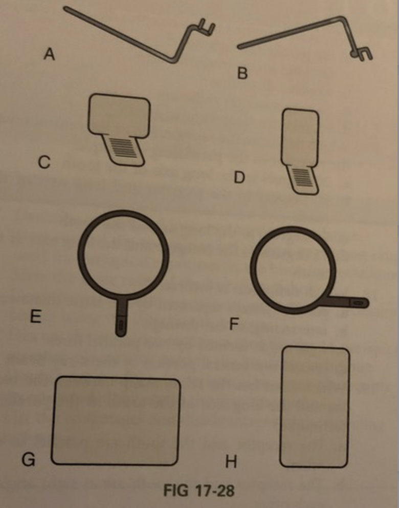
Rinn XCP aiming ring, posterior
F

Size 2 receptor
G

Size 1 receptor
H
What happens to the image when the object-receptor distance is increased?
image magnification, loss of definition
What piece of equipment is required to hold the receptor parallel to the long axis of the tooth in the paralleling technique?
film holder
What do the letters X, C, and P refer to?
X = extension
C = cone (PID)
P = Paralleling
What size receptor is used with the anterior XCP instrument?
size 1
What size receptor is used with the posterior XCP instrument?
size 2
What beam alignment devices are recommended for use with the paralleling technique to reduce radiation exposure of the patient?
rinn XCP
How is a patients head positioned before exposing receptors?
Maxillary arch parallel to the floor; and midsagital line perpendicular to the floor
Why is an increased target-receptor distance required in the paralleling technique?
a. to avoid image magnification
b. to avoid distortion
c. to reduce scatter radiation
d. to improve receptor placement
to avoid image magnification
which of the following describes the relationship of the central ray to the receptor in the paralleling technique?
a. 20 degrees to the long axis of the tooth
b. 90 degrees to the receptor and long axis of the tooth
c. 75 degrees to the long axis of the tooth
d. 15 degrees to the receptor and the long axis of the tooth
90 degrees to the receptor and long axis of the tooth
which of the following definitions is INCORRECT?
a. parallel: always separated by the same distance
b. interesting: to cut through
c. right angle: formed by two parallel lines
d. central ray: central portion of the x-ray beam
right angle: formed by two parallel lines
which of the following describes the relationship between the receptor and the long axis of the tooth in the paralleling technique?
a. the receptor and the tooth are parallel to each other
b. the receptor and the tooth are at right angle to each other
c. the receptor and the tooth are perpendicular to each other
d. the receptor and the tooth are interesting each other
the receptor and the tooth are parallel to each other
which of the following describes the distance between the receptor and the tooth in the paralleling technique?
a. the receptor is placed as close as possible to the tooth
b. the receptor is placed away from the tooth and toward the middle of the oral cavity
c. either a or b
d. none of the above
the receptor is placed away from the tooth and toward the middle of the oral cavity
which of the following about receptor placement is correct
1. anterior receptors are placed horizontally
2. anterior receptors are placed vertically
3. posterior receptors are placed horizontally
4. posterior receptors are placed vertically
a. 1, 2, and 3
b. 2, 3, and 4
c. 2 and 3
d. 1 and 4
2 and 3
Which of the following about the exposure sequence for periapical receptors is incorrect?
a. anterior receptors are always exposed before posterior receptors
b. either anterior or posterior receptors may be exposed first.
c. in posterior quadrants, the pre molar receptor is always exposed before the molar receptor.
d. when exposing anterior receptors, work from the patients right to left in the maxillary arch and then work form left to right in the mandibular arch.
either anterior or posterior receptors may be exposed first.
Which of the following about the lack of parallelism between the receptor and the long axis of the tooth is correct.
a.If the lack of parallelism is greater than 30 degrees, the image is generally acceptable.
b. If the lack of parallelism is less than 20 degrees, the image is generally acceptable.
c. If the lack of parallelism is less than 50 degrees, the image is generally acceptable.
d. If the lack of parallelism is greater than 50 degrees, the image is generally acceptable.
If the lack of parallelism is less than 20 degrees, the image is generally acceptable.
Which are advantages of the paralleling technique?
1. increased accuracy
2. simplicity of use
3. ease of duplication
4. ease of receptor placement
a. 1, 2, 3, and 4
b. 1, 2, and 3
c. 2, 3, and 4
d. 1, 3, and 4
1, 2, and 3
T/F: The advantages of the paralleling technique outweigh the disadvantages.
true
State the basic principle of the paralleling technique.
1. The receptor is placed in the mouth parallel to the long axis of the tooth being radiographed.
2. The central ray of the x-ray beam is directed perpendicular (at a right angle) to the receptor and the long axis of the tooth.
3. A beam alignment device must be used to keep the receptor parallel with the long axis of the tooth. The patient cannot hold the receptor in this manner.
Describe why a beam alignment device must be used in the paralleling technique.
The paralleling technique requires the use of a beam alignment instrument or a receptor holding device to position the receptor parallel to the long axis of the tooth. Beam alignment devices are used to position an intraoral receptor in the mouth and maintain the receptor in position during exposure of the tooth.
State the five rules of the paralleling technique.
1. Receptor placement. The receptor must be positioned to cover the prescribed area of teeth to be examined. Specific placements are detailed in the procedure sections of this chapter.
2. Receptor position. The receptor must be positioned parallel to the long axis of the tooth. The receptor and beam alignment device must be placed away from the teeth and toward the middle of the oral cavity (see Figure 17-3).
3. Vertical angulation. The central ray of the x-ray beam must be directed perpendicular (at a right angle) to the receptor and the long axis of the tooth (see Figure 17-3).
4. Horizontal angulation. The central ray of the x-ray beam must be directed through the contact areas between teeth (Figure 17-6).
5. Receptor exposure. The x-ray beam must be centered on the receptor to ensure that all areas are exposed. Failure to center the x-ray beam results in a partial image on the receptor or a "cone-cut." Cone-cuts can be produced with either a round PID or a rectangular PID (Figure 17-7). Cone-cuts are discussed in Chapter 20.
Discuss the patient and equipment preparations that must be completed before using the paralleling technique.
Patient preparation
o Following infection control procedures and preparation of the tx area and supplies, the pt should be seated.
Equipment preparation
o After pt is prepared equipment must be prepared before the exposure of any receptors.
Discuss the exposure sequence for 15 periapical receptor placements using the paralleling technique.
Anterior:
1. Assemble the anterior Rinn XCP instrument.
2. Begin with the maxillary right canine (tooth #6).
3. Expose the maxillary anterior teeth working from the patient's right to the patient's left.
4. End with the maxillary left canine (tooth #11).
5. Next, move to the mandibular arch.
6. Begin with the mandibular left canine (tooth #22).
7. Expose all the mandibular anterior teeth working from the patient's left to the patient's right.
8. Finish with the mandibular right canine (tooth #27).
Posterior:
1. Begin with the maxillary right quadrant.
2. Assemble the posterior Rinn XCP instrument for this
area.
3. First, expose the premolar receptor (teeth #4 and 5),
then expose the molar receptor (teeth #1, 2, and 3).
4. Without reassembling the posterior Rinn XCP
instrument, move to the mandibular left quadrant.
5. First, expose the premolar receptor (teeth #20 and 21), then expose the molar receptor (teeth #17, 18, and
19).
6. Reassemble the posterior Rinn XCP instrument over
a covered work surface.
7. Next, move to the maxillary left quadrant.
8. First, expose the premolar receptor (teeth #12 and
13), then the molar receptor (teeth #14, 15, and 16).
9. Finish with the mandibular right quadrant.
10. First, expose the premolar receptor (teeth #28 and 29), and end with the exposure of the molar receptor
(teeth #30, 31, and 32).
Discuss the exposure sequence for 15 periapical receptor placements using the paralleling technique.
Summarize the guidelines for periapical receptor positioning with the paralleling technique
1. If using film, the white side of the film always faces the teeth ("white in sight"). When using a sensor, position the sensor toward the x-ray tube according to manufacturer directions.
2. The anterior receptors are always placed vertically.
3. The posterior receptors are always placed horizontally.
4. If using film, the identification dot is always placed in the slot of the bite-block, toward the occlusal end of the film. ("Place the dot in the slot.")
5. When placing the receptor in the mouth, always lead with the apical end of the receptor, and then rotate the beam alignment device.
6. When positioning the beam alignment device, always place the receptor away from the teeth and toward the middle of the oral cavity.
7. When positioning the beam alignment device, always center the receptor over the area to be examined (as defined in the prescribed placements).
8. When positioning the beam alignment device, ask the patient to "slowly close" on the bite-block. Always make certain that the bite-block is stabilized by the teeth and not the lips.
Explain the modifications in the paralleling technique that are used for a shallow palate, bony growths, or a sensitive premolar region.
Cotton rolls. To position the receptor parallel to the long axis of the tooth, two cotton rolls can be placed, one on each side of the bite-block (Figure 17-22). As a result, however, periapical coverage is reduced.
Vertical angulation. To compensate for the lack of parallelism, the vertical angulation can be increased by 5 to 15 degrees more than the Rinn XCP instrument indicates. However, image distortion occurs as a result.
a type of beam alignment device that is used with the paralleling technique; these include plastic bite blocks, plastic aiming rings, and metal indicator arms
Rinn XCP Instruments
an intraoral imaging technique used to exposed periapical receptors; the receptor is placed parallel to the long axis of the tooth; a beam alignment device must be used to keep the receptor parallel to the long axis of the tooth
Paralleling Technique
something that responds to a stimulus; a recording medium; examples: x-ray film or digital sensors
receptor
intersecting at or forming right angles
perpendicular
an imaginary line that divides a tooth longitudinally into two equal halves
long axis
a bony growth in the oral cavity
torus
central portion of the primary beam of x-radiation
central ray
moving or lying in the same plane; always separated by the same distance and not intersecting
parallel
receptor is parallel / perpendicular to the CR
perpendicualr
receptor is parallel / perpendicular to the long axis of tooth
parallel
receptor is parallel / perpendicular to the biting surface of bite block
perpendicular
receptor is parallel / perpendicular to the opening of the PID
parallel
CR is parallel / perpendicular to the receptor
perpendicular
CR is parallel / perpendicular to the long axis of the tooth
perpendicular
CR is parallel / perpendicular to the biting surface of bite block
parallel
CR is parallel / perpendicular to the opening of the PID
parallel