VS331: Module 19: Cardiovascular system - Heart
1/6
There's no tags or description
Looks like no tags are added yet.
Name | Mastery | Learn | Test | Matching | Spaced | Call with Kai |
|---|
No analytics yet
Send a link to your students to track their progress
7 Terms
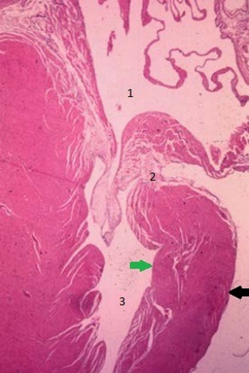
Green arrow = endocardium
Black arrow = epicardium
Thick muscle layers between = Myocardium
Space 1 = Atrium
Space 3 = Ventricle
2 = AV valve
Identify the 3 layers in the heart indicated by the green & black arrows. Identify the spaces indicated by 1 & 3. Identify the structure indicated by 2.
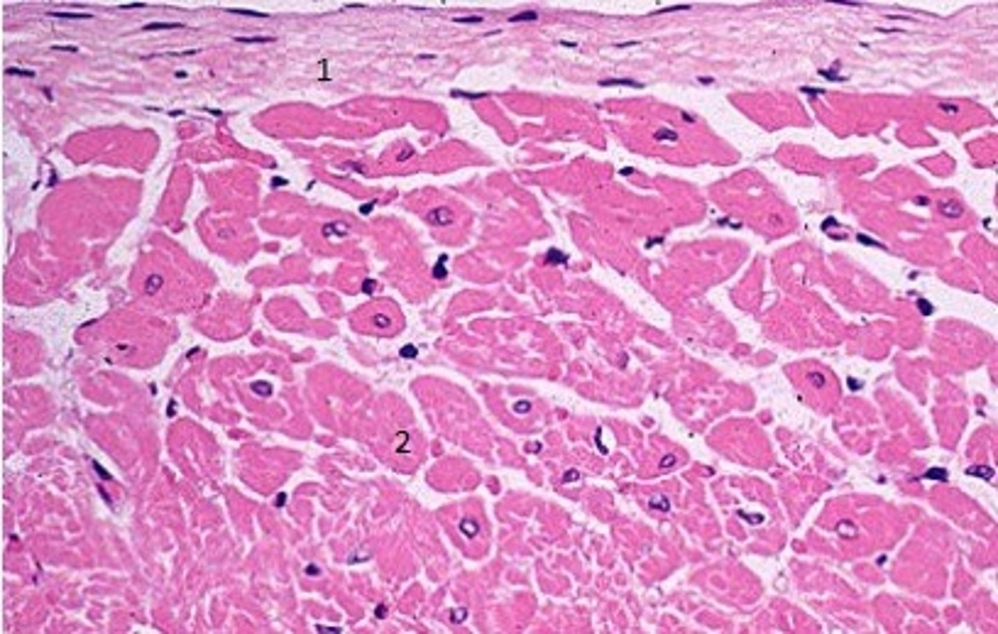
1. Endocardium - made of endothelium and underlying connective tissue
2. Myocardium - made up of cardiac myocytes
Identify the layers indicated by 1 & 2.
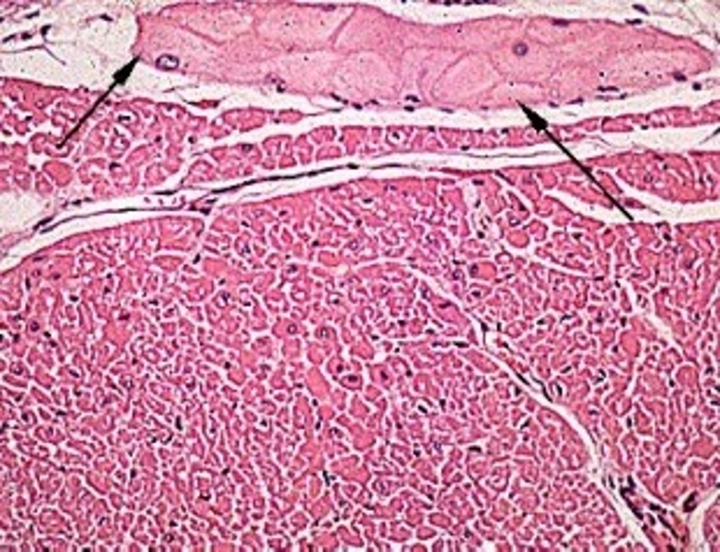
These are Purkinje cells/fibers. They are modified cardiac muscle cells that make up the impulse conducting system in the heart.
Identify the cell indicated by the black arrows and describe their function
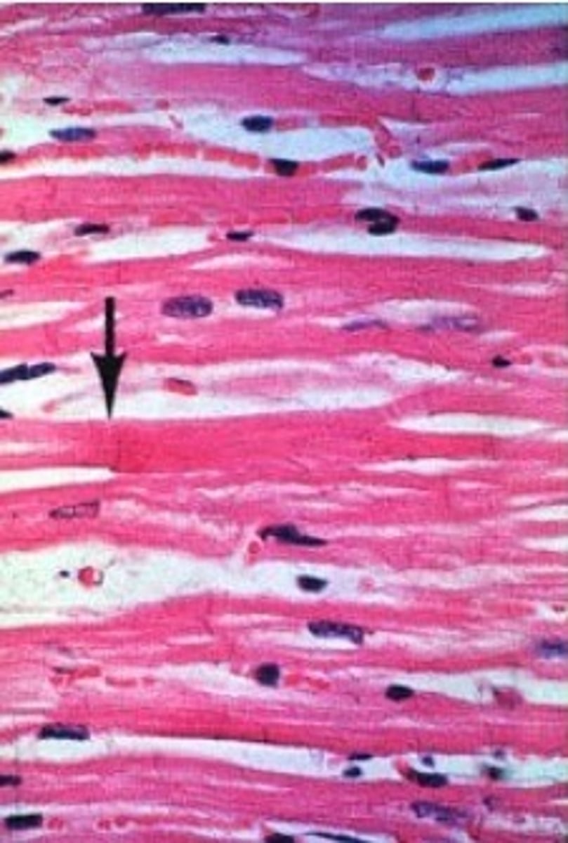
Cardiac muscle
- Characteristics: Striations (less so that skeletal muscle), Branching, and nuclear halos.
Arrow = Intercalated disc
Identify the tissue, list common characteristics and identify what the arrow is pointing to.
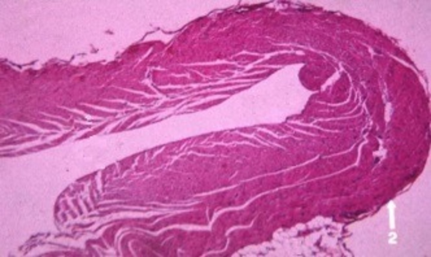
Epicardium = Mesothelium & thin layer of connective tissue
Identify the tissue indicated by #2 and give a second name for the tissue.
1. SA node: initiates the regular heartbeat = Pacemaker
2. AV node: Delays impulse from SA node allowing the emptying of the atria
3. Purkinje fibers: form "tracts" to assist in the conduction of action potentials and leads to contraction of the ventricles
Identify the flow of action potentials through the conduction system of the heart starting with the Sinoatrial node (SA node).
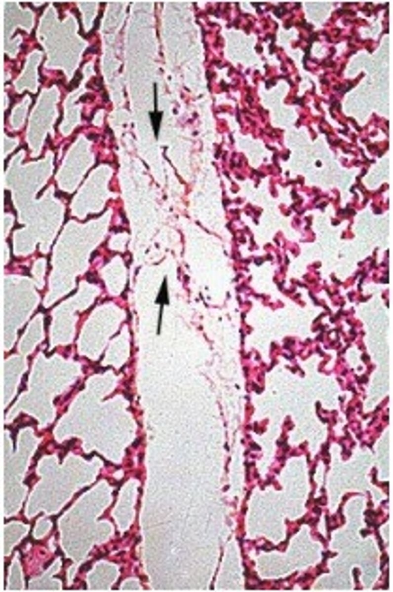
Lymph vessel
- Function to return excess tissue fluids to the venous system for recirculation
-Characteristics: "Leaky" or very fenestrated, very thin vessels, minimal smooth muscle, endothelium, and one-way valves
Identify the vessel, describe their function and common characteristics.