A&P Chapter 9: Joints and Articulations
1/54
Earn XP
Description and Tags
Name | Mastery | Learn | Test | Matching | Spaced | Call with Kai | Chat |
|---|
No analytics yet
Send a link to your students to track their progress
55 Terms
Joint Design and Movement - Overview
Overview
the adult human body has 206 bones, and all are connected together (except hyoid)
because bones are inflexible, movement can only occur at articulations or joints, where two bones connect
each joint reflects compromise between need for strength and mobility = differ in amount of movement permitted (range of motion/ROM)
anatomical structure of a joint determines type and amount of movement that may occur

Structural Classification of Joints
structural classification is based on anatomical components that make up joint
fibrous joints: held together by fibrous connective tissues but lack cartilage and possess no cavity between bones. they are either SYNARTHROTIC or AMPHIARTHROTIC
cartilaginous joints: held together by fibrous connective tissues such as ligaments, but also possess hyaline/fibrocartilage. cartilaginous joints lack a joint cavity and are either SYNARTHROTIC or AMPHIARTHORIC
synovial joints: held together by fibrous connective tissues, hyaline cartilage and/or fibrocartilage, and possess a joint cavity. all synovial joints are DIARTHORTIC. they are complex in structure + the most numerous joint type. they permit greatest range of motion.

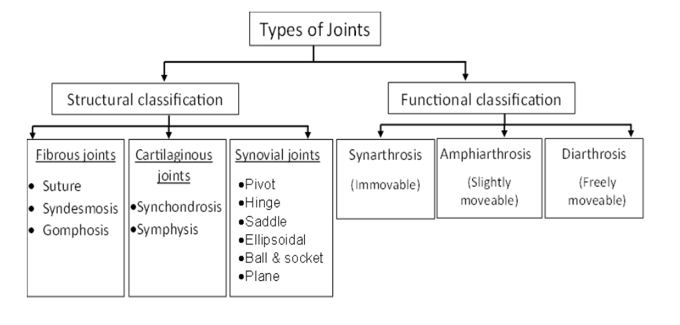
Functional Classifications of Joints
functional classifications are based on range of motion allowed
synarthrosis: NO MOVEMENT IS PERMITTED. the body edges are close together/interlocking. these strong joints are located where movement between bones must be prevented.
sutures, gomphoses (teeth), synchondroses
amphiarthrosis: SLIGHT MOVEMENT IS ALLOWED. permits more movement than synanthrotic joints, but is much stronger than freely moveable joints
intervertebral discs, pubic symphysis, tibiofibular joint, sternomanubrial joint
diarthrosis: FREELY MOVEABLE JOINT (aka synovial joint). provide wide range of motion, typical in appendages
shoulder/hip, elbow/knee, atlantoaxial, wrist/knuckles, carpal bones

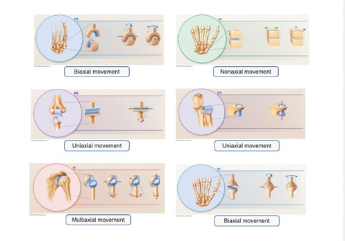
Axis of motion
axis of motion: the movements of diarthrotic joints are divided along the three planes: frontal, transverse, and sagittal
non-axial motion: LINEAR movements where bones SLIDE (vertebrocostal joints, sacroiliac joint, and intercarpal joints)
uniaxial motion: movement in ONE planes (interphalangeal joints, elbow, knee)
biaxial motion: movement in TWO planes (metacarpophalangeal joint, occipital condyles to atlas)
multi-axial motion: movement in THREE planes (shoulder + hip joint)
![<p>[Fibrous Joints]</p><p>Sutures</p>](https://assets.knowt.com/user-attachments/5af42cb4-3674-4cc4-af2c-17ba6b4f08f6.png)
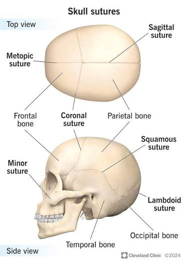
[Fibrous Joints]
Sutures
synarthrotic joint located only between the bones of skull
edges of bone are interlocked + bound together at the suture by dense fibrous connective tissue
in newborns/infants: areas of connective tissue is wider and called fontanelles (soft spots)
in adulthood: sutures of skull transform to synostoses (where bones have fused together + tissue turned to bone)
abnormal fusion of bones = synostosis that shouldn’t exist (ex: premature ossification of cranial sutures limits growth of brain) (ex 2: radio-ulnar synostosis)
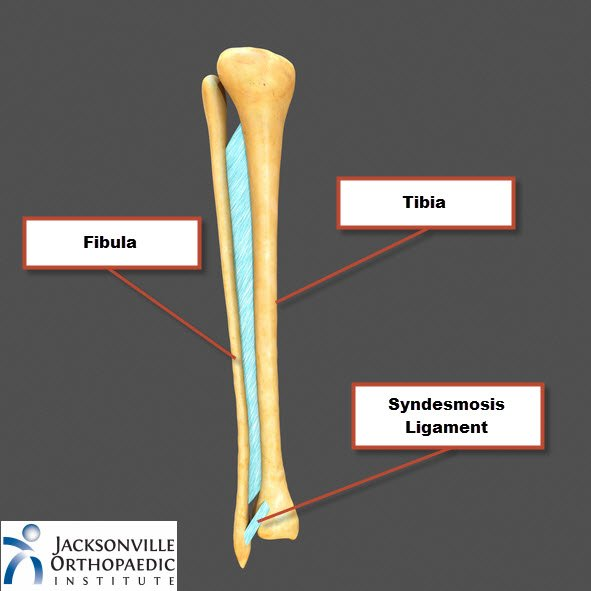
![<p>[Fibrous Joints]</p><p>Syndesmosis</p>](https://assets.knowt.com/user-attachments/5bd1ae07-16fc-41a8-a5f6-8073c59e803f.png)
[Fibrous Joints]
Syndesmosis
bones are connected by ligaments or broad/sheet-like membrane called an interosseous membrane
they are amphiarthrotic
most common example: distal articulation between the tibia and fibula called the tibiofibular joint
example: middle radio-ulnar joint, but NOT THE proximal/distal ends of the radius and ulna because those are synovial/diarthrotic
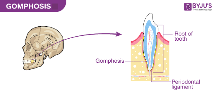
![<p>[Fibrous Joints]</p><p>Gomphosis</p>](https://assets.knowt.com/user-attachments/f0d23ca2-6451-4f9a-92a9-38ce8c715848.png)
[Fibrous Joints]
Gomphosis
found in the maxillae and mandible where teeth are fixed securely in sockets of the alveolar margins
fibrous connective tissue between a tooth and its socket is a periodontal ligament
a synarthrotic joint is sometimes called a “peg-in-socket” joint
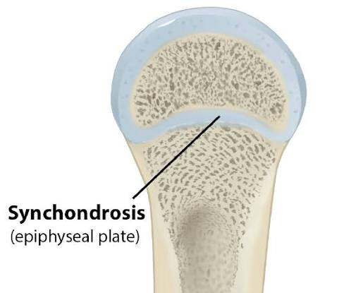
![<p>[Cartilaginous Joints]</p><p>Synchondrosis</p>](https://assets.knowt.com/user-attachments/42d94857-0273-4877-a1db-e18e16dcbfc5.png)
[Cartilaginous Joints]
Synchondrosis
rigid, hyaline cartilage bridge units the bones
example: cartilaginous joint found between ends of the first pair of ribs + manubrium of the sternum (all other ribs are synovial joints)
example: epiphyseal plate found holding the epiphysis of a long bone to the diaphysis
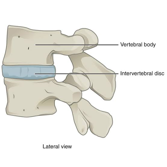
![<p>[Cartilaginous Joints]</p><p>Symphysis</p>](https://assets.knowt.com/user-attachments/548e0253-3485-4399-9305-e32fc2e4019e.png)
[Cartilaginous Joints]
Symphysis
articulating bones are separated by a wedge or pad of fibrocartilage
example: between vertebrae where thick pad of fibrocartilage forms the intervertebral disc
example: two pubic bone (pubic symphysis)
it is an AMPHIARTHROTIC joint = slight movement
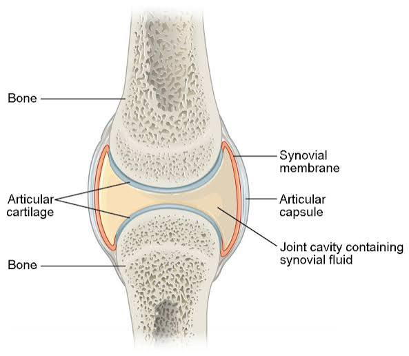
![<p>[Synovial Joints]</p><p>Structural features of a synovial joint</p>](https://assets.knowt.com/user-attachments/eb643f0d-54d2-40ac-a22d-0d7763a739ca.png)
[Synovial Joints]
Structural features of a synovial joint
joint cavity: space between articulating bones
joint capsule: layers of connective tissue enclose the cavity to house the fluid
articular capsule: thick outer layer = strength/stability
synovial membrane: inner soft tissue, leak plasma to produce synovial fluid
synovial fluid: derived from blood + egg consistency. small amount of liquid. three primary functions:
lubrication: reduces friction, called weeping lubrication
nutrient distribution: provides nutrients/waste disposal for chondrocytes. circulates when joint moves
shock absorption: provides cushion against shock
articular cartilages: line the surfaces of articulating bones; composed of hyaline cartilage. provides slick, smooth surface = less friction
accessory organs (in complex synovial joints like the knee)
ligaments: support, strengthen, reinforce (intrinsic: parallel bundles within the joint capsule) (extrinsic: separate from joint capsule, can pass outside or inside joint capsule)
bursa: small, fluid-filled pocket inside connective tissue. contains synovial fluid + lined by membrane. form where tendon/ligament rub against each other = shock absorbers and reduce friction
tendon sheath: smaller bursa, where tendon crosses a joint
fat pads: localized masses of adipose tissue. protects articular cartilages + packing material
meniscus: pad of fibrous cartilage between opposing bones withina joint. can channel flow of fluid.
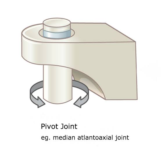
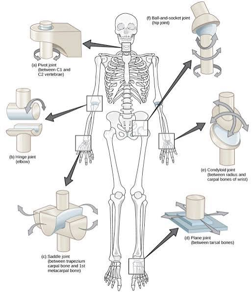
[Types of Synovial Joints]
Pivot Joints
rounded end of one bone protrudes into a sleeve or ring composed of bone/ligament; uniaxial
proximal radio-ulnar joint, neck or dens of axis to atlas
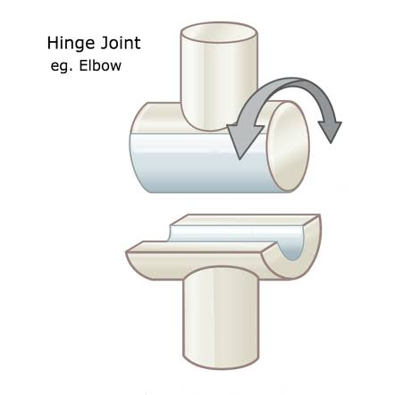
[Types of Synovial Joints]
Hinge Joints
cylindrical projection of one bone fits into a trough-shaped surface on another bone; uniaxial
elbow joint (this is both a pivot and hinge), knee joint, ankle joint, and interphalangeal joints
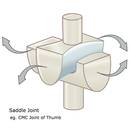
[Types of Synovial Joints]
Saddle Joints
articular surfaces have a concave area on one that fits with the convex area of the other; biaxial
first carpometacarpal joint in the thumb
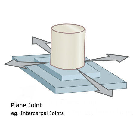
[Types of Synovial Joints]
Plane Joints
AKA gliding joints
articular surfaces are flat and only allow for short gliding movements; multi axial
intercarpal and intertarsal joints, sacro-iliac joint, vertebrocostal joint, acromioclavicular and sternoclavicular joints, and between superior/inferior articulating processes of vertebrae)
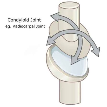
[Types of Synovial Joints]
Condyloid Joints
AKA ellipsoid joint
oval articular surface of one bone fits into a complementary depression in another; biaxial
metacarpophalanges 2-5 (knuckles), radiocarpal joints, and metatarsophalangeal joints
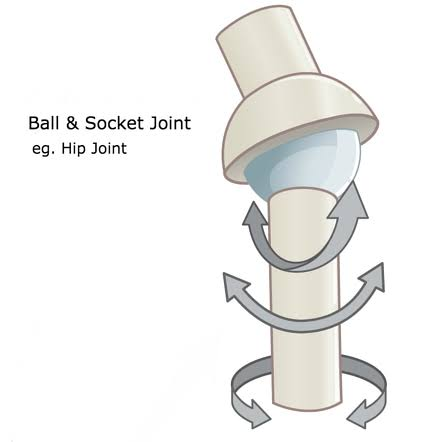
[Types of Synovial Joints]
Ball and Socket Joints
the spherical end of one bone articulates with cup-like socket of another bone; multi-axial
shoulder joint and hip joints

How does joint movement correlate to joint strength?
a joint cannot be both highly mobile and very strong
the greater the range of motion —> the weaker it becomes
a synarthrotic joint, the strongest type of joint, permits no movement
a diarthrotic joint (ex: shoulder), is far weaker, but permits a broad range of motion
[Body Movement]
Flexion
bending a joint to decrease the angle
detail: basically going upwards with a limb. happens in the anterior-posterior plane (front-to-back)
example: lifting a dumbbell in a biceps curl
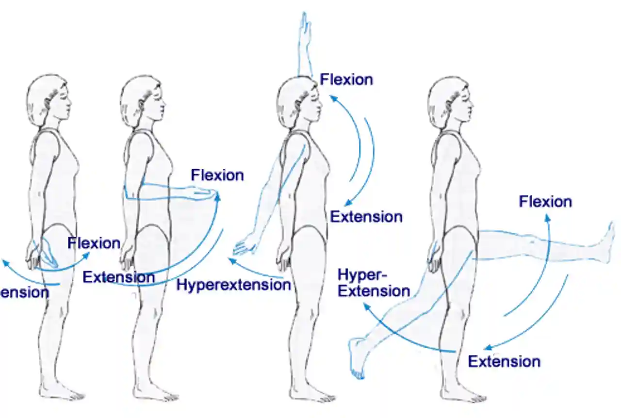
[Body Movement]
Extension
straightening a joint to increase the angle
detail: basically going backwards or back down with a limb. in the anterior-posterior plane (front-to-back)
in anatomical position, all major joints (except ankle) are at full extension
example: lowering a dumbbell back down after a bicep curl

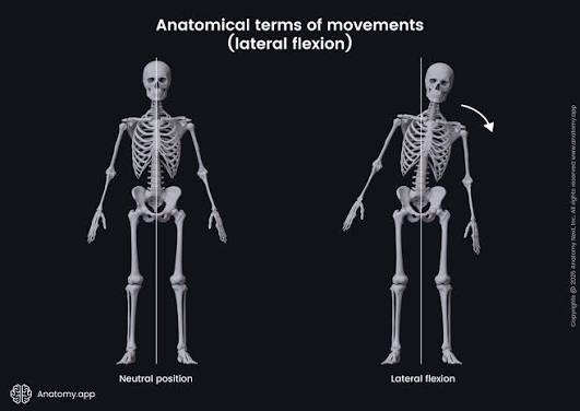
[Body Movement]
Lateral Flexion
tilting your upper body or head to the side
detail: bending the neck or body to the left or right
[Body Movement]
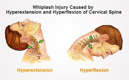
Hyperextension and Hyperflexion
moving a joint too far past its normal limit
detail: moving it past the anatomical position. they are a common cause of injury in the joints like the knee or elbow
example: looking up at the stars hyperextends the neck
[Body Movement]
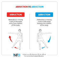
Abduction
moving a body part outward, away from your center line
detail: happens in the lateral-medial plane (side-to-side). moves away from the longitudinal axis (center line)
example: raising arms for a jumping jack, spreading fingers, or cocking the wrist
memory trick: “ABDUCT” means to take away (away from body)

[Body Movement]
Adduction
bringing a body part inward, toward the center line
detail: opposite of abduction, lateral-medial plane (side-to-side), moves toward the longitudinal axis (center line)
example: bringing arms down in a jumping jack, bringing fingers together, or snapping the wrist
[Body Movement]
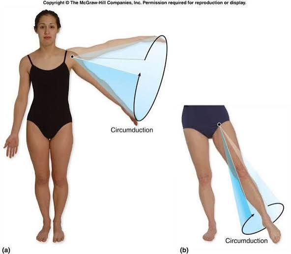
Circumduction
moving a limb so the end of it draws a circle
detail: moving a limb in a circle creates a cone in space
example: doing windmill exercises with your arm
[Body Movement]
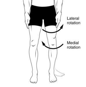
Medial (internal) Rotation
twisting a limb inward toward your center line
detail: the anterior (front) surface of the limb turns toward the body’s midline
example: standing pigeon-toed (toes pointed inward)
[Body Movement]
Lateral (external) Rotation
twisting a limb outward away from your center line
detail: anterior (front) surface turns away from the midline
example: a ballerina’s first position (heels together, toes out)

[Body Movement]
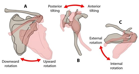
Superior and Inferior Rotation (image just shows scapula but doesn’t necessarily match)
tilting your shoulder blade up or down
details: these are movements of the scapula. they are tracked by the direction the glenoid cavity faces
[Body Movement]
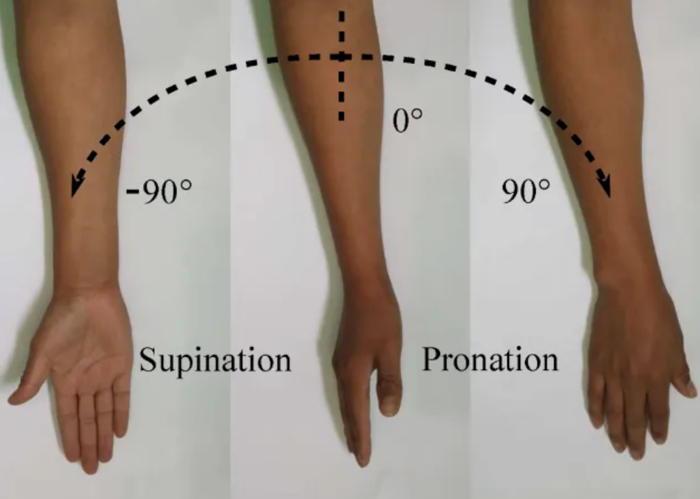
Supination
turning the forearm so the palms face forwards/up
detail: in anatomical position, the forearm is supinated, the radius/ulna are parallel, and the palms face forward (anteriorly)
example: holding a bowl of soup
[Body Movement]
Pronation
turning the forearm so the palms face backwards/down
detail: the radius rotates and crosses over the ulna. the palms face backward (posteriorly)
example: basketball players pronate to dribble a ball

[Body Movement]
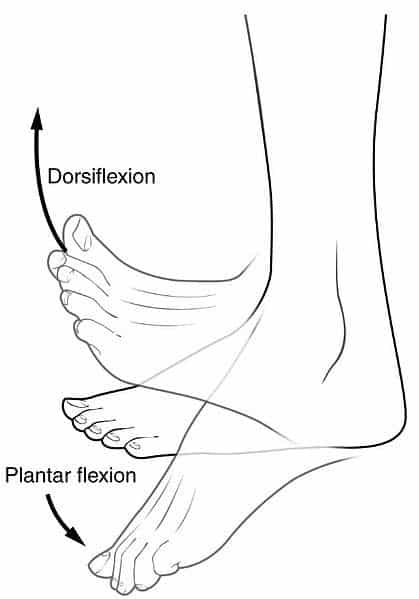
Dorsiflexion
pulling your foot up at the ankle
details: flexion at the ankle joint that elevates the sole
example: rocking back onto your heels
[Body Movement]
Plantar Flexion
pushing your foot down at the ankle
detail: opposite of dorsiflexion, extends the ankle and elevates the heel
example: standing on your tippy toes

[Body Movement]
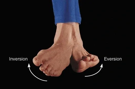
Inversion
tilting the bottom of your foot inward
detail: the sole turns inward, lifting the medial/inner edge of the sole
example: club foot
[Body Movement]
Eversion
tilting the bottom of your foot outward
detail: the sole turns outward, lifting the lateral/outer edge of the sole

[Body Movement]
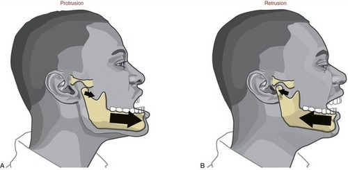
Protraction
pushing a body part forward horizontally
detail: moving a part anteriorly (forward) in the horizontal plane
example: sticking your jaw out into an under-bite

[Body Movement]
Retraction
pulling a body part backward horizontally
detail: opposite of protraction, moving a part posteriorly (backwards) in the horizontal plane
example: pulling your jaw back into an overbite
[Body Movement]
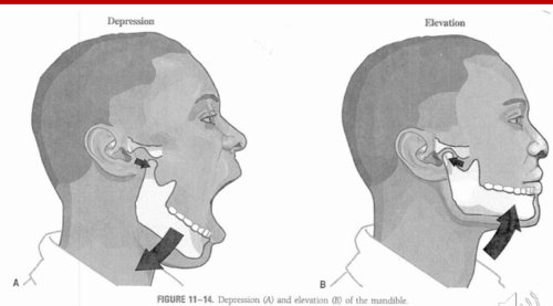
Depression
moving a body part downward
detail: structure moves inferiorly/downwards
example: opening your mouth (the jaw)

[Body Movement]
Elevation
moving a body part upwards
detail: structure moves superiorly/upward
example: closing your mouth (the jaw)
[Body Movement]
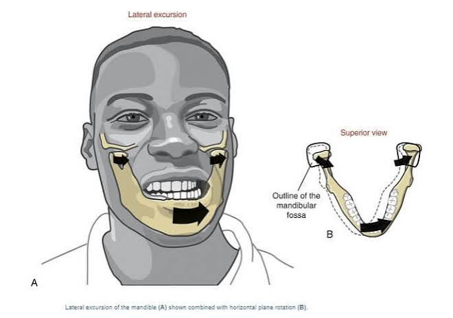
Excursion
shifting your jaw side-to-side
detail: specific to the mandible (jawbone)
[Body Movement]
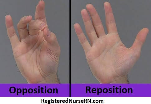
Opposition
touching your thumb to your fingers
details: moving the thumb towards the palm or the pads of other fingers
example: snapping your fingers
[Body Movement]
Reposition
putting your thumb back to original position (away from pointer finger)
detail: returning thumb to original position after opposition

![<p>[Anatomy of Selected Synovial Joints]</p><p>Vertebral Column</p>](https://assets.knowt.com/user-attachments/8c94d00c-9485-4326-add7-56d789d84669.png)
[Anatomy of Selected Synovial Joints]
Vertebral Column
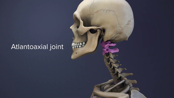
held together by intervertebral discs + adjacent vertebrae also articulate with each other at synovial joints formed between superior/inferior articular processes called zygapophysial joints
atlanto-occipital joint is formed by the articulations between the superior articular processes of the atlas and occipital condyles on base of skull
the atlantoaxial joint is located between the atlas and axis
![<p>[Anatomy of Selected Synovial Joints]</p><p>Temporomandibular Joint (TML)</p>](https://assets.knowt.com/user-attachments/f6092375-c6cd-46ff-bdc5-1fd7a484ea7e.png)
[Anatomy of Selected Synovial Joints]
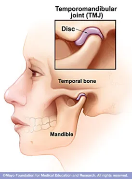
Temporomandibular Joint (TML)
the joint that allows for opening (mandibular depression) and closing (mandibular elevation) of the mouth
also side-to-side and protraction/retraction motions of lower jaw
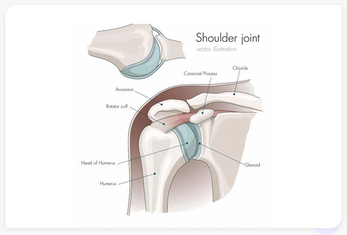
![<p>[Anatomy of Selected Synovial Joints]</p><p>Shoulder Joint</p>](https://assets.knowt.com/user-attachments/937e3a3f-537b-4a99-b494-b3366e0e7955.png)
[Anatomy of Selected Synovial Joints]
Shoulder Joint
the shoulder joint is called the glenohumeral joint
it is a ball-and-socket joint formed by head of humerus and glenoid cavity of scapula
glenoid labrum: small lip of fibrocartilage, extends around outer margin and deepens socket
coracohumeral ligament: structural support for the joint, thickenings of articular capsule wall
glenohumeral ligament: three ligaments, anterior side
rotator cuff: thickening of capsule formed by fusion of four muscle tendons
subacromial burse and subscapular bursa: help prevent friction between rotator cuff muscle tendons and scapula
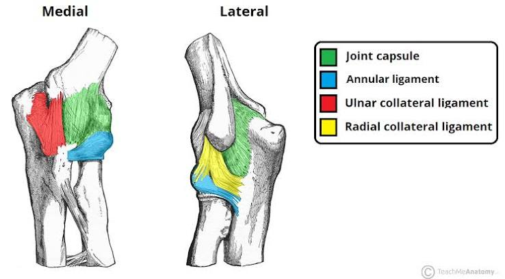
![<p>[Anatomy of Selected Synovial Joints]</p><p>Elbow Joint</p>](https://assets.knowt.com/user-attachments/7e520856-d836-474a-b277-4d4438a09183.png)
[Anatomy of Selected Synovial Joints]
Elbow Joint
uniaxial hinge joint formed by the humeroulnar joint
it is the articulation between the trochlea of the humerus and the trochlear notch of the ulna
also: humeroradial joint and the proximal radioulnar joint
on the medial side: ulnar collateral ligament
on the lateral side: radial collateral ligament
the annular ligament encircles the head of the radius
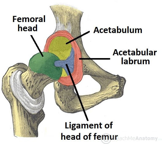
![<p>[Anatomy of Selected Synovial Joints]</p><p>Hip Joint</p>](https://assets.knowt.com/user-attachments/fbc9257a-a2b3-4f7c-b17c-ae73c53eb853.png)
[Anatomy of Selected Synovial Joints]
Hip Joint
multiaxial ball-and-socket joint between head of femur and the acetabulum of the hip bone
the acetabulum is deepened by the acetabular labrum (fibrocartilage lip)
ligaments: iliofemoral ligament, pubofemoral ligament, and ischiofemoral ligament (all spiral around head/neck of femur)
the ligament of the head of femur spans between acetabulum and femoral head
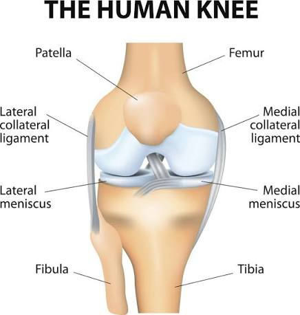
![<p>[Anatomy of Selected Synovial Joints]</p><p>Knee Joint</p>](https://assets.knowt.com/user-attachments/8893b7e3-6ce6-48eb-882a-f43c1090b624.png)
[Anatomy of Selected Synovial Joints]
Knee Joint
the knee joint is the largest joint of the body
three articulations:
femoropatellar joint: between patella and distal femur
medial tibiofemoral joint and lateral tibiofemoral joint: between medial and lateral condyles of femur/tibia
patellar ligament: continuing from patella to tibia
fibular collateral ligament: lateral side + spans from lateral epicondyle of femur to head of fibula
tibial collateral ligament: medial knee runs from medial epicondyle of femur to tibia
anterior cruciate ligament and posterior cruciate ligament: inside the knee, two intracapsular ligaments
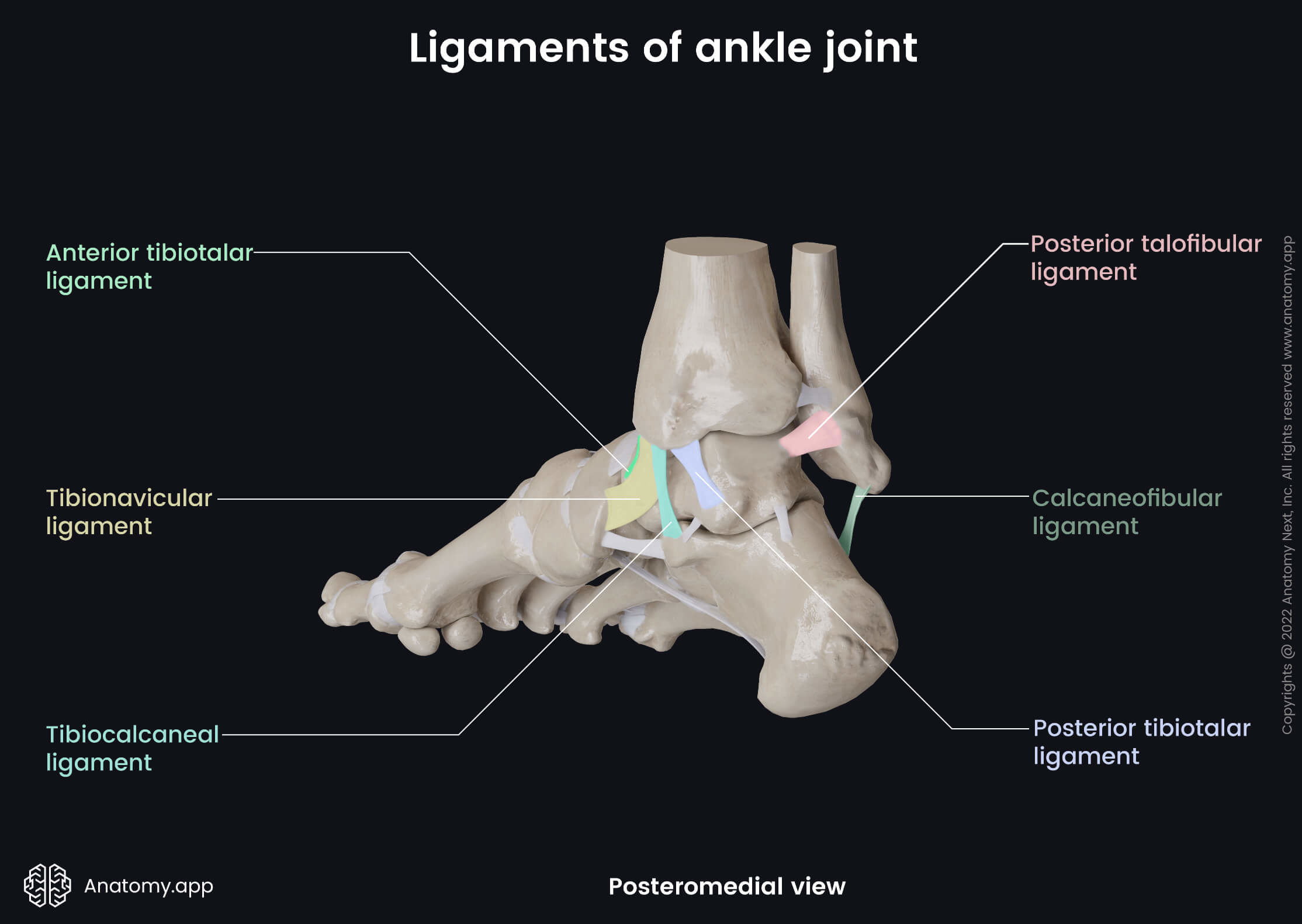
![<p>[Anatomy of Selected Synovial Joints]</p><p>Ankle and Foot Joints</p>](https://assets.knowt.com/user-attachments/098ec962-43bd-4e89-a681-98e787562da6.png)
[Anatomy of Selected Synovial Joints]
Ankle and Foot Joints
the ankle is formed by the talocrural joint
consists of articulations between talus bone of foot and distal ends of tibia/fibula
deltoid ligament: on the medial side; supports the ankle joint and resists excessive eversion
anterior talofibular ligament and posterior talofibular ligament: small ligaments, span between talus bone and malleolus of fibula
calcaneofibular ligament: located between calcaneus bone (heel) and fibula
Development of Joints
joints form during embryonic development in conjunction with the formation and growth of associated bones
mesenchyme: the embryonic tissue that gives rise to all bones, cartilages, and connective tissues of the body
[Common Joint Injuries]
Sprain
stretching or tearing of a ligament across the joint capsule
[Common Joint Injuries]
Strain
stretching or tearing of a tendon attaching muscle to bone
[Common Joint Injuries]
Dislocation
also known as luxation
reinforcing structures cannot protect a joint from extreme stresses = articulating surfaces are forced out of position
displacement damages articular cartilages, tear ligaments, and distorts the joint capsule
even though inside of joints have no receptors, nerves monitor the capsule, ligaments, and tendon = painful
subluxation: a partial dislocation
[Common Joint Injuries]
Bursitis
inflammation of the bursa
[Common Joint Injuries]
TMJ Disorder
a type of temporomandibular disorder/TMD
can cause pain in your jaw joint and in the muscles
can be due to: genetics, arthritis or jaw injury
they tend to clench or grind their teeth (bruxism), but doesn’t have to be related
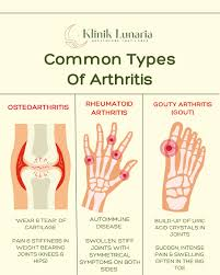
![<p>[Common Joint Injuries]</p><p>Arthritis</p>](https://assets.knowt.com/user-attachments/2caaefb0-322f-48e1-9107-17b7af6f2a79.png)
[Common Joint Injuries]
Arthritis
inflammation/degenerative disease of the joint where synovial membranes thicken (pannus) and fluid production decreases = friction and pain
arthroscopic surgery may be necessary to treat injuries + install artificial joints
osteoarthritis (AKA degenerative arthritis/joint disease): affects individuals 60+ normally. can result from wear and tear or genetic factors (collagen). 25% of women and 15% of men over age 60 show signs of this
rheumatoid arthritis: an autoimmune disease. can occur at any age but more common in middle age + women. infection, genes, and hormone changes can cause this. usually impacts joints on both sides of body equally. most common parts: wrists, fingers, knees, feet, and ankles.
gouty arthritis: gout is caused by too much uric acid in the blood = form hard crystals in joints. this causes burning pain, stiffness, and swelling, especially in big toe. more common in men.