unit 3.1, 3.2, 3.5 muscles intro and specifics on skeletal + smooth
0.0(0)
Studied by 7 peopleCard Sorting
1/42
Earn XP
Description and Tags
Last updated 3:09 AM on 11/4/22
Name | Mastery | Learn | Test | Matching | Spaced | Call with Kai | Chat |
|---|
No analytics yet
Send a link to your students to track their progress
43 Terms
1
New cards
what is a muscle?
a tissue specialized to convert biochem reactions into mech work
2
New cards
two main functions of a muscle
to generate motion and force
- can also generate heat and contribute to body temp homeostasis
- can also generate heat and contribute to body temp homeostasis
3
New cards
restrictions on movement of muscle
- can only contract and relax
- cannot expand unless when externally pulled by another muscle group (stretching for example)
- cannot expand unless when externally pulled by another muscle group (stretching for example)
4
New cards
3 types of muscles (intro to each)
skeletal
- attach to bones of the skeleton
- control body movement
- can only contract when there is a somatic motor neuron signal (meaning it CANNOT be influenced by hormones or contract on its own)
- very striated and organized
cardiac
- found only in the heart
- controls blood movement
- striated, but not as organized as skeletal
smooth
- primary muscle of internal organs and tubes (stomach, vessels, bladder, etc)
- controls material movement throughout body
- no striations or order
5
New cards
skeletal muscle characteristics
- responsible for positioning and movement of the skeleton
- attached to bones via TENDONS
- makes up ~half of body weight
- attached to bones via TENDONS
- makes up ~half of body weight
6
New cards
tendons
- connects muscle to bone
- composed of dense regular connective tissue called collagen (a protein arranged in parallel alignment)
- composed of dense regular connective tissue called collagen (a protein arranged in parallel alignment)
7
New cards
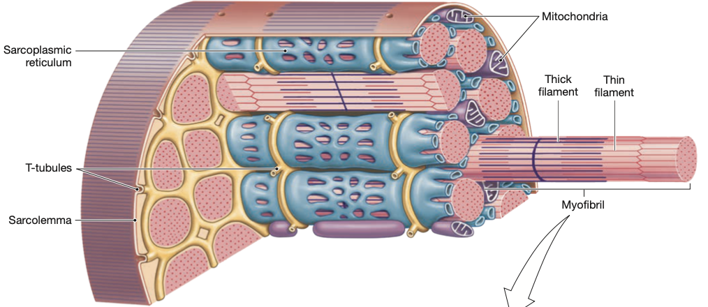
5 components of a skeletal muscle
epimysium, perimysium, fascicle, endomysium, myofibrils
8
New cards
epimysium
the outermost connective tissue layer of a muscle
- contains bundles of muscle tissue (so outer layer of groups of groups)
- contains nerves and blood vessels within
- epi = above, myo = muscle
- contains bundles of muscle tissue (so outer layer of groups of groups)
- contains nerves and blood vessels within
- epi = above, myo = muscle
9
New cards
perimysium
connective tissue that covers a single bundle of muscle fibres
- peri = around, myo = muscle
- specifically covers fascicles
- peri = around, myo = muscle
- specifically covers fascicles
10
New cards
fascicle
a bundle of muscle cells/fibres
- covered by perimysium
- covered by perimysium
11
New cards
functional unit of skeletal muscle
myofibril, which is in every muscle fibre
- they are contractile and elastic protein bundles
- they are contractile and elastic protein bundles
12
New cards
endomysium
innermost connective tissue layer
- sheath around exactly one muscle fibre
- endo = inner, myo = muscle
- sheath around exactly one muscle fibre
- endo = inner, myo = muscle
13
New cards
why do muscle cells not have many organelles?
there are so many myofibrils that there's very little room for organelles
14
New cards
what's in the cytosol of a muscle cell?
- glycogen granules (energy storage)
- mitochondria (ATP synthesis)
- mitochondria (ATP synthesis)
15
New cards
general structure of muscle fibre
long and cylindrical cell with hundreds of nuclei on the surface
16
New cards
what is the cell membrane of a muscle cell called?
sarcolemma
- sarco = flesh, lemma = shell
- sarco = flesh, lemma = shell
17
New cards
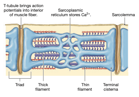
what is the endoplasmic reticulum of a muscle cell called?
sarcoplasmic reticulum
- associated with this SR are a series of branching tubes: t-tubules
- stores a lot of Ca ions
- associated with this SR are a series of branching tubes: t-tubules
- stores a lot of Ca ions
18
New cards
t-tubules
aka transverse tubules
- indentations of membrane with lumen (aka space within a tube) that is full of ECF (extracellular fluid)
- closely associated with terminal cisternae (aka "large vessel"), which are the ends of an SR
- allow for rapid AP diffusion into the muscle fibre (necessary for proper movement)
- indentations of membrane with lumen (aka space within a tube) that is full of ECF (extracellular fluid)
- closely associated with terminal cisternae (aka "large vessel"), which are the ends of an SR
- allow for rapid AP diffusion into the muscle fibre (necessary for proper movement)
19
New cards
what is the cytoplasm of a muscle cell called?
sarcoplasm
20
New cards
triad in muscle cell
one t-tubule with one terminal cisternae on each side

21
New cards
4 general terms that have different names in muscle context
muscle cell = muscle fibre
cell membrane = sarcolemma
cytoplasm = sarcoplasm
endoplasmic reticulum = sarcoplasmic reticulum
cell membrane = sarcolemma
cytoplasm = sarcoplasm
endoplasmic reticulum = sarcoplasmic reticulum
22
New cards
3 types of proteins in a myofibril
contractile, regulatory, accessory
23
New cards
contractile proteins in myofibrils
CAM
- specifically myosin and actin
- "can the cell contract?": responsible for the physical component of a contraction
- specifically myosin and actin
- "can the cell contract?": responsible for the physical component of a contraction
24
New cards
regulatory proteins in myofibrils
RTT
- specifically troponin and tropomyosin
- "will the cell contract?": determines whether a muscle will actually contract or not
- specifically troponin and tropomyosin
- "will the cell contract?": determines whether a muscle will actually contract or not
25
New cards
accessory proteins
ATN
- specifically titin and nebulin
- specifically titin and nebulin
26
New cards
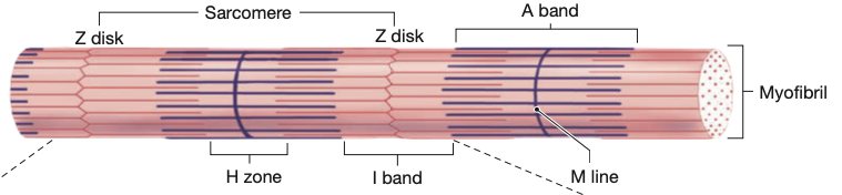
sarcomere
one repeated pattern of myofibrils
27
New cards
sarcomere structure (has 5 parts)
ZIAHM:
z line
i band
a band
h zone
m line
z line
i band
a band
h zone
m line
28
New cards
what causes striations in muscle cells?
the organization of protein components (thick and thin filaments; myosin and actin respectively) of the myofibrils
29
New cards
myosin
- motor protein that consists of two coiled protein molecules/chains, each consisting of two parts: a head and tail
- head and tail are stiff and connected by a flexible hinge, which allows heads to pivot (this is important for generating contractions)
- are thick filaments due to how many molecules coil together
- heads are always toward the ends of the SR/toward Z
- head and tail are stiff and connected by a flexible hinge, which allows heads to pivot (this is important for generating contractions)
- are thick filaments due to how many molecules coil together
- heads are always toward the ends of the SR/toward Z
30
New cards
actin
- subunits of G-actin (G for globular)
- polymerize to form a chain, called F-actin (F for filamentous)
- two F-actins coil to form the basis of thin filament: associates with regulatory proteins (RTT) and cause contractions when interacting
- completed thin filament is F-actins + troponin + tropomyosin
- myosin heads directly interact with F-actin (interactions are called CROSSBRIDGES)
31
New cards
z-line
- think z for zigzag
- discs that act as site of attachment for thin filaments
- signify the ends of a sarcomere (one full sarcomere is two z-discs and the filaments between them)
- discs that act as site of attachment for thin filaments
- signify the ends of a sarcomere (one full sarcomere is two z-discs and the filaments between them)

32
New cards
i band
- contains only thin filaments
- one z disc will run through the middle of an i band so that an i band is split in half: one half to one sarcomere, the other to a separate sarcomere
- one z disc will run through the middle of an i band so that an i band is split in half: one half to one sarcomere, the other to a separate sarcomere

33
New cards
a band
- contains thick and thin filaments: very dense (hence the darker color of it)!
- thick and thin overlap at outer edges of a band
- center consists of thick ONLY...h band
- thick and thin overlap at outer edges of a band
- center consists of thick ONLY...h band

34
New cards
h band
- center of a band, has only thick filaments
- lighter than outer edges of a band
- lighter than outer edges of a band
35
New cards
m line
- m line = midline; the center of the sarcomere
- site of attachment for thick filaments
- site of attachment for thick filaments

36
New cards
cross-sections of sarcomeres and the thin/thick ratio
- every thin filament is surrounded by 3 thick filaments
- every thick filament is surrounded by 6 thin filaments
- every thick filament is surrounded by 6 thin filaments
37
New cards
titin
- largest known protein
- elastic protein that stretches from one z disc to an m line in the next sarcomere
- stabilizes myosin, which are contractile filaments
- returns stretched muscles to their resting length (think titin tightens back)
- elastic protein that stretches from one z disc to an m line in the next sarcomere
- stabilizes myosin, which are contractile filaments
- returns stretched muscles to their resting length (think titin tightens back)
38
New cards
nebulin
- non-elastic and attaches to z disc
- guides actin filaments in the sarcomere to properly align them
- guides actin filaments in the sarcomere to properly align them
39
New cards
if you took a cross-section of a sarcomere through the outer edge of the a band, what would you see?
both myosin thick filaments and actin thin filaments
40
New cards
smooth muscle arrangement
has two distinct arrangements
1) single unit
- muscle cells are coupled by gap junctions
- don't need electrical stimulation for each individual cell
- think lining of organs: network of cells
2) multi unit
- no gap junctions
- every cell works on its own, innervated separately
- think of iris of eyes
1) single unit
- muscle cells are coupled by gap junctions
- don't need electrical stimulation for each individual cell
- think lining of organs: network of cells
2) multi unit
- no gap junctions
- every cell works on its own, innervated separately
- think of iris of eyes
41
New cards
general differences between smooth and skeletal
- contraction of smooth changes shape and length, skeletal only changes length
- smooth develops tension/force slowly
- smooth muscle doesn't fatigue as quickly due to contraction (think bladder: it's constantly adjusting, as that's its function)
- smooth develops tension/force slowly
- smooth muscle doesn't fatigue as quickly due to contraction (think bladder: it's constantly adjusting, as that's its function)
42
New cards
cellular differences between smooth and skeletal
- smooth muscle fibres are much smaller
- no sarcomere arrangement in smooth, so no striations
- actin is anchored at dense bodies of cell membrane, unlike skeletal which is attached to z lines
- no t-tubules for smooth and not much SR (instead, smooth has caveolae vesicles within the sarcolemma to store calcium)
- force of contraction in smooth is related to amount of calcium released
43
New cards
what is the effect of not having t-tubules for smooth muscles?
no direct coupling of AP to release calcium from SR through dhp-ryr receptor coupling (skeletal). instead, Ca entering sarcolemma results in Ca release from SR.