Topic 7-Run For Your Life
1/113
There's no tags or description
Looks like no tags are added yet.
Name | Mastery | Learn | Test | Matching | Spaced | Call with Kai |
|---|
No analytics yet
Send a link to your students to track their progress
114 Terms
What is a Skeletal Muscle?
Muscles that are attached to bones by Tendons.
What do Ligaments do?
Attach bones to other bones.
What do Skeletal Muscles do?
Contract and Relax to move bones at a joint.
What are Antagonistic Pairs?
Muscles that work together to move a bone.
What are the bones of the lower arms attached to and with what?
Biceps
Triceps
By Tendons
What happens when the bicep contracts?
Tricep relaxes
This pulls the bone so your arm bends.
What is a Flexer?
A muscle that bends a joint when it contracts.
Eg.Bicep
What happens when the Tricep contracts?
Bicep Relaxes.
This pulls the bone so the arm straightens (extends).
What is an Extensor?
A muscle that straightens a joint when it contracts.
Eg.Tricep.
What is Skeletal Muscle made from?
Muscle Fibres.
What is the Cell Membrane of Muscle Fibres cells called?
Sarcolemma
What is made when parts of the Sarcolemma folds inwards and sticks to the sarcoplasm?
Forms Transverse(T) Tubules.
What do T Tubules do?
Help spread electrical impulses throughout the sarcoplasm, so they reach all parts of the muscle fibre.
What does the Sarcoplasmic Reticulum do?
Stores and releases calcium ions that are needed for muscle contraction.
Why do Muscle Fibres have many Mitochondria?
To provide ATP for muscle contraction.
What are Muscle Fibres made of?
Myofibrils
What 2 kinds of Myofilaments do Myofibrils contain?
Thick Myofilaments-Protein Myosin
Thin Myofilaments-Protein Actin.
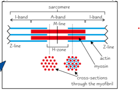
What are Darks bands that appear on electron microscopes when you look at a myofibril?
Dark bands contain the thick myosin filaments and some overlapping thin myosin filaments.-Called A Bands.
What are the Light Bands that appear when you look at a myofibril under an electron microscope?
Thin Actin filaments only-called I Bands.
What are Myofibrils made up of?
Scarcomeres
Draw a diagram of a Scarcomere
How does the Sarcomere contract?
Myosin and Actin filaments slide over one another.
Draw a relaxed and contracted Sarcomere
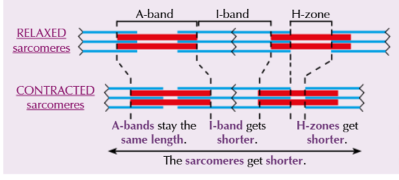
When Sarcomeres contract do Myosin and Actin Filaments stay the same length?
Yes
Describe the Process of muscle Contraction(First Stage)
1.Action Potential is sent to a muscle down a neurone.
2.Signal reaches the neural muscular junction and passes into the sarcolemma of the muscle fibre cell.
3.Signal travels from the Sarcolemma deep into the muscle fibre cell down the T Tubules, then enters the Sarcoplasmic Reticulum.
4.Action potential causes the Sarcoplasmic Reticulum to release Ca2+ ions.
5.Ca2+ bind to Troponin, which pulls Tropomyosin along to reveal the Myosin Binding Sites.
Describe Sliding Filament Theory(Second stage of Muscle Contraction)
1.ATP binds to the Myosin head, causing Myosin to detach from Actin.
2.ATP is hydrolysed into ADP+Pi, Causing the Myosin head to change angle.
3.Myosin head binds to actin forming a cross bridge.
4.ADP+Pi released causes Myosin head to change angle pulling Actin along-Power Stroke.
5.A new ATP binds, allowing Myosin to detach, cycle is repeated.
Describe what happens when Excitation Stops
Ca2+ leave their binding sites on the Troponin molecules and are moved by active transport back into the Sarcoplasmic Reticulum.
Troponin molecules return to their original shape, pulling the attached Tropomyosin with them. So, the Actin-Mysin binding sites are blocked again.
Actin filaments slide back into a relaxed position, which lengthens the sarcomere.
What are Slow Twitch Muscle Fibres?
Muscle fibres that contract slowly.
What are Slow Twitch Muscle Fibres used for?
Endurance Activities-Long Distance Running.
Describe how Slow Twitch Muscles are adapted for their function
-Can work for a long time without getting tired.
-Energy released through Aerobic Respiration.
-Lots of Mitochondria.
-Lots of blood vessels supply muscles with oxygen.
-Lots of red myoglobin, muscles appear red.
What are fast Twitch Muscle Fibres?
Muscles that contract very quickly.
What are Fast Twitch Muscles Used for?
Short bursts of Speed and Power-Sprinting.
Describe how Fast Twitch Muscle Fibres are adapted for their function
-Get tired very quickly.
-Energy released through Anaerobic Respiration.
-Few Mitochondria.
-Few blood vessels.
-Little Red Myoglobin, so can’t store much oxygen-whitish colour.
What is the overall equation for Aerobic Respiration?
C6H1206 + 6O2 → 6CO2 +6H2O + Energy
What is Aerobic Respiration?
Process where a large amount of energy is released by splitting glucose into CO2 and H2.
What is the energy released in Aerobic Respiration used for?
To Phosphorylate ADP to ATP.
What are the 4 stages of Aerobic Respiration?
Glycolysis
Link Reaction
Krebs Cycle
Oxidative Phosphorylation
Where does Glycolysis occur?
Cytoplasm
Where do the Link Reaction, Krebs Cycle and Oxidative Phosphorylation occur?
Mitochondria.
What is enzyme with the slowest activity called?
Rate Limiting-Determines the overall rate of respiration.
What do NAD and FAD do?
Transfer hydrogen from one molecule to another- meaning it can reduce of oxidise a molecule.
What does Coenzyme A do?
Transfers acetate between molecules.
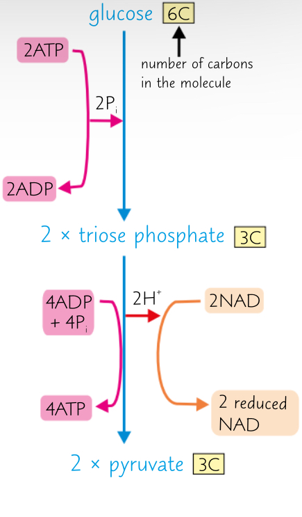
Stage 1: Explain Glycolysis
Part 1: Phosphorylation
Glucose is Phosphorylated by adding 2 phosphates, from 2 molecules of ATP.
This creates 2 molecules of triose phosphate and 2 molecules of ADP.
Part 2: Oxidation
Triose Phosphate is oxidised (loses oxygen), forming 2 pyruvate.
NAD collects the hydrogen ions, forming 2 reduced NAD.
4 ATP produced, but 2 are used in Stage 1 so there’s a net gain of 2 ATP.
What happens to the products produced in Glycolysis?
2 Reduced NAD are used in Oxidative Phosphorylation.
2 Pyruvate go into matrix of Mitochondria for Link Reaction.
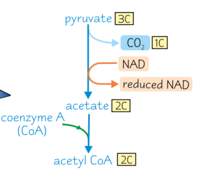
Stage 2: Link Reaction
Pyruvate is decarboxylated in the form of CO2.
NAD is reduced- collecting Hydrogen from pyruvate, changing pyruvate into acetate.
Acetate is combined Coenzyme A to form Acetyl Coenzyme A.
No ATP produced.
How many times does the Link Reaction occur for every Glucose Molecule?
Twice
How many Pyruvate molecules are made for each glucose molecule?
2
Describe what happens for each glucose molecule after the Link Reaction
2 Acetyl Coenzyme A go into Krebs Cycle.
2 CO2 molecules are released as a waste product of respiration.
2 reduced NAD are formed and used in the last stage (oxidative phosphorylation).
Stage 3: Krebs Cycle
Part 1:
Acetyl CoA from Link Reaction combines with oxaloacetate to form citrate.
Coenzyme A goes back to Link Reaction to be used again.
Part 2:
6C citrate molecule is converted to a 5C molecule.
Decarboxylation occurs CO2 is removed.
Dehydrogenation occurs.
Hydrogen is used to produce Reduced NAD from NAD.
Part 3:
5C molecule converted to 4C molecule.
Decarboxylation and Dehydrogenation occur, producing 1 reduced FAD and 2 Reduced NAD.
ATP is produced by direct transfer of phosphate group from an intermediate compound to ADP.
When a phosphate group is directly transferred from 1 molecule to another it’s called Substrate-level Phosphorylation.
Citrate has been converted to oxaloacetate.
What are the products of the Krebs Cycle used for?
1 Coenzyme A- reused in the next link reaction.
Oxaloacetate- Regenerated for use in the next Krebs Cycle.
2 CO2- Released as waste product.
1 ATP- Used for energy.
3 Reduced NAD- to Oxidative Phosphorylation.
1 Reduced FAD- to Oxidative Phosphorylation.
What is the outcome of Oxidative Phosphorylation?
The energy carried by electrons, from reduced Coenzymes (Reduced NAD and Reduced FAD), is used to make ATP.
What 2 processes does Oxidative Phosphorylation involve?
Electron Transport Chain.
Chemiosmosis.
Stage 4: Oxidative Phosphorylation
H are released from Reduced NAD and Reduced FAD, as they’re oxidised to NAD and FAD. The H atoms split into H+ and e-.
e- move down the electron transport chain , losing energy at each carrier.
Energy is used by the electron carrier to pump protons from the mitochondrial matrix into the intermembrane space.
Concentration of protons is now higher in the intermembrane space than in the mitochondrial matrix - forming an electrochemical gradient.
Protons move down the electrochemical gradient, back in the mitochondrial matrix, via the enzyme ATP Synthase. Movement drives the synthesis of ATP from ADP+Pi.
Movement of H+ ions across a membrane, which generates ATP is called Chemiosmosis.
In the Mitochondrial Matrix, at the end of the transport chain, the protons, electrons and O2 combine to form water.
Oxygen is the final electron acceptor.
How many ATP are made from 1 Glucose Molecule?
38
What is the SAN?
It sets the rhythm of the heartbeat by sending out regular waves of electrical activity to the atrial walls, like a pacemaker.
Describe how the Cardiac Muscle controls the regular beating of the heart
Process starts in the Sino-Atrial Node, which is in the wall of the right atrium.
It causes the right and left atria to contract at the same time.
A band of non-conducting collagen tissue prevents the waves of electrical activity from being passed directly from the atria to the ventricles.
Instead, the waves of electrical activity are transferred from the SAN to the AVN.
The AVN is responsible for passing the waves of electrical activity on to the bundle of His. But, there’s a slight delay before the AVN reacts, to make sure the ventricles contract after the atria have emptied.
The bundle of His is a group of muscle fibres responsible for conducting the waves of electrical activity to the finer muscle fibres in the right and left ventricle walls, called Purkyne fibres.
Purkyne fibres carry the waves of electrical activity into the muscular walls of the right and left ventricles, causing them to contract simultaneously, from the bottom up.
What is an electrocardiograph?
A machine that records the electrical activity of the heart.
When does the heart depolarise?
When the heart contracts
When does the heart repolarise?
When it relaxes.

What is a P Wave?
Caused by the contraction (depolarisation) of the atria.
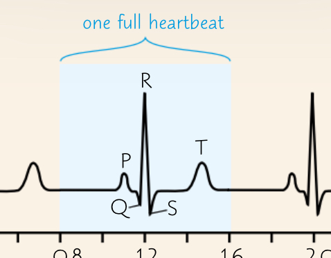
What is the main peak of the heartbeat called?
The QRS complex, caused by contraction (depolarisation) of the ventricles.
What causes a T Wave?
Due to relaxation (repolarisation) of the ventricles.
What does the height of the wave indicate?
It indicates how much electrical charge is passing through the heart- a bigger wave means more electrical charge, so a bigger wave means a stronger contraction.
What word describes the heart beating too fast?
Tachycardia-around 120bpm.
What word describes the heart beating too slow?
Bradycardia- below 60bpm
Define Ectopic Heartbeat
An extra heartbeat.
Define Fibrillation
An irregular Heartbeat
What symptoms may Fibrillation cause?
Chest pain.
Fainting.
Lack of pulse/death.
Describe how the body does more aerobic respiration during exercise
Increasing breathing rate and depth- to get more oxygen and remove more carbon dioxide.
Increasing heart rate- to deliver oxygen and glucose to the muscles faster and to remove extra carbon dioxide produced by the increased rate of respiration.
Which part of the brain controls the breathing rate?
Medulla Oblongata
What 2 ventilation centres does the Medulla have?
Inspiratory centre.
Expiratory centre.
Explain how the Medulla Oblongata controls breathing rate
Inspiratory centre in the Medulla Oblongata sends nerve impulses to the intercostal and diaphragm muscles to contract.
Increasing the volume in the lungs lowers the pressure. Nerve impulses are also sent to the expiratory centre to inhibit its action.
Air enters the lungs due to the pressure difference between the lungs and the air outside.
As lungs inflate, stretch receptors in the lungs are stimulated. Stretch receptors send nerve impulses back to the Medulla Oblongata. This inhibits the inspiratory centre.
Expiratory centre sends nerve impulses to the diaphragm and intercostal muscles to relax. Causing the lungs to deflate, expelling air. As lungs deflate, stretch receptors become inactive.
Inspiratory centre is no longer inhibited and the cycle starts again.
Explain how Exercise triggers an increase in Breathing Rate by Decreasing Blood pH
During Exercise, level of CO2 in the blood increases which decreases blood pH.
Chemoreceptors in the Medulla Oblongata, aortic bodies and carotid bodies that are sensitive to changes in blood pH.
If Chemoreceptors detect a decrease in blood pH, they send nerve impulses to the Medulla Oblongata, which sends more frequent nerve impulses to the intercostal muscles and diaphragm. Increasing rate and depth of breathing.
Causes gaseous exchange to speed up. CO2 level drops and extra O2 is supplied for the muscles- the pH returns to normal and breathing rate decreases.
Define Ventilation Rate
The volume of air breathed in or out in a period of time.
What happens to Ventilation Rate during exercise.
Breathing rate and depth increases.
Describe how pressure receptors and chemical receptors detect stimuli in the blood
Pressure receptors called baroreceptors in the aortic an carotid bodies- stimulated by high an low pressure.
Chemoreceptors in the aortic and carotid bodies and in the Medulla Oblongata monitor oxygen level in the blood.
What is the Sympathetic Nervous System?
Triggers Fight or Flight- helps increase heart rate during exercise.
What is the Parasympathetic Nervous System?
Triggers Rest and Digest by calming the body down- helps decrease the heart rate after exercise.
How does the Heart respond to High Pressure?
Baroreceptors detect high blood pressure.
Impulses are sent to the cardiovascular control centre, which sends impulses along parasympathetic neurones.
They secrete Acetylcholine (neurotransmitter) which binds to receptors on SAN.
SAN fires impulses less frequently to slow heart rate and reduce blood pressure to normal levels.
How does the Heart respond to Low Blood Pressure?
Baroreceptors detect low blood pressure.
Impulses are sent to the cardiovascular control centre, which sends impulses along sympathetic neurones.
They secrete noradrenaline (neurotransmitter)m which binds to receptors on the SAN.
SAN fires impulses more frequently to increase heart rate and increase blood pressure to normal.
How does the Heart respond to High blood O2, Low CO2 or High pH levels?
Chemoreceptors detect chemical changes in the blood.
Impulses are sent to the cardiovascular control centre, which sends impulses along parasympathetic neurones.
They secrete Acetylcholine (neurotransmitter), which binds to receptors on the SAN.
SAN fires impulses less frequently to decrease heart rate and return O2, CO2 and pH levels back to normal.
How does the Heart respond to Low blood O2, High CO2 or Low pH levels?
Chemoreceptors detect chemical changes in the blood.
Impulses are sent to the cardiovascular control centre, which sends impulses along sympathetic neurones.
They secrete noradrenaline (neurotransmitter), which binds to receptors on the SAN.
SAN fires impulses more frequently to increase heart rate and return O2, CO2 and pH levels back to normal.
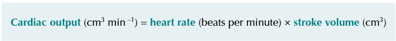
Define Cardiac Output
The total volume of blood pumped by a ventricle every minute.
What is the Equation for Cardiac Output?
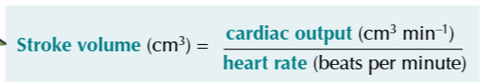
Define Stroke Volume
The volume of blood pumped by one ventricle each time it contracts.
What is the Equation for Stroke Volume?
What do Homeostatic use to reverse a change?
Negative Feedback
Describe how Homeostatic use Negative Feedback to reverse a change?
Receptors detect when a level is too high or low an the information is communicate via the nervous system or hormonal system to effectors.
Effectors respond to counteract the change.
When may Negative Feedback not work?
When the change is too big.
Effectors may not be able to counteract it.
What do Homeostatic Systems use to Amplify a change from normal levels?
Positive Feedback.
How do the effectors respond to stimuli in a Positive Feedback System?
Respond to further increase the level away from the normal level.
What is Positive Feedback useful for?
To rapidly activate something, eg.blood clot after an injury.
In what other way might a Positive Feedback System be triggered?
When a Homeostatic System Breaks down.
Describe the Mechanisms used to Reduce Body Temperature
Sweating-Water in sweat evaporates from the surface of the skin and takes heat from the body.
Hairs lie flat-Erector pili muscles relax so the hairs lie flat. Less air is trapped, so skin is less insulated and heat can be lost easier.
Vasodilation- arterioles near the surface of the skin dilate. More blood flows through the surface layers of the dermis. Meaning more heat can be lost from the skin by radiation.
Describe Mechanisms to Increase Body Temperature
Shivering- Muscles contract in spasms and more heat is produced from increased respiration.
Less Sweat-Reduced heat lost.
Hairs Stand Up- Erecter pill muscles contract, trapping more air and preventing heat loss.
Vasoconstriction- Arterioles near the surface of the skin constrict, so less blood flows through the capillaries in the surface layers of the dermis, reducing heat loss.
Hormones- Body releases adrenaline and thyroxine. This increases metabolism and more heat is produced.
What is Body Temperature in mammals controlled by?
Hypothalamus
How does the Hypothalamus work during Thermoregulation?
Hypothalamus receives information from thermoreceptors.
Thermosreceptors send impulses along sensory neurones to the Hypothalamus, which sends impulses along motor neurones to effectors.
Effectors respond to restore body temperature to normal.(eg.Vasoconstriction,sweating)
What are Transcription factors that increase the rate of Transcription called?
Activators.
What are Transcription factors that decrease the rate of Transcription called?
Repressors.