AP Bio ch 25, 6, 7
CHAPTER 25
There is an overarching connection between biology, geology, and chemistry that provided the opportunity for “living cells” to come about.
Indexing fossils - being able to put the fossils in order based on age. The fossils found closer to the surface are likely to be younger. Deeper fossils are likely older.
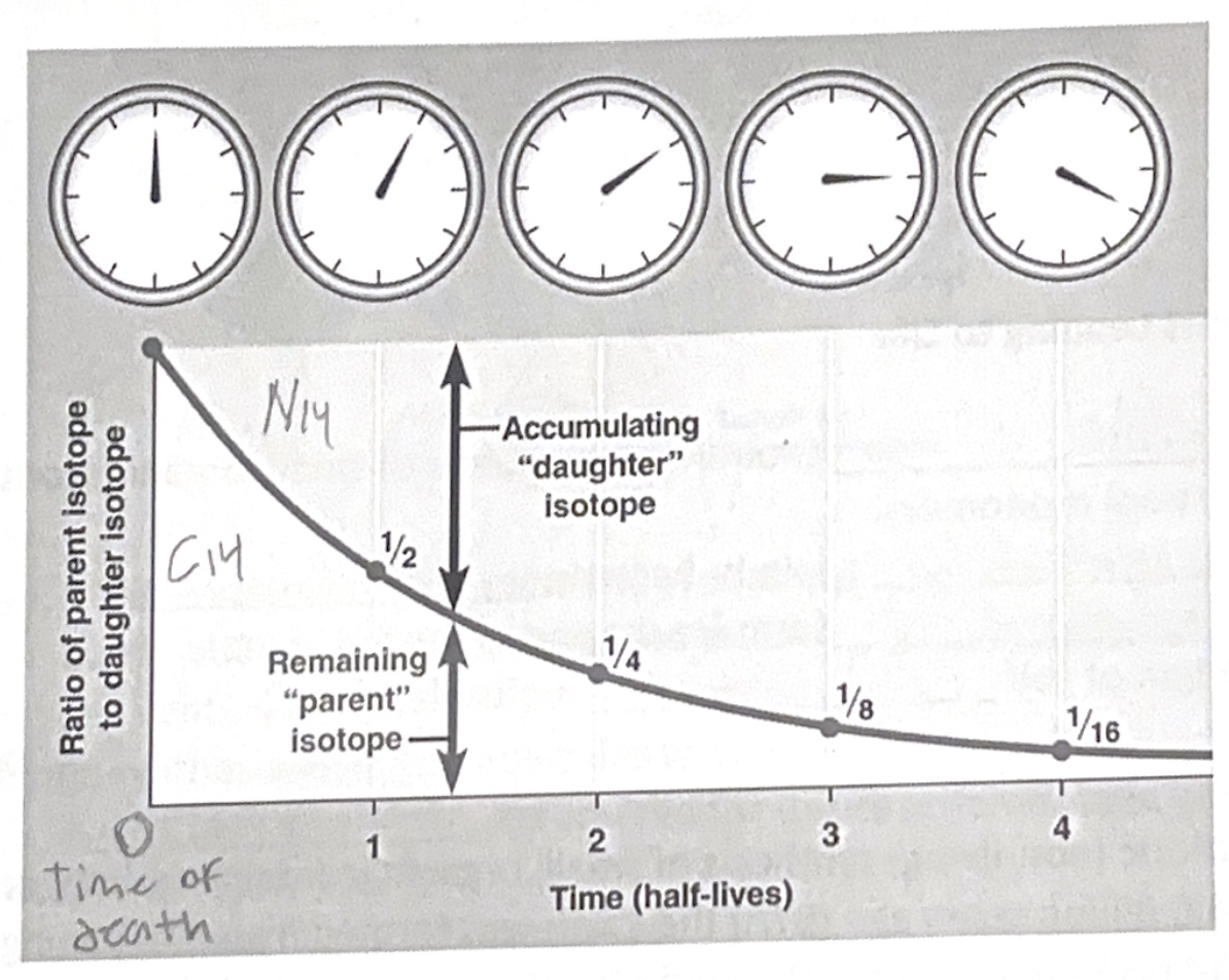
Radiometric dating - Measures the decay of radioactive isotopes
- By knowing the half-life of each element (the amount of time in which 50% of the original amount decays) we can estimate ages of fossils (and rocks)
- For once living objects we would use the isotope Carbon 14 because it will decay into Nitrogen 14
- For non-living objects we would use Potassium 40 because it will decay into Argon 40
Geologic record -
Mass extinctions - Earth's history has had multiple mass extinctions including one that is thought to have destroyed 96% of all ocean life. Cause? Asteroids or comets.
Snowball Earth - The term used to describe an ice age period that caused the oceans to freeze over.
Magnetic reversals - The magnetic poles have reversed many times in the history of earth. We know this because we can look at the orientation of metals in rocks and sediments.
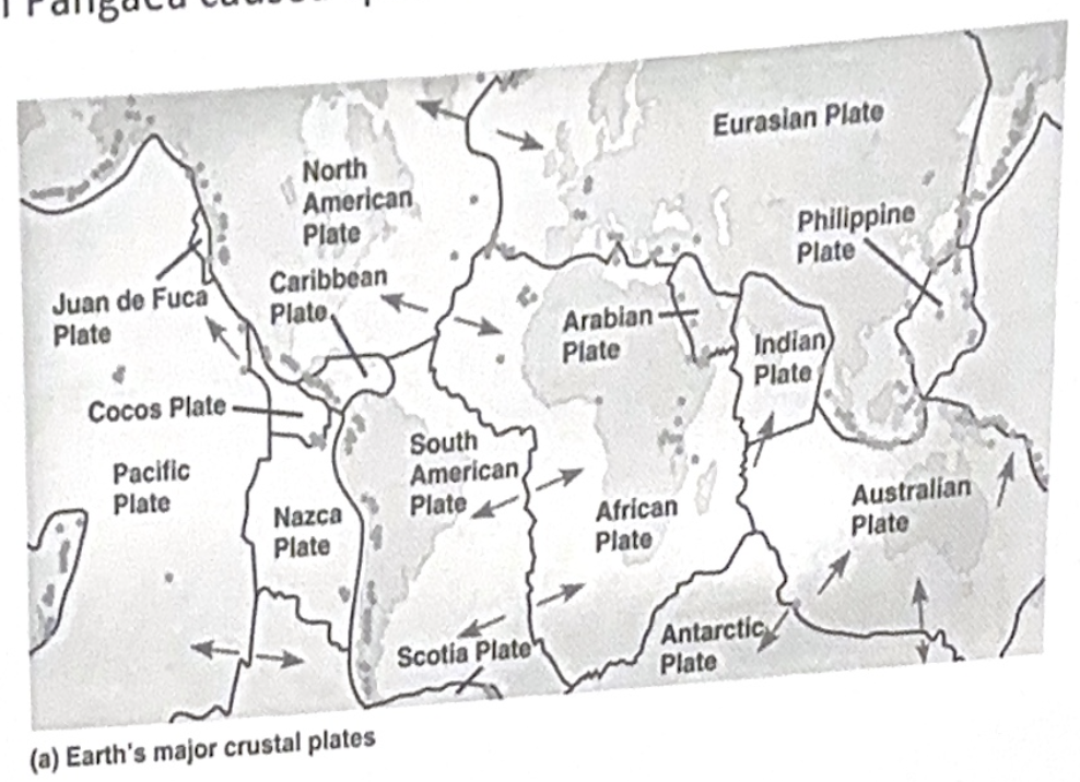
Continental drift - The tectonic plates are separating or colliding causing different continents to move at the rate of centimeters per year (in some cases).
- Pangaea - It is thought that ar one time all land on earth was connected. This “super-continent” was known as pangaea. The split of pangaea caused speciation.
Order of Events Leading to Life
Abiotic (non-living) synthesis of small, organic (contains carbon) monomers.
Monomers join to become polymers
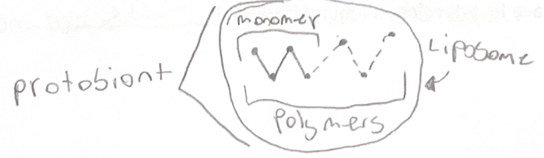
Protobionts form (packagung of the polymers)
Origin of self replicating molecules aka RNA and DNA
Details
Abiotic (non-living) synthesis of small, organic (containing carbon) monomers.
- 4.6 billion years ago the earth was formed from the joining of dust and rocks from space. This early “earth” was constantly being bombarded by debris. This caused all of the water to evaporate.
- 3.9 billion years ago the atmosphere was thick with water vapor, carbon dioxide, nitrogen oxides, methane, hydrogen (and sulfides), and ammonia. The earth began to cool and the water began to cool into oceans/lakes.
1920’s - Haldane and Oparins hypothesis stated that the atmosphere was reducing (adds electrons) and by the input of energy (lightning), small monomers (such as amino acids) could be build (mainly in the oceans)
- Miller and Ures tested this hypothesis by creating lab conditions similar to this. Amino acids constructed themselves.
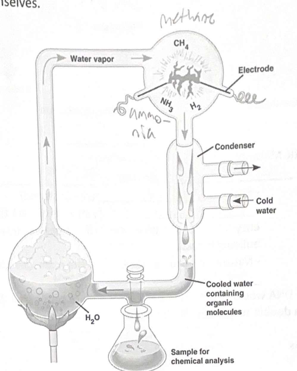
- Prime atmospheric conditions probably did not exist in all locations on earth, but were likely to be found around volcanoes and deep sea vents.
Abiotic synthesis of larger molecules known as polymers.
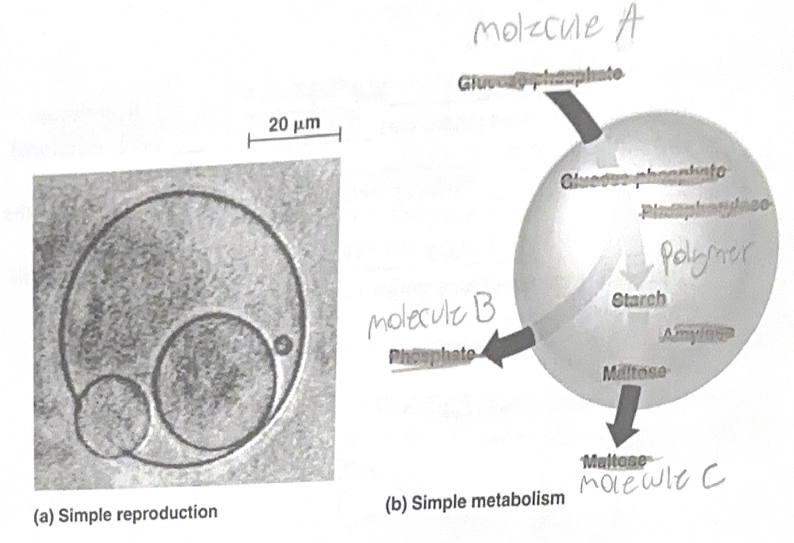
- Heat is required for this reaction to take place.Protobionts - Early Life?
- Two main characteristics of life
1. Accurate replication2. Metabolism (chemical reactions)
- It is thought that liposomes (membrane like structures) surrounded some of the polymers present in the area. These liposomes would be selectively permeable and could possibly carry out a very simple metabolic pathway.
Genetic Material
- RNA originated before DNA
- RNA is simpler (single strand)
- Robozymes - Simple strands of RNA that act as enzymes and can make a copy of themselves (given the correct building blocks)
- Natural selection at the level of the molecule - the most stable survive.
- DNA would come about because it is more stable than RNA because it is a double strand and can hold more information
Living Organisms
- The oldest known fossils are 3.5 billion years old and are called stromatdites. They are a mixture of bacteria and sediment forming mat-like structures in shallow ocean water.
- Autotroph (no oxygen required) were first and then gave rise to the heterotrophs.
- Photosynthesis developed early in order to “build food”
- These organisms that photosynthesized released oxygen into the ocean.
- This oxygen reacted with dissolved iron to form iron oxide (which can be clearly seen in the geologic record)
- After this reaction, oxygen entered the atmosphere and reacted with rocks on land.
- In fact, there was too much oxygen (oxygen can be amazingly reactive at high concentrations).
- This concentration of oxygen killed many of the early prokaryotes
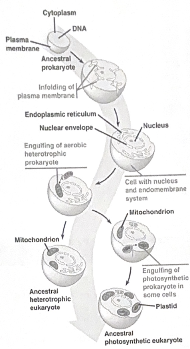
- The rise of the Eukaryote (Euks)
- Thought to have first developed around 2.1 billion years ago
- Endosymbionts by Serial Endosymbiosis
- First Euks were thought to be predators of small Proks
- These prok cells are thought to be the organelles (mitochondria and plastids) now found in all Euk cells.
Evidence
- Both the Mitochondria and Chloroplast have their own DNA that is in a loop, exactly like that of a Prok cell.
- They became dependent on one another.
- Early Euks gave rise to the first multicellular Euk organisms.
- 1.5 billion years ago colonies of unicellular organisms grouped together. Each cell had a special role in the colony.
- Colonization of land - 500 million years ago.
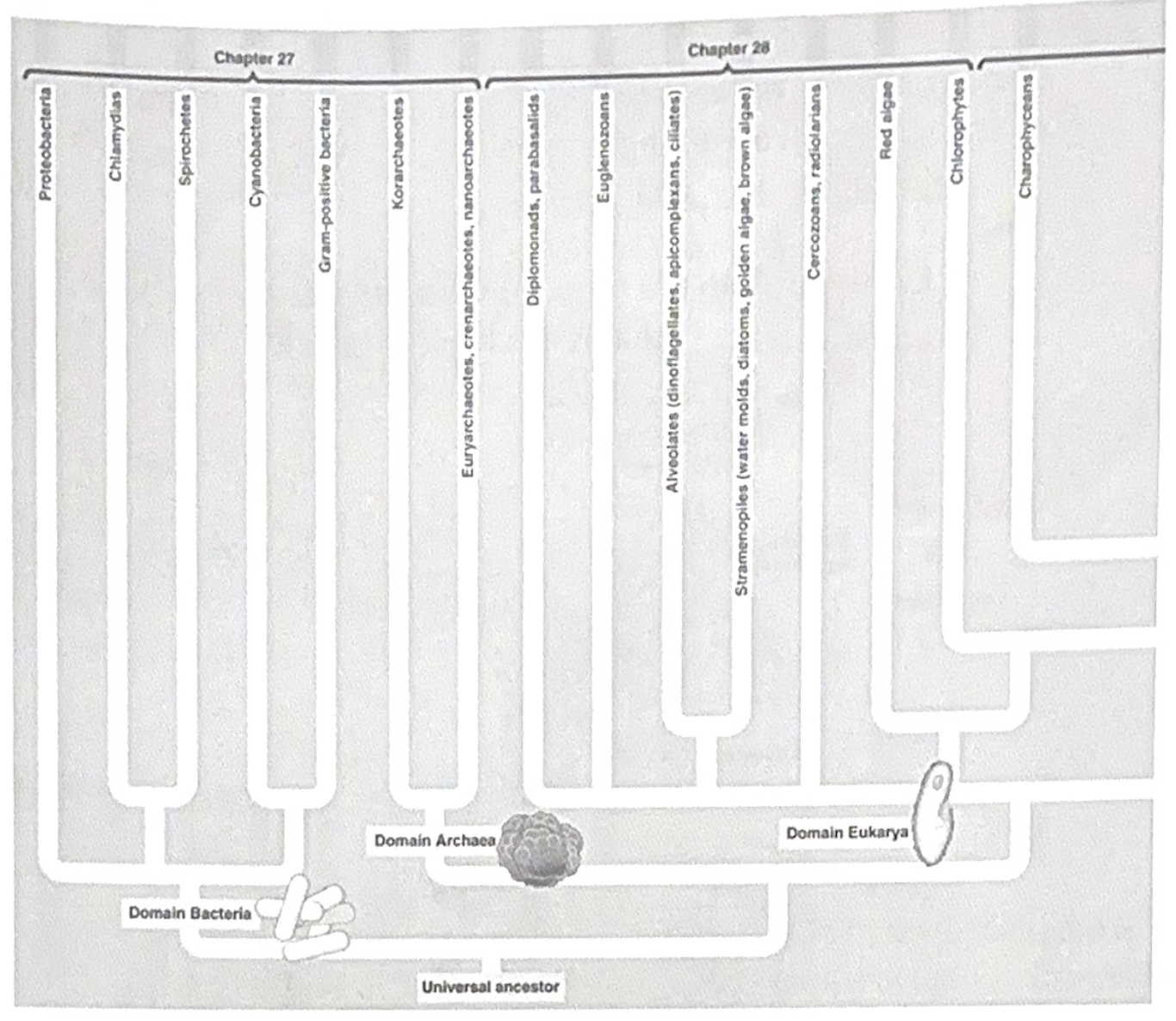
Previous taxonomic systems -
The two kingdom system (Plants and Animals)
The five kingdom system (Animals, Plants, Fungi, Protista, Monera)
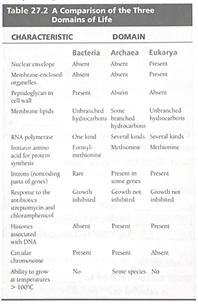
The three domain system build by molecular and genetic evidence (Bacteria, Archaea, Eukaryotes)
- We as Euks are more closely related to archaea than archaea is related to bacteria.
CHAPTER 6
Cell = The simplest collection of matter that can live independently
Tools to study the cell - Microscopy
- Light microscope (what we use the most)
- Light is passed through the specimen
- Magnification - Ratio of image to actual size (usually a 1000x max)
- Resolution - Clarity of image
- Can usually resolve objects down to .2 um or 200 nm (about the size of bacteria)
-Electron microscopes
-Resolve down to 1 nm
-Scanning electron (SEM) - Scans surface of object
Transmission Electron (TEM) - Transmits electrons through object
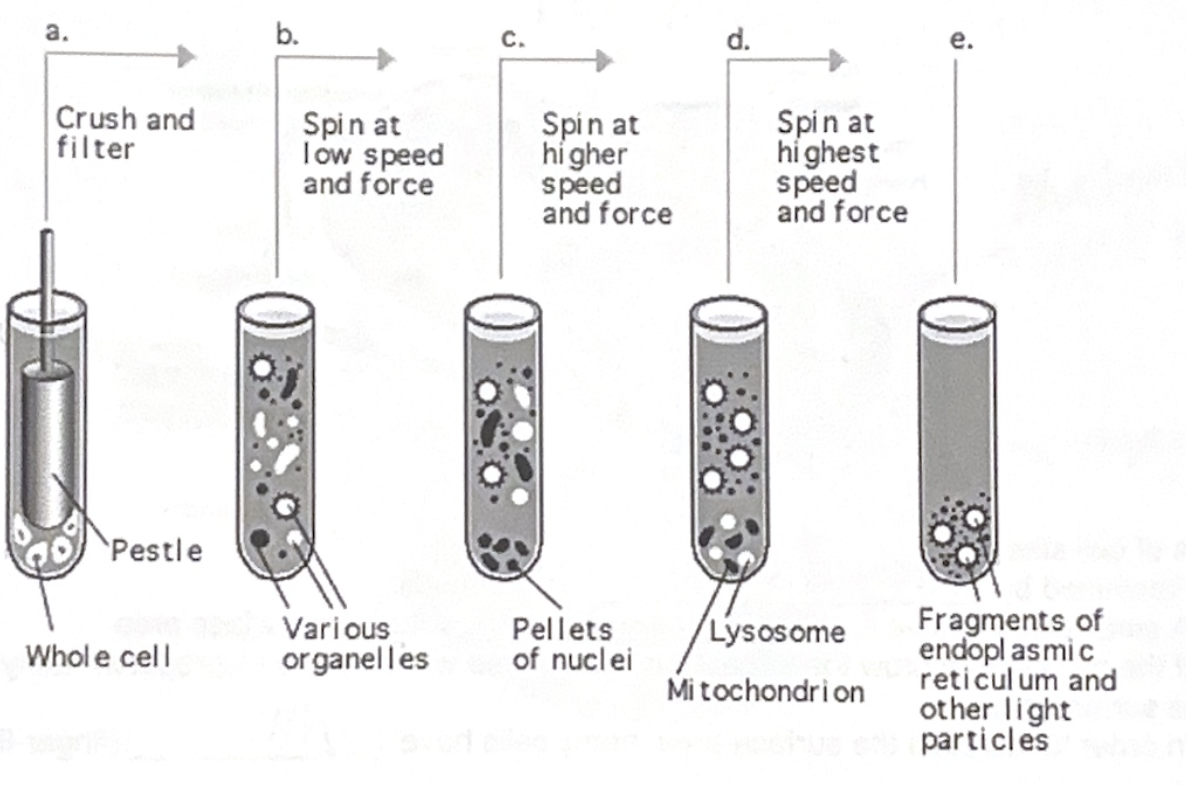
- Cell fractionation - Take the cell apart and separate the organelles by weight.
- Completed by using an (ultra)centrifuge
- The heaviest organelles settle out first, the lightest settle out last.
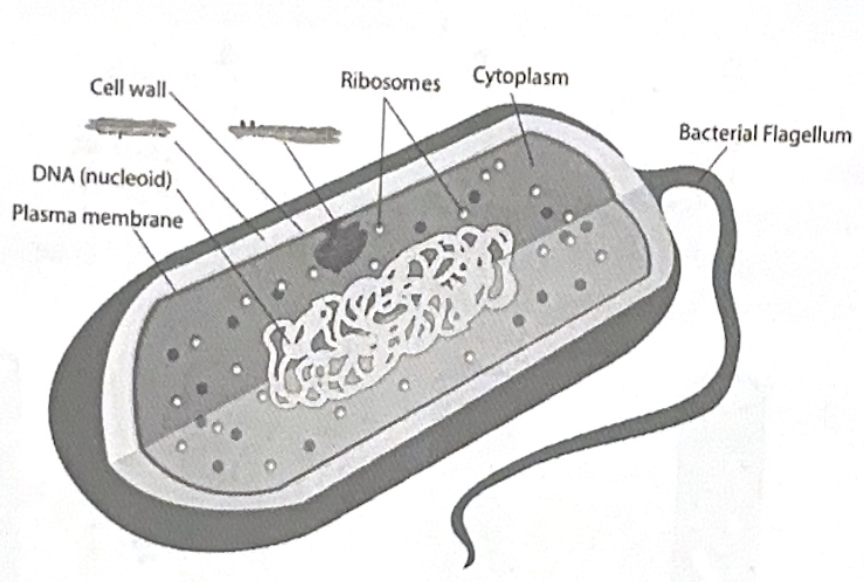
ALL cells have
- Chromosomes (genetic material aka DNA)
- Ribosomes
- Cytoplasm
- Membrane
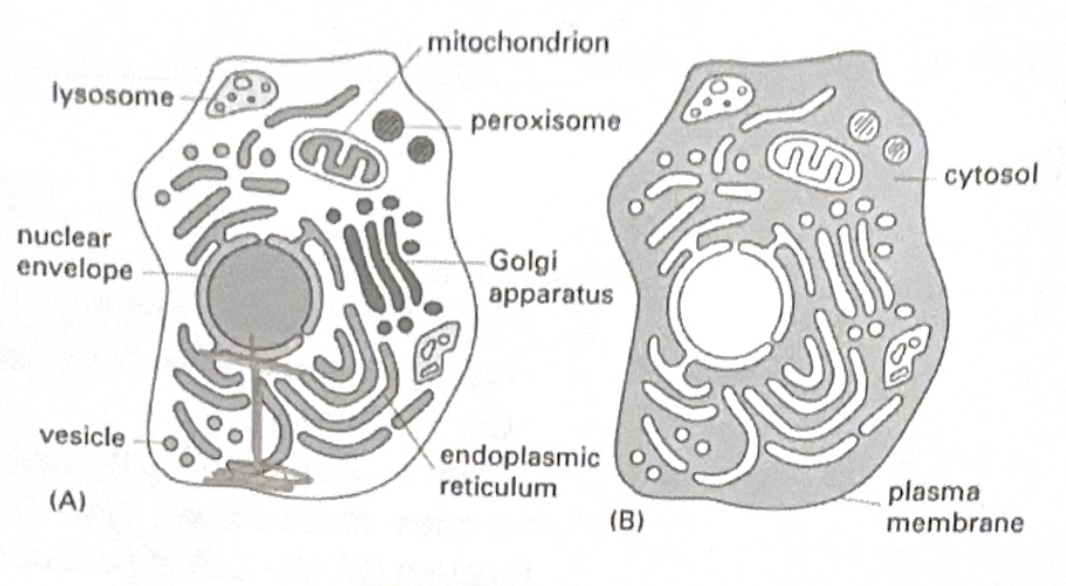
Eukaryotes - “True kernel” - Has a nucleus. Large (10-100 um)
- Internal membrane for compartmentalizing functions
- Bound by plasma membrane
- Fluid inside of the cell is called cytosol
- The region between the nucleus and plasma membrane is the cytoplasm
Prokaryotes - “Before kernel” - No nucleus. Small (1-10 um)
- Bound by plasma membrane
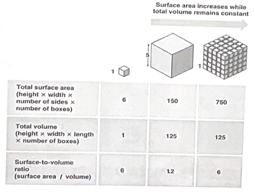
Limitations of cell size
- Restricted by metabolic requirements
- A small cell will have proportional volume to surface area
- If the cell were to grow too large, then the volume will increase disproportionately (larger) than the surface area.
- In order to increase the surface area, many cells have villi (finger-like projections)
Internal membranes separate areas of separating metabolic purposes
The plasma membrane is composed of a phospholipid bilayer
- Many proteins are embedded in the plasma membrane
ORGANELLES
Nucleus - Library of the cell because it contains MOST of the cells genes
- A nuclear envelope that encloses the nucleus
- Double membrane
- Has pores that are regulated by pore complexes
Nuclear lamina - A net-like structure on the inside of the nucleus that provides mechanical support (constructed of proteins)
Nuclear matrix - Framework inside of the nucleus for additional structural support
Nucleolus - RNA is synthesized here
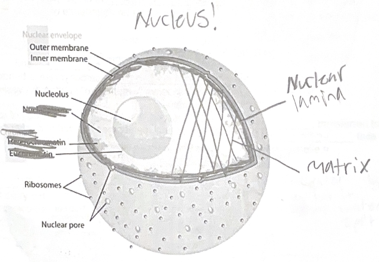
Ribosomes - Protein synthesis
- Two types
1. Free ribosomes - In cytosol
- Proteins constructed by these are usually used in the cytosol
2. Bound ribosomes - Attached to the outside of the endoplasmic reticulum and nuclear envelope
- Proteins constructed by these usually incorporated into membranes and used for packaging and export.
- Free ribosomes and bound ribosomes can switch back and forth from one to another depending on cellular needs.
Endomembrane system - includes the following:
- Smooth Endoplasmic Reticulum
- Rough Endoplasmic Reticulum
- Golgi Apparatus
- Lysosomes
- Vacuoles
- Has close ties to plasma membrane
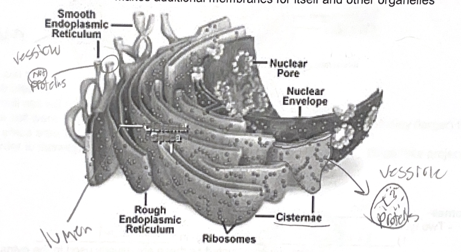
Endoplasmic Reticulum - Biosynthetic factory. “Plasmic” = within cytoplasm, “Reticulum” = little net
- Accounts for more than half of the total membrane in many Euks.
- Network containing tubules and sacks called “cisternae” (reservoir)
- Internal compartments of the cisternae are called the “Lumen” (cavity)
- Two types
Smooth ER - no ribosomes
- Synthesis of lipids (oils, steroids, phospholipids)
- Metabolism of carbs
- Detoxification of drugs/poisons (especially in liver cells)
- There will be an abundance of smooth ER when many toxins are present
- Storage of calcium ionsRough ER - ribosomes
- Construction of proteins and glycoproteins (a carbohydrate is bonded to a protein)
- Proteins can leave “wrapped up” in transport vesicles and travel to Golgi Apparatus
- Transport vesicles come from a piece of the rough ER
- Makes additional membranes for itself and other organelles
Golgi Apparatus - Shipment, protein, modification
- Transport vesicle arrive; contents are dropped off and then shipped out again
- Common in secretion cells
- Composed of flat sacks (cisternae) in stacks
- The two opposing sides of the Golgi bodies have distinct polarities
- Cis - nearest the ER and it transport vesicles fuse to this side and then incorporate good
- Trans - Vesicles pinch off and move around cell
- Modification of goods in Golgi apparatus happens during transit from the cis to the trans side
-Cisternal maturation model - actually cisternae progress from cis to trans side
- Goods are shipped with “tags” that will only bind to specific sites

Lysozomes - Empty compartments for intracellular digestion (protein)
- Contains enzymes that digest macromolecules
- Membranes of the lysozome are made by the Rough ER, transferred to the Golgi apparatus, and are then “budded” off of the trans face
- Can digest the host cells organic waste of entire non-functioning organelle = autophagy and then re-use organic goods.
Vacuoles - Storage cell can have one or many
- Can carry out hydrolysis
- Food vacuoles come from phagocytosis
- Contractile vacuoles pump out excess water
- Central (or sap for AICE Bio) vacuole - plants
- Surrounded by a membrane called the tonoplast
- Can hold reserves of organic goods
- Repository of inorganic waste
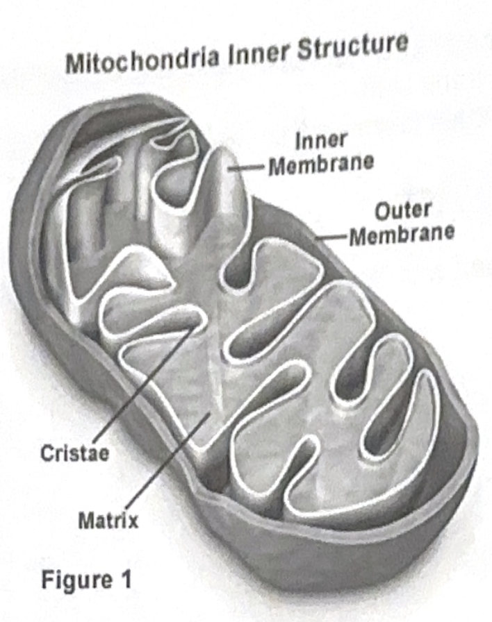
Mitochondria/Chloroplasts - Converts energy from one form to another
- Enclosed by membrane, but often still uses proteins from the cytosol (proteins constructed by nuclear transcription)
Mitochondria - Energy, proteins (they house some ribosomes)
- Has two membranes, each is a phospholipid bilayer
- Outer membrane is smooth
- Inner membrane is heavily folded (folds are called cristae)
- Increases the surface area, increases the amount of ATP produced
Mitochondrial matrix - the inside of the inner membrane
- Where majority of “work” takes place
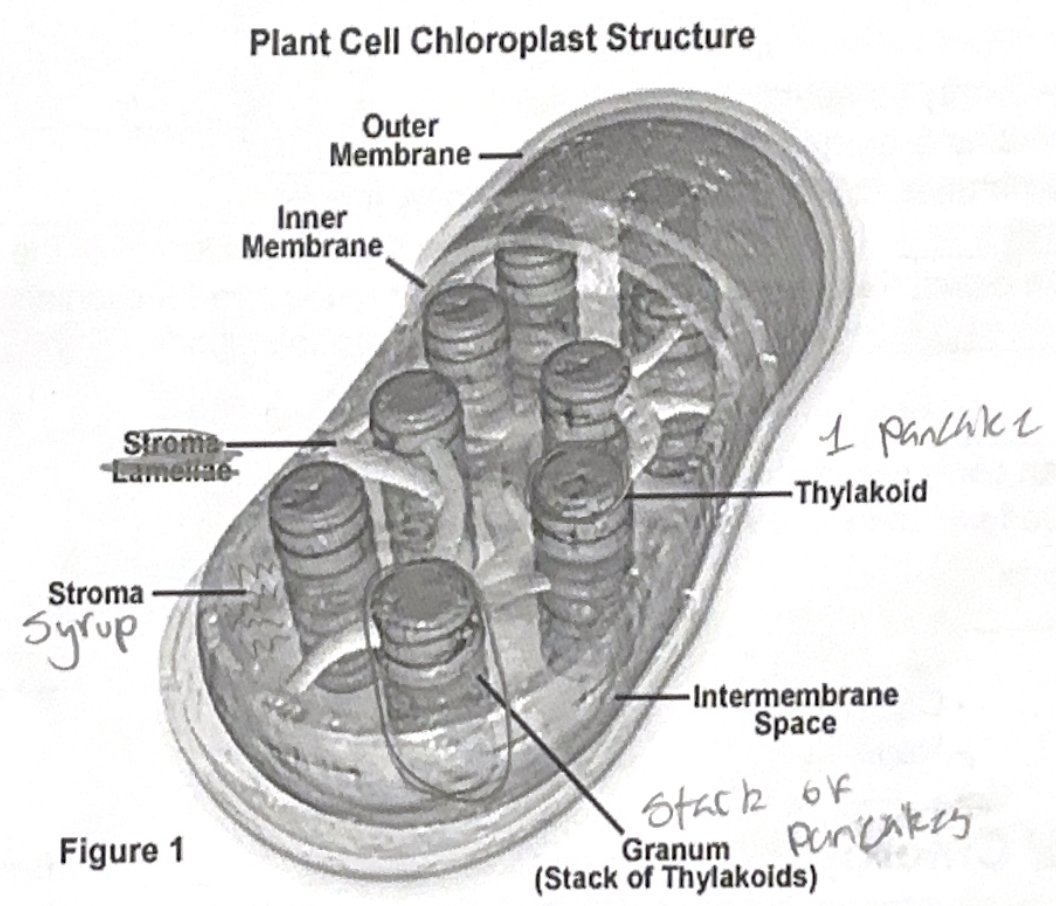
Chloroplast - Light to sugar energy. A plastid
- Two membranes
- Inner membrane connected to sacks = Thylakoids
- Stack of Thylakoids = Grana
- Fluid outside of Grana = Stroma (contains DNA/Ribosomes)
Peroxisomes - Oxidation (breaks things down, deals mostly with toxic waste)
- Metabolic component bound by a single membrane
- Contains enzymes that break down molecules and produce hydrogen peroxide (H2O2)
- Hydrogen peroxide is toxic to cells, but a local enzyme breaks down H2O2 to water
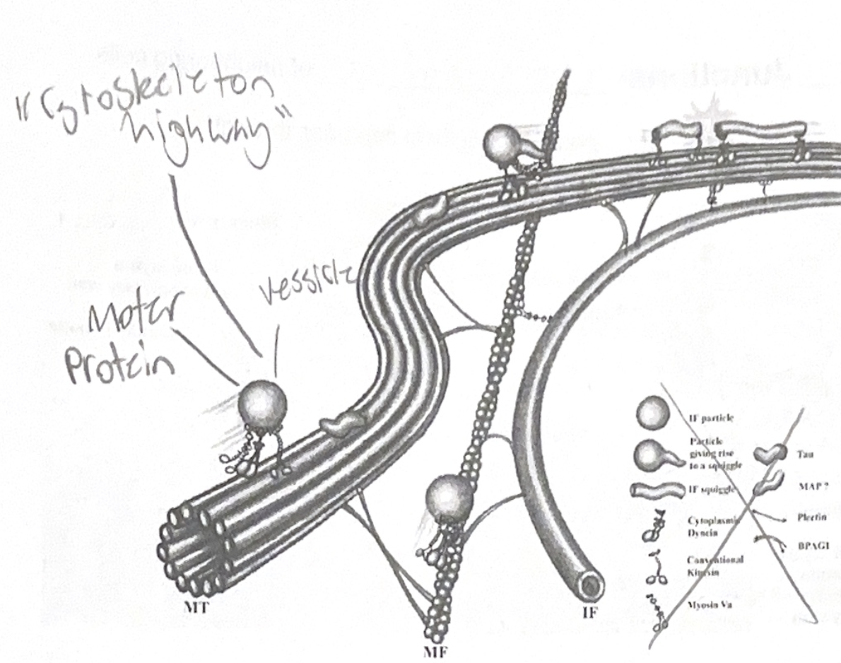
Cytoskeleton - Framework that organize structures and activities
- Mechanical support/shape
- Motilits
-Motor proteins used to help cell change shape
-Can act like a “monorail” and transport goods
- Regulation of biochemical activities
- Can transmit an outside force to the inside of the cell allowing for re-arrangement of organelles
Three types:
Microtubules - In all Euks (not in proks) and are crush resistant
- Hollow rods but fairly thick
- Composed of globular protein = Tubulin (2 types)
- Alpha tubulin and Beta tubulin
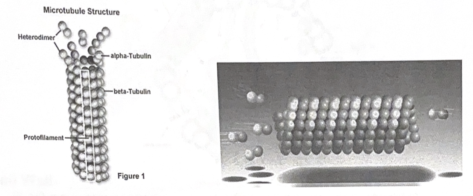
- Proteins are added to ends in order for elongation to take place
Centrosomes and Centriols
- Microtubules originate from a centrosome (the centrosome is a REGION, and there is a PAIR of centriols in the centrosome region)
- Centriols replicate when cell replicates
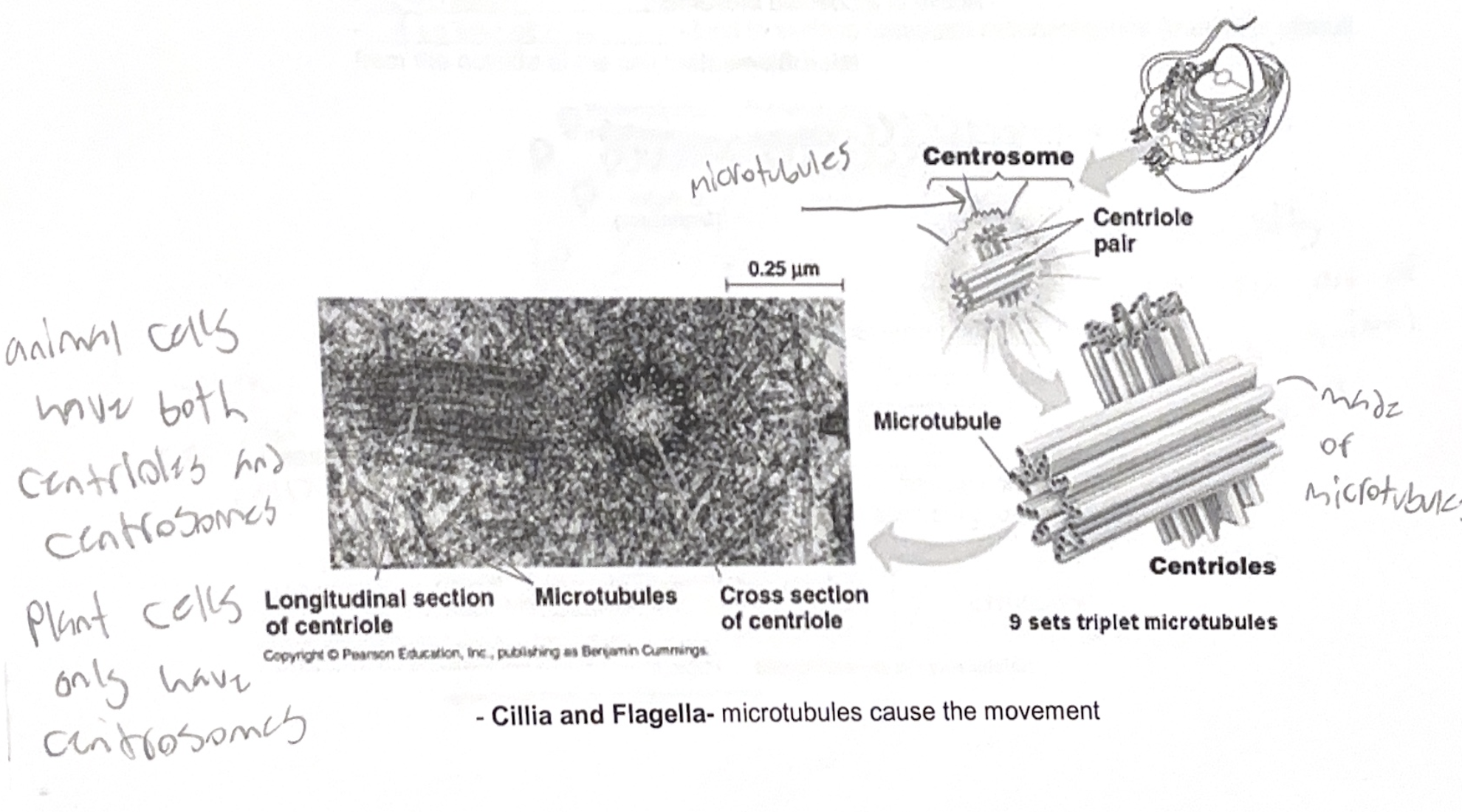
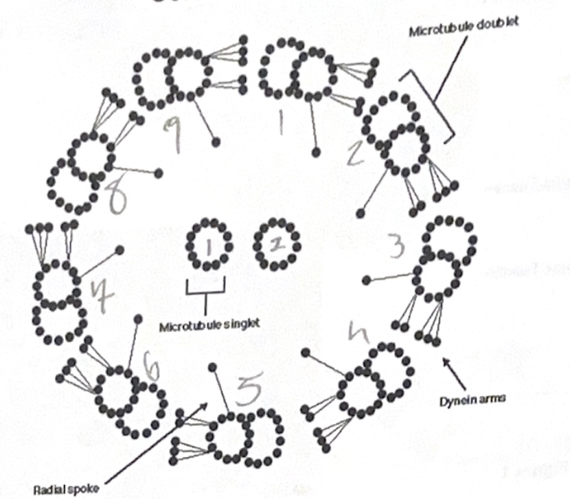
Cillia and Flagella - microtubules cause the movement
9+2 arrangement in Euks
- 9 doublets or microtubules in a ring and 2 single microtubules in the center
- Motor proteins BETWEEN the doublets (called dynein) are what actually cause the movement with input of ATP
- Proteins hold structure together
- Connected to cell at the basal body
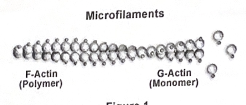
Microfilaments - also called actin filaments (composed of actin proteins)
- Solid rods, but small diameter
- Twisted, double chain of actin
- Made to bear tension (ie: pull)
- Main component of muscle
- Interwoven with another protein called myosin (motor protein)
- Active in amoeboid movement
Intermediate Filaments - somewhere between the size of microtubules and microfilaments
- Tension bearers
- Composed of fibrous proteins of all sorts
- A more permanent structure than the previous structures (microtubules and microfilaments)
- Reinforces the cells shape
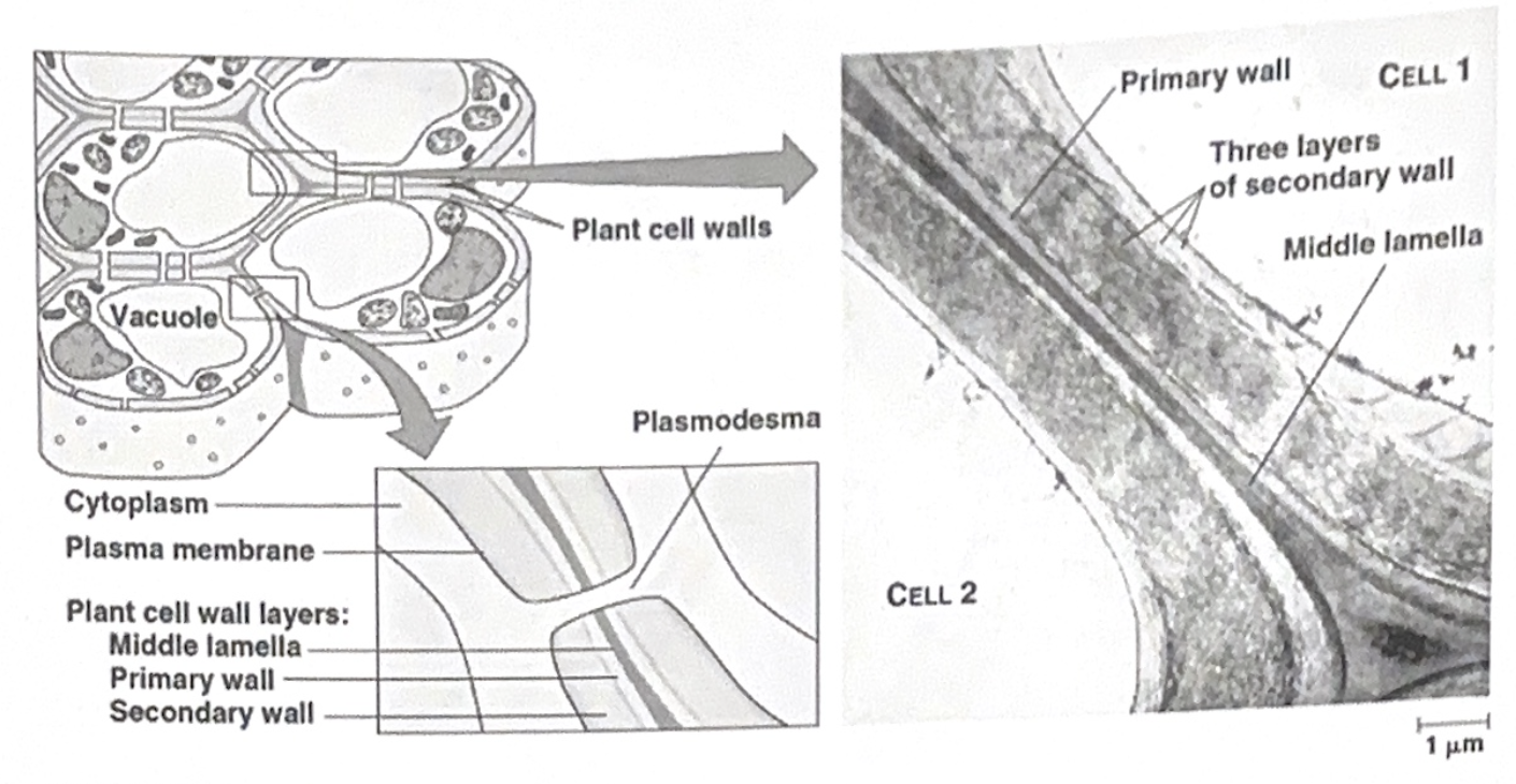
Cell wall - Protects, gives shape and keeps cell “water tight”
- Composed of cellulose matrix (with proteins) like sugar (salad)
- Primary cell wall build when cell is “young”, but eventually it will either harden or a secondary cell wall will be build
- Middle lamella is a sticky substance that holds the cells together
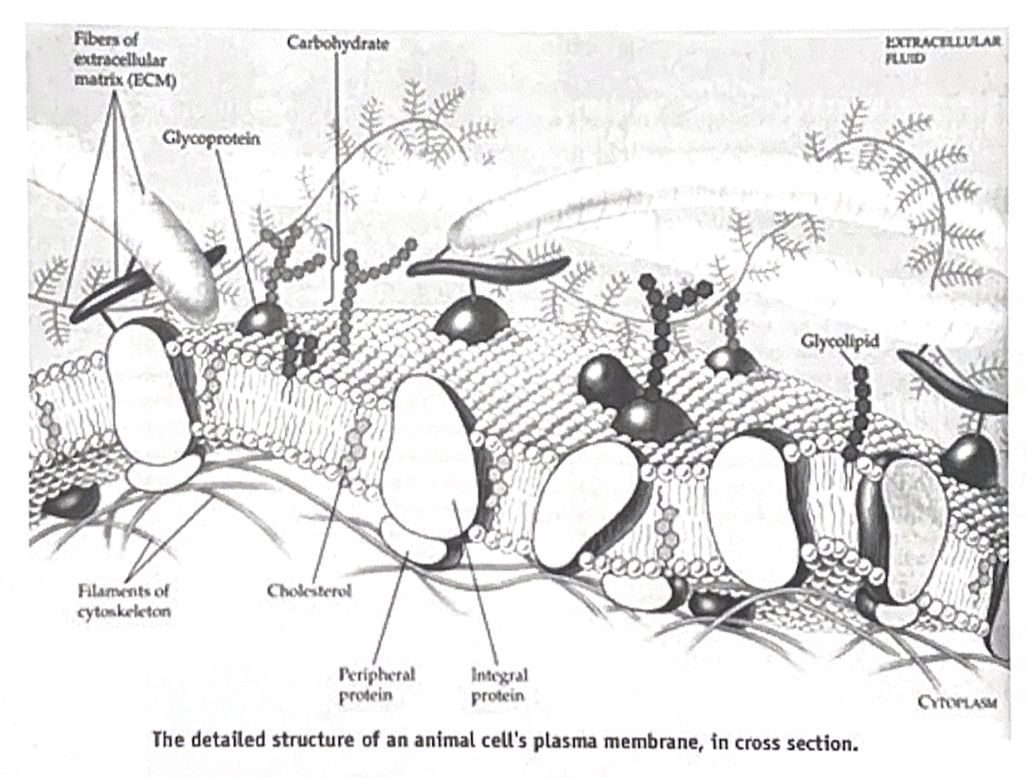
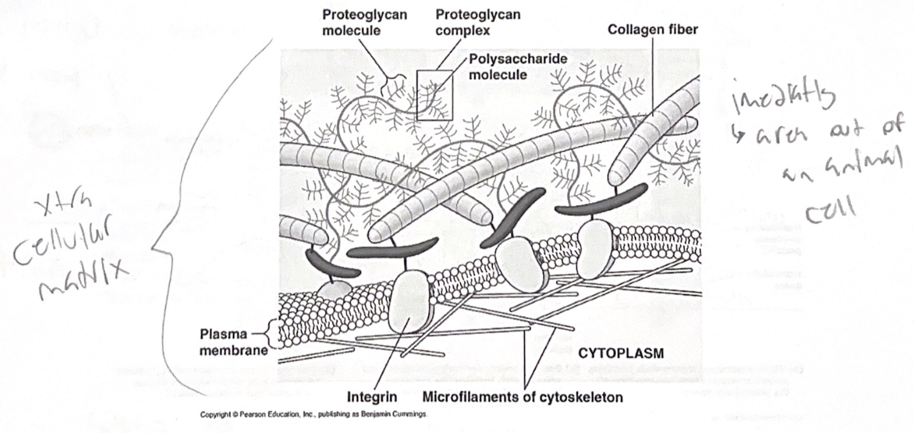
Extracellular matrix of animal cells - immediate outside of cell
- Composed of Glycoproteins
Collogen - Strong fibers
Fibronectin - bind to surface receptors called integrins (transmits stimuli from the ourside of the cell to the inside)
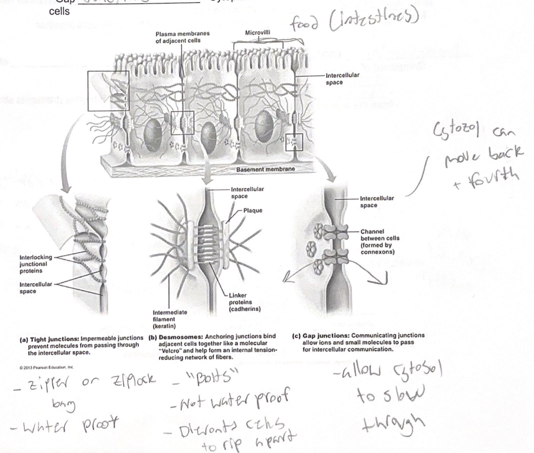
Intercellular Junctions - communication of neighboring cells
- Plants - Plasmodesmata pores that cyrosol can pass through
- Animals
- Tight junctions - prevents “leaking” from between cells
- Desmosomes - “Rivets” that hold the cells together
- Gap junctions - cytoplasmic channels for communication between cells
Chapter 7
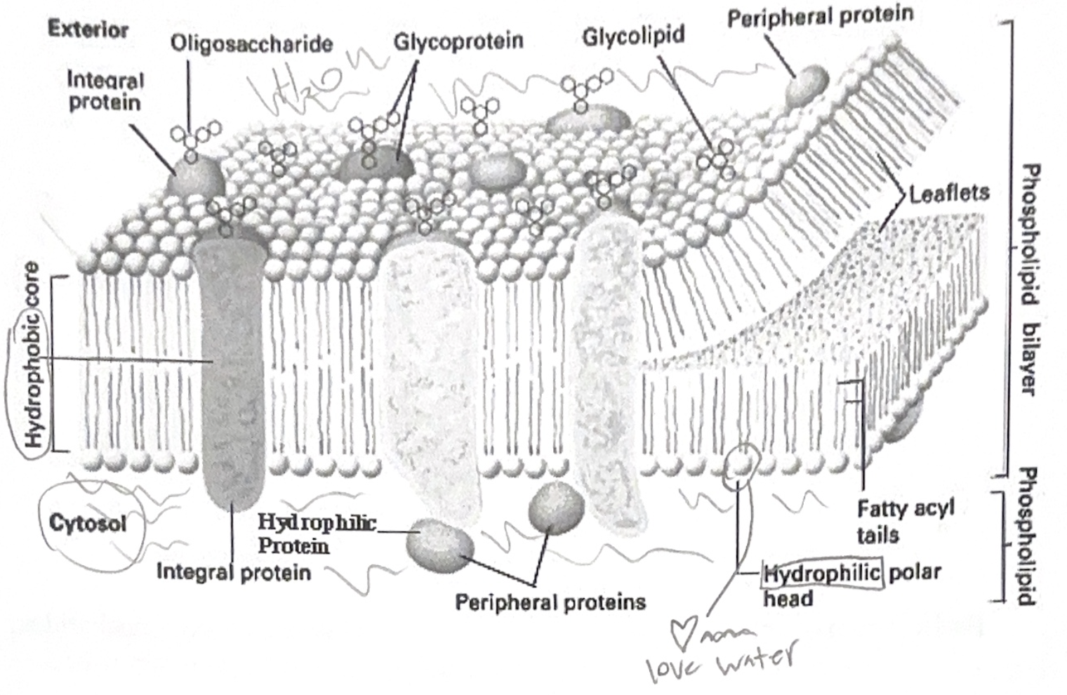
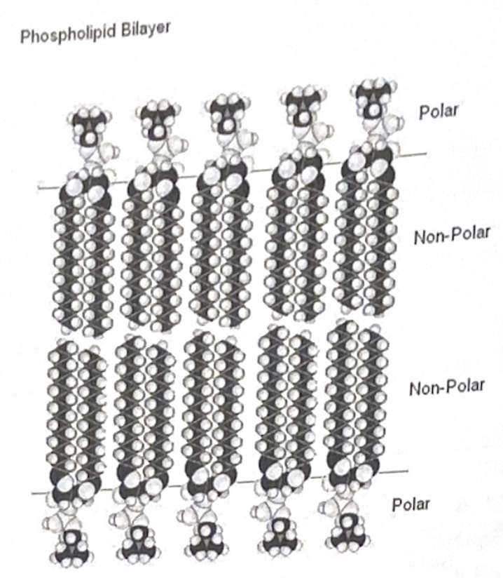
Cell membrane
- 8 nm thick and controls entry/exit of molecules
- Selectively permeable
- Fluid mosaic of proteins/lipids - “Fluid mosaic model”
- Phospholipids most abundant lipid in membrane
- Amphipathic molecules - has a hydrophobic and hydrophilic region
- Proteins are embedded in phospholipid bilayer (singer, nicolson. 1972)
Movement of lipids and proteins in membrane
- Lipids move in a lateral manor frequently
- Proteins sometimes move, sometimes don’t
Fluidity - Similar to oil
- Will remain a fluid until it becomes to cold that the phospholipids closely pack together
- Cholesterol is an animal’s membrane “buffer”. Helps keep membrane fluid at high and low temperatures
Membrane proteins - define specified functions of a membrane (different cell = different proteins)
- Integral proteins - penetrate hydrophobic core of lipid bilayer
- Peripheral proteins - not embedded at all - “appendages”
Functions -
- Transport - can be channels that are selective and change shape
- Enzymatic activity - part of an enzymatic pathway
- Signal transduction - can pass on exterior chemical changes, ie: hormones
- cell-cell recognition - identifies foreign or similar cells
- Intercellular joining - gap/tight junctions
- Attachment to cytoskeleton + extracellular membrane
Membrane Carbohydrates - Essential for cell-cell recognition, ie: blood type
- Important in tissue development
- Recognition of foreign bodies, ie: organ transplants
- Most are glycoproteins
- Glycoproteins/glycolipids - start in the inside of a membrane and end up on the outside (Rough ER to Golgi Bodies to Incorporation)
Selective Permeability - Controlled by “fluid mosaic”
- Hydrocarbons, carbon dioxide, oxygen all dissolve into cell without aid of proteins
- Polar molecules such as glucose, water, and some sugars cross very slowly
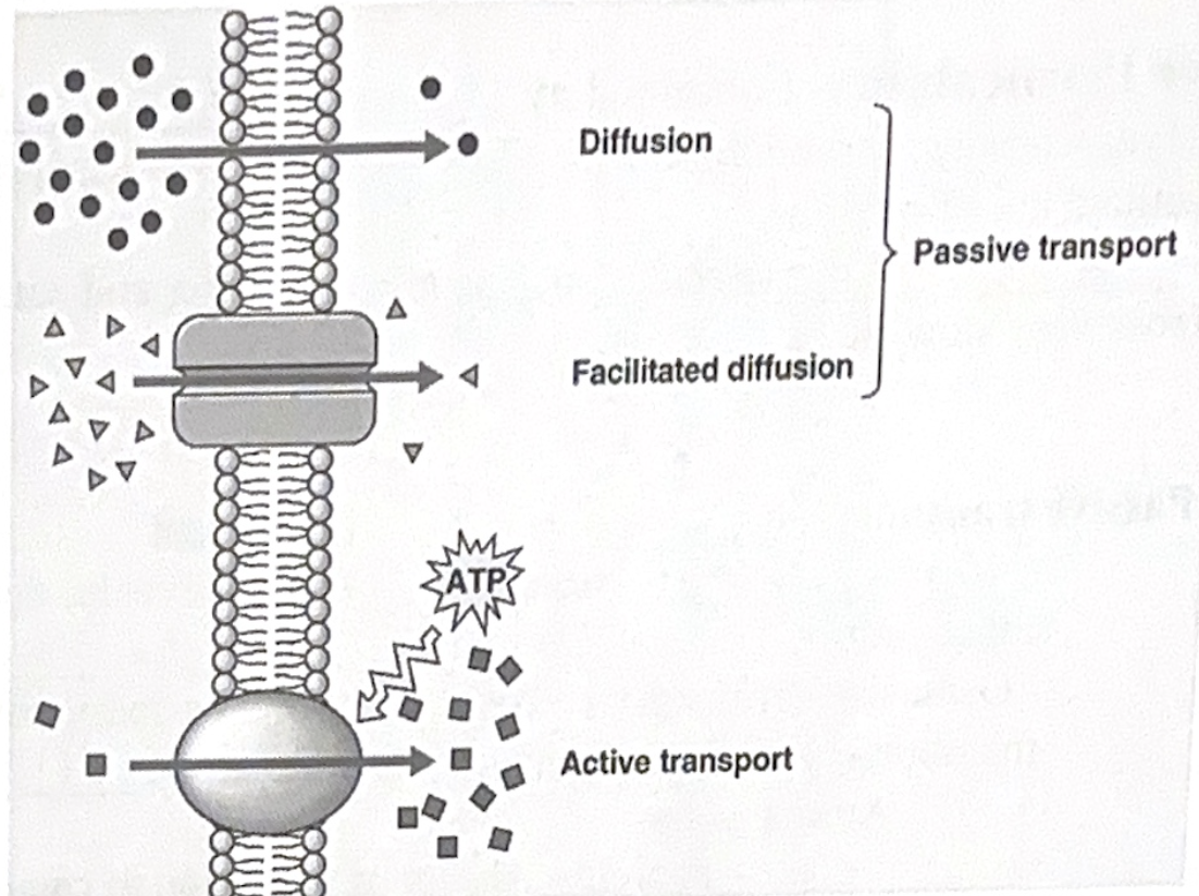
Passive transport - NO energy investment
- Diffusion - Molecules spread out evenly, down concentration gradient. Striving for equilibrium
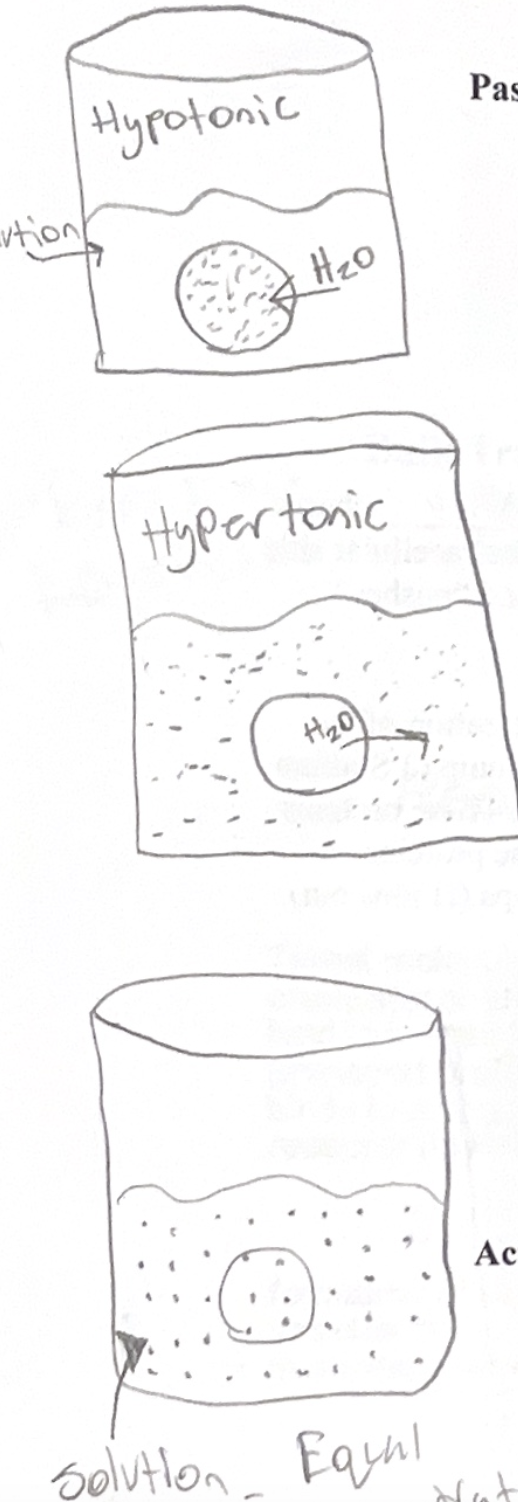
- Osmosis - Diffusion of “free” water across a semipermiable membrane until the solutes on both sides are at equality
Animal cells -
- Tonicity - Ability of a solution to cause a cell to gain/lose water
- Isotonic - No net movement of water, volume of animal cell remains the same
- Hypertonic solution - More non-penetrating solutes outside of cell. Cell will lose water to environment (ie: freshwater organism in saltwater)
- Hypotonic solution - less non-penetrating solutes outside of cell. Cell will fill, and maybe even lyse (burst)
Adaptations for osmoregulation - Contractile
Plant, fungi, protist cells
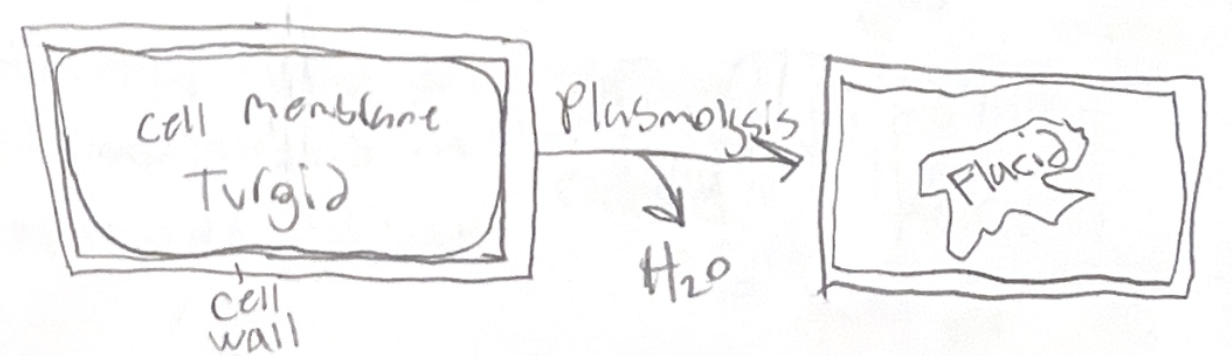
- Cell wall helps regulate pressures
- Turgid cells - full and “happy”
- Flacid cells - limp
- Plasmolysis - Shrinking of membrane, pulls away from cell wall, cell dies
Hypertonic environments
Facilitated diffusion - passive transport aided by proteins
- Channel proteins - “corridors”, but specific
- Aquaporins - ion channels (gates) that allow water to pass through
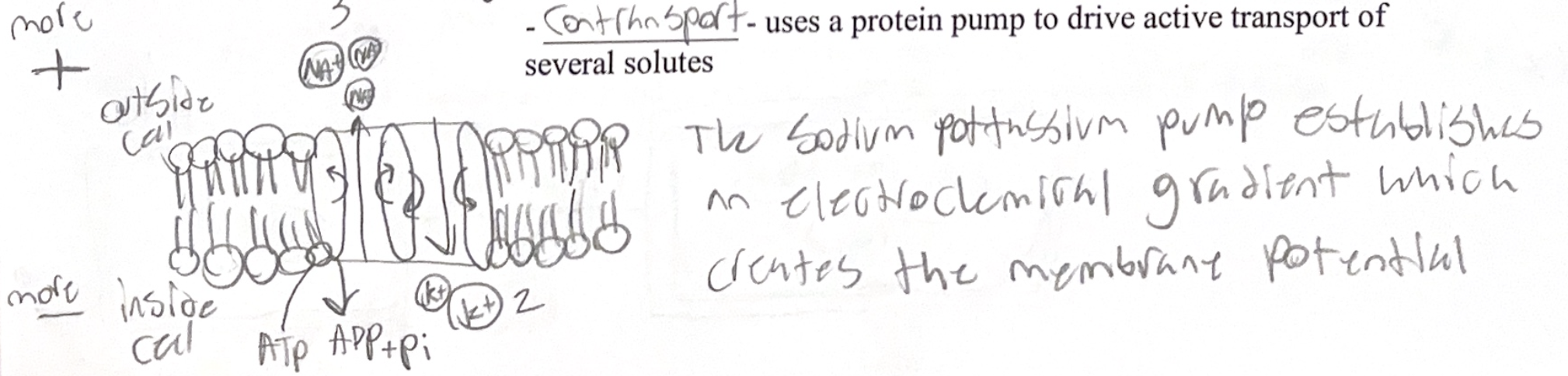
Active transport - Against concentration gradient
-Requires input of energy
- Carrier proteins
- Example - Sodium/Potassium pump
- Exchanges sodium for potassium
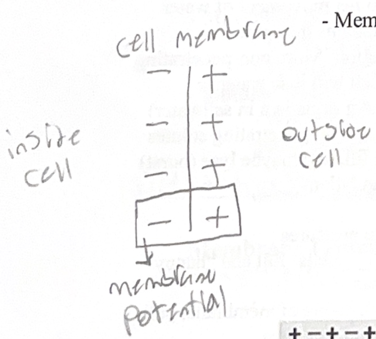
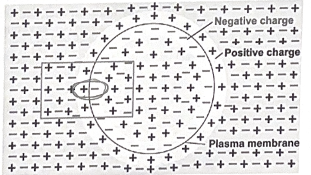
Membrane potential - voltage across membrane
- Intracellular side of membrane has negative charge in comparison to positive charge of extracellular side
- Favors the passage of cations (positive) and “pushes” anions (negative) out of cell
- Electrochemical gradient
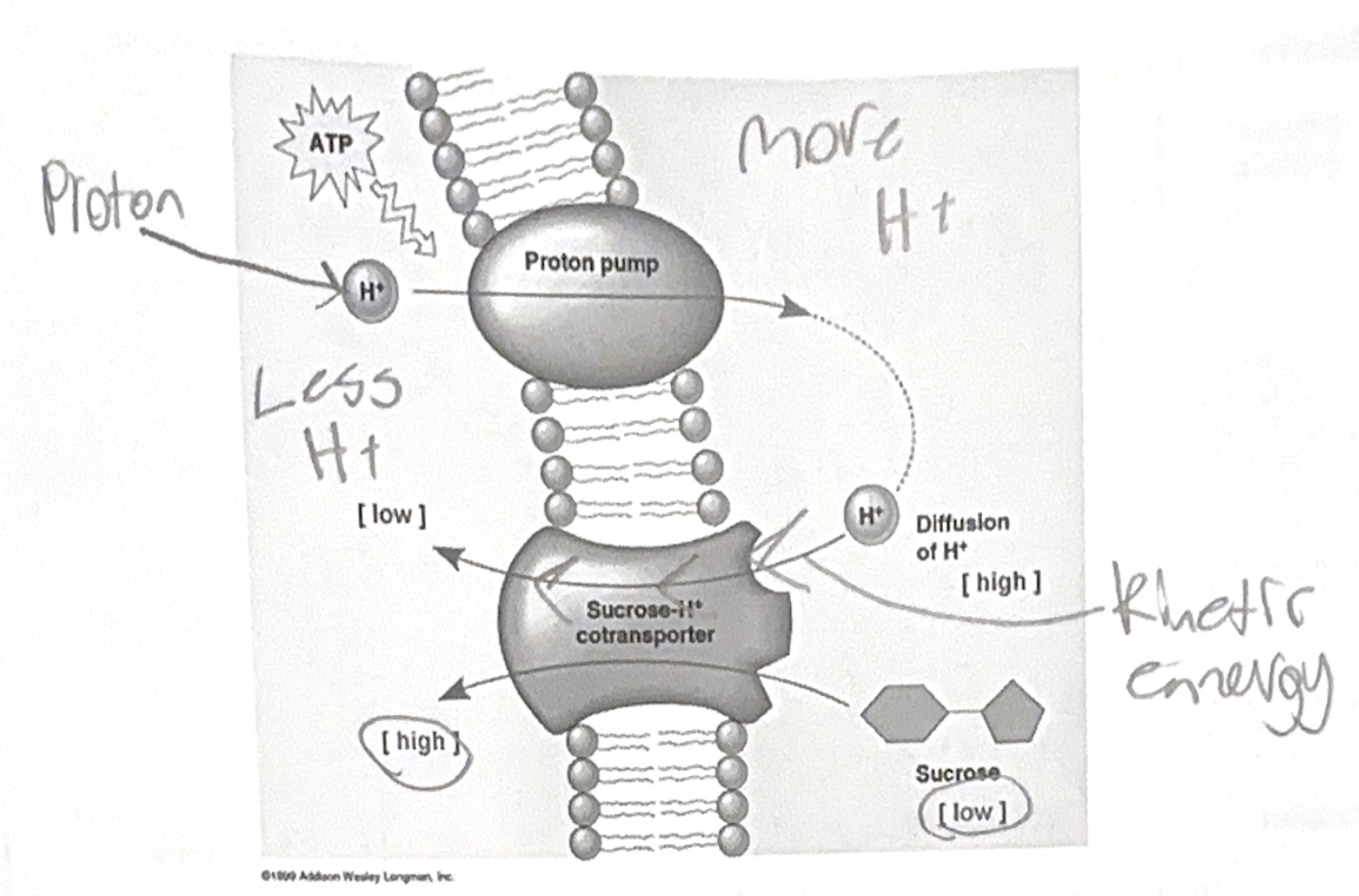
- Some membrane proteins help drive the creation of the charge different ie: Sodium/Potassium pump (3 Sodium exchanged for 2 Potassium - stores energy). These proteins in animals called electrogenic pumps. These proteins in plants, fingi, bacteria called proton pumps (H ions out)
Contransport - Uses a protein pump to drive active transport of several solutes
Bulk Transport - Transport of large molecules (proteins, polysaccharides)
Exocytosis - Fusion of vesicles with membrane in order to dump wastes
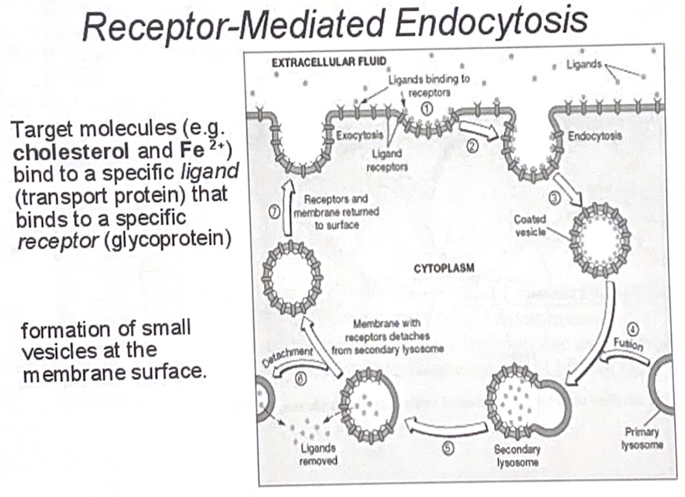
Endocytosis - Formation of vesicles
- Phagocytosis - eating
- Pinocytosis - drinking
- Receptor mediated endocytosis - proteins “collect” what they need (molecules that they “need” - ligands)