Bio Exam 3
1/138
There's no tags or description
Looks like no tags are added yet.
Name | Mastery | Learn | Test | Matching | Spaced | Call with Kai |
|---|
No analytics yet
Send a link to your students to track their progress
139 Terms
Name the three types of cytoskeleton, their main function and their location.
Intermediate filaments: helps cells withstand mechanical stress
Microtubules:Highways for intracellular transport and segragation of chromosomes
Actin: Cortex stabilization, cell movement, and muscle contraction
Describe the structure, assembly and function of IFs.
Structure: Rope-like fibers and each protein has a central α-helical rod domain with flexible ends
Assembly:
Monomer → single IF protein
Dimer → two monomers twist together (coiled-coil)
Tetramer → two dimers align in opposite directions (anti-parallel)
Protofilament → filament → tetramers pack together
Function: Mechanical support, Structural integrity, Tissue strength
Name an intermediate filament protein
Keratin, Vimentin, Neurofilaments, and nuclear lamins
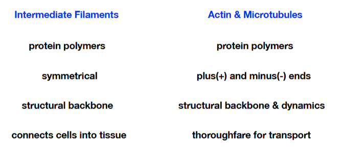
How are IFs different from actin and microtubules structurally?
IFS are rope-like solid fibers while actin are thin two-stranded helical chains and microtubules are Hollow tubes
What is the function of nuclear lamins?
Lamins that make up the nuclear lamin protect the nuclear envelope
Describe the structure, assembly and function of microtubules.
Structure: Hollow tubes
Assembly: Ends are distinct from each other, ends are called + and - ends because of the different rates of adding dimers
Function: highways for intracellular transport and segregation of chromosomes
What are the protein subunits of microtubules called?
Tubulin
What do we mean by plus end of microtubule?
Plus end = “adding end” (fast-growing/adds tubulin)
What is an MTOC?
Microtubule-Organizing Center ( organizes the location/number/orientation of the microtubules)
What is meant by dynamic instability of microtubules?
Microtubules switch from polymerization to depolymerization
How does GTP hydrolysis control dynamic instability?
GTP-tubulin is added faster than GTP is hydrolyzed
The GTP cap is maintained
Microtubule stays straight and continues growing
What do microtubule capping proteins do?
Regulates growth or shrinkage
Describe the structure, assembly and function of Actin.
Structure: two-stranded helical chains
Assembly: Actin filaments are thin and flexible. Simple assembly formed from a twisted chain of monomers and has plus and minus ends for assembly & transport
Function: Cortex stabilization; cell movement; muscle contraction
What do we mean by actin treadmilling?
Gain subunits at + end and lose subunits at - end
How can actin structure be modified?
Directed growth using the addition of subunits
What is the role of myosin? Which direction does it move on in actin?
Myosin is a motor protein that converts ATP into movement
Usually moves toward the plus (+) end
Compare and contrast the three cytoskeleton proteins.
What are the two motor proteins that walk along microtubules to move cargo?
Kinesin and dynein
Compare dynein and kinesin.
Kinesin: Stretches the ER along microtubules like a net (plus end directed)
Dynein: Pulls golgi towards the nucleus (minus end directed)
What powers movement of motors?
ATP
Which domain of the motor protein binds cargo, which domain binds cytoskeleton?
Head domain binds actin, tail binds cargo
Give an example of how Kinesin and Dynein help position organelles
Kinesin → out (to cell edge)
Dynein → in (to cell center)
What is the motor protein that walks along actin?
Myosin
What are the functions of myosin?
hydrolyzes ATP to fuel cycles of binding, release, rebinding
Provide a step wise description of how an action potential leads to muscle contraction.
Step 1: Electrical signal travels from neuron to muscle
Step 2: Action potential in muscle cell releases calcium into the cytoplasm of the muscle cell
Step 3: Ca2+ binding proteins move, exposing myosin binding sites on actin
Step 4:Myosin moves along actin filaments
What is the role of calcium in muscle contraction?
Calcium moves tropomyosin away from actin, exposinh the myosin-binding sites
Other than calcium what else is needed for muscle contraction?
Myosin, troopnin, actin, and topomysoin
What functions do organelles serve?
Organelles compartmentalize the cell
This means they:
Separate different processes
Create optimal environments for reactions
Prevent interference between functions
What is the endomembrane system? Whats in the system?
The endomembrane system is a group of organelles inside a cell that are physically connected or communicate via vesicles
Included:
Nuclear Envelope, ER, Golgi, Vesicles, Lysosomes, and Plasma membrane
Name the process used to bring molecules into a cell?
Name the process used to move molecules out of a cell?
Endocytosis — the cell membrane folds inward to form a vesicle that brings substances into the cell.
What are the three mechanisms by which proteins are sorted in a cell?
Gated transport - Movement of proteins through nuclear pores between the nucleus and cytosol
Transmembrane transport - Proteins move across a membrane through protein channels, often requiring unfolding
Vesicular transport - Proteins are transported in vesicles between membrane-bound organelles
What determines where a protein goes in a cell? How did scientists
figure this out?
Signal sequences (short amino acid sequences) determine this, they direct proteins to specific locations.
Scientists attached signal sequences to proteins and observing changes in their location
How do large molecules enter/exit the nucleus?
Through the nuclear pore complex using transport receptors.
What is the nuclear pore? Describe its structure.
A large protein complex that regulates transport between the nucleus and cytoplasm.
Its structure is a large channel made of multiple proteins (nucleoporins) with FG-repeat regions forming a selective barrier.
What determines if a protein gets sorted to the nucleus? How can this be tested?
A nuclear localization signal (NLS).
Add/remove NLS from a protein and observe if it enters the nucleus (e.g., fluorescence microscopy).
What are nuclear import receptors (importins)?
Proteins that bind NLS-containing cargo and transport them into the nucleus.
Describe the process of nuclear import.
Importin binds cargo → moves through NPC → Ran-GTP binds importin → cargo released in nucleus.
How is directionality of nuclear import/export maintained?
By a gradient of Ran-GTP (nucleus) and Ran-GDP (cytoplasm).
Which form of Ran is in high concentration in the nucleus and why?
Ran-GTP, because a GEF (guanine exchange factor) in the nucleus converts Ran-GDP to Ran-GTP.
Which form of Ran is in high concentration in the cytoplasm and why?
Ran-GDP, because a GAP (GTPase activating protein) converts Ran-GTP to Ran-GDP.
Which form of Ran has higher affinity to importins?
Ran-GTP
What is immunoprecipitation? What is it used for?
A technique using antibodies to isolate a specific protein from a mixture. Used to study protein presence and protein–protein interactions.
What does it mean when two proteins to immuno-precipitate together?
They likely interact or are part of the same complex.
What are some good control to use in IP experiments?
No-antibody control, non-specific antibody control, and input sample control
What is rough ER?
ER with ribosomes attached; site of protein synthesis for secreted and membrane proteins.
What is the role of SRP in ribosome docking on rough ER?
Signal Recognition Particle binds signal sequence and pauses translation to target ribosome to ER.
How does SRP help ER protein into translocator?
SRP binds ribosome → docks to SRP receptor on ER → ribosome transferred to translocator.
Once inside translocator what happens for:
a. ER lumen/soluble secreted proteins
b. Single pass membrane proteins
c. Double pass membrane proteins
A. They fully enter the ER lumen and are released after signal sequence is cleaved.
B. A stop-transfer sequence anchors them in the membrane with one transmembrane domain.
C. Multiple start/stop sequences insert them into the membrane twice, forming two transmembrane domains.
What proteins remain in the ER and what go on to Golgi?
ER-resident proteins have retention signals (e.g., KDEL) and stay in ER; secreted and membrane proteins move to Golgi.
How are membrane vesicles formed? What is the role of coat proteins?
By budding from membranes using coat proteins that bend the membrane into vesicles. They shape the membrane, select cargo, and help form a transport vesicles
How do membrane vesicles move from one organelle to another?
Along cytoskeletal tracks using motor proteins
What do SNARE proteins help with?
They mediate vesicle docking and membrane fusion between vesicle and target membrane.
What is cis vs trans golgi?
Cis-Golgi = receiving side (from ER); trans-Golgi = shipping side (to cell destinations).
What is regulated secretion?
Secretion only occurs when a signal triggers vesicle fusion
Describe role of constitutive secretion.
Continuous secretion of proteins without regulation.
Define exocytosis.
Fusion of vesicles with plasma membrane to release contents outside the cell.
Compare Phagocytosis, Pinocytosis and Receptor mediated endocytosis
Phagocytosis = large particles (“cell eating”)
Pinocytosis = fluid uptake (“cell drinking”)
Receptor-mediated = specific ligand uptake via receptors
List the pathway of an internalized molecule via an endosome/phagosome.
Plasma membrane → early endosome → late endosome → lysosome (or recycling back to membrane).
Compare early endosome to late endosome
Early endosome: sorting hub, recycling occurs
Late endosome: more acidic, matures into lysosome
What is the structure and function of a lysosome?
Acidic membrane-bound organelle containing hydrolytic enzymes that degrade macromolecules.
How is the low pH of lysosomes maintained?
Proton pumps (H⁺ ATPases) actively pump H⁺ into lysosome.
What is meant by receptor recycling?
Receptors are returned to plasma membrane after delivering cargo.
How do lysosomal proteins like degradative enzymes get to lysosomes?
Why do they not degrade there?
Tagged in Golgi (mannose-6-phosphate) → sent via vesicles to endosomes/lysosomes. They are inactive at neutral pH and only activated in acidic lysosome.
What is the primary symptom of FH patients?
High LDL cholesterol in blood → early atherosclerosis.
In normal cells, what happens to cholesterol biosynthesis when there is no LDL?
Cholesterol biosynthesis increases.
In normal cells, what happens to cholesterol biosynthesis when there is LDL?
Cholesterol biosynthesis decreases (feedback inhibition).
In FH cells, what happens to cholesterol biosynthesis when there is no LDL?
Cholesterol synthesis stays high (cells “think” no cholesterol is present because LDL can’t enter).
In FH cells, what happens to cholesterol biosynthesis when there is LDL?
LDL cannot enter cells → no feedback inhibition → cholesterol synthesis remains high.
What did Brown and Goldstein find about cholesterol metabolism in normal people?
LDL is taken up by receptor-mediated endocytosis, and cells regulate cholesterol synthesis based on intracellular cholesterol levels.
What did Brown and Goldstein find about cholesterol metabolism in FH patients?
FH patients lack functional LDL receptors, so cells cannot take up LDL → cholesterol builds up in blood and cells keep making cholesterol.
What is the cell cycle?
Ordered series of events where a cell grows, duplicates DNA, and divides into two daughter cells.
Are all eukaryotic cell cycle durations the same?
No, cell cycle length varies widely between cell types.
What are the four main stages of a cell cycle and explain what happens at each stage?
G1 (growth), S (DNA replication), G2 (preparation), M (mitosis + division)
What stages make up interphase?
G1, S, and G2.
What divisions make up M phase?
Mitosis (nuclear division) + cytokinesis (cell division).
What is G0 stage?
A resting state where cells exit the cycle and do not divide.
DNA replication begins and completes during which phase?
S phase.
How are large genomes replicated quickly?
Multiple origins of replication fire simultaneously.
Which phases of the cell cycle does the mass of the rest of the cell increase?
Mainly during G1 and G2 phases.
How do mitochondria replicate?
By fission
How do ER, Golgi, cell membrane increase in size?
By membrane lipid synthesis and vesicle addition.
When does centrosome duplicate?
During S phase.
What is the function of the centrosome in the cell cycle?
Organizes microtubules and forms the spindle poles during mitosis.
State two application of flow cytometry.
Cell cycle analysis and identifying specific cell populations
What are the two types of proteins required for controlling cell cycle?
Cyclins and cyclin-dependent kinases (CDKs).
Which cell cycle regulatory protein is always present but activated by another protein?
CDKs.
How are cyclin levels regulated?
They are synthesized and degraded in cycles.
How can cyclins and CDKs be detected in cells?
Western blotting or fluorescent tagging.
What do we mean by cell cycle arrest?
Temporary stopping of the cell cycle due to checkpoints or damage.
How can one control the activity of Cyclin-CDK complexes to arrest cell cycle?
CDK inhibitors or checkpoint signaling pathways block CDK activation.
What happens if there is DNA damage detected during G1/S checkpoint?
Cell cycle is paused for repair or apoptosis is triggered.
What is the role of p53 in the cell cycle?
Tumor suppressor that halts cell cycle or induces apoptosis after DNA damage.
What will a G1/S checkpoint arrest look like by flow cytometry?
Accumulation of cells with 2N DNA content (G1 peak increases).
Why is p53 called a tumor suppressor?
It prevents damaged cells from dividing.
What are the two crucial parts of M phase?
Mitosis and cytokinesis.
What happens in metaphase and anaphase?
Metaphase = chromosomes align; Anaphase = sister chromatids separate.
What is the function of kinetochores?
Attach chromosomes to spindle microtubules.
What two components are essential to generate tension between sister kinetochores?
Spindle microtubules from opposite poles pulling on chromosomes.
When is cohesin loaded onto chromosomes? When is it removed?
Loaded in S phase; removed at anaphase.