Cell Junctions and the Extracellular Matrix
1/47
There's no tags or description
Looks like no tags are added yet.
Name | Mastery | Learn | Test | Matching | Spaced | Call with Kai |
|---|
No analytics yet
Send a link to your students to track their progress
48 Terms
what is the Epithelium and Connective tissue?
how are mechanical stresses transmitted?
what does the extracellular matrix do?
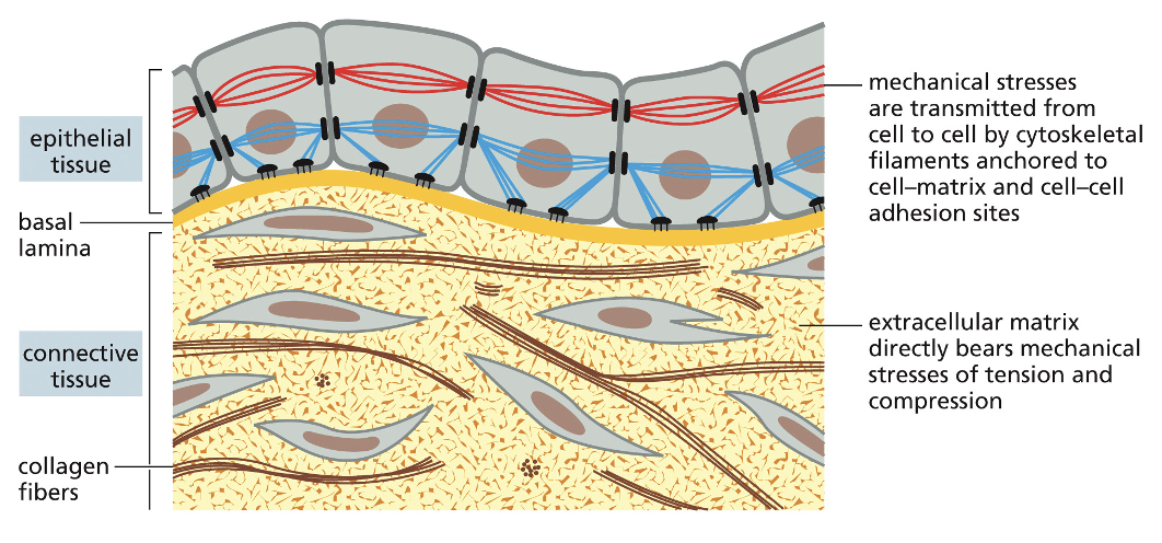
Epithelium: sheets of cells that are polarized, with discrete functional domains at opposite ends (apical and basal) of the cells
Connective Tissue: more loosely organized, in which cells are attached to each other, a rigid scaffold, or both
mechanical stresses are transmitted from cell to cell by cytoskeletal filaments anchored to cell-matrix and cell-cell adhesion sites
extracellular matrix directly bears mechanical stresses of tension and compression
Cell-Cell Junctions
what do multicellular organisms do and form?
what are cell-cell junctions?
Multicellular organisms have means of joining cells in long-term associations to form tissues and organs
the specialized structures where two cells come together are called cell-cell junctions
Animal VS Plant Cell-Cell Junctions
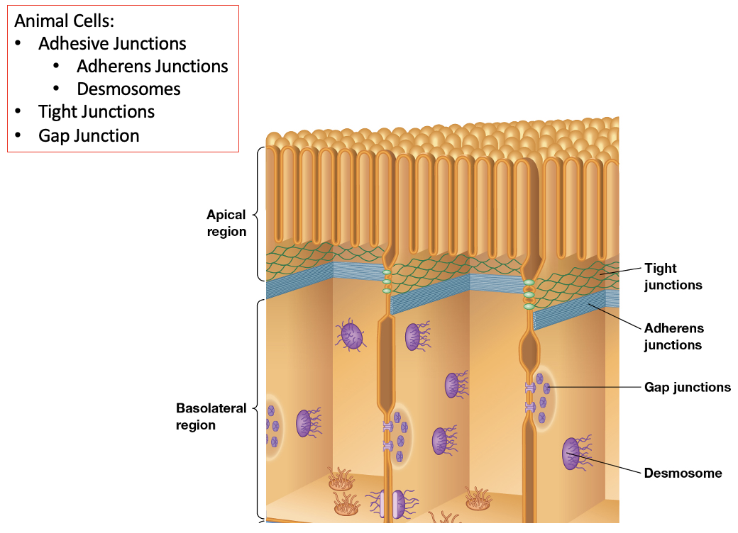
Animal Cells
1) Adhesive Junctions
adherens junctions
desmosomes
2) Tight Junctions
Gap Junctions
Plant Cells
Plasmodesmata
Animal Cell Cell-Cell Junction Image
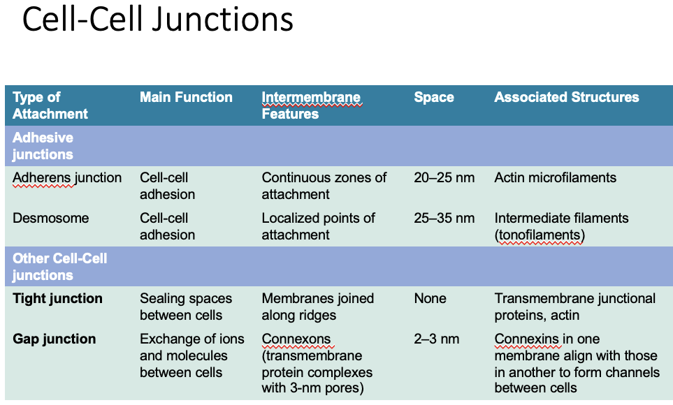
Cell-Cell Junction Chart
Adherens Junctions and Desmosomes
what do they rely on?
what are their traits?
adhesion proteins are focal points for what?
what is cell adhesion coordinated with?
Rely on:
intracellular attachment proteins to link the junction to the cytoskeleton
Cadherins on the outer surface to bind cells to each other
Traits:
hold similar cells together
basis for tissue formation
adhesion proteins are focal points for signaling complexes and cytoskeletal structures
many adhesion proteins are continuously recycled through endocytic and exocytic pathways
cell adhesion is coordinated with cell signaling, cell movement, cell proliferation, and cell survival
Adherens Junctions
what are they? what do they interact with?
what are cadherins characterized by? (3)
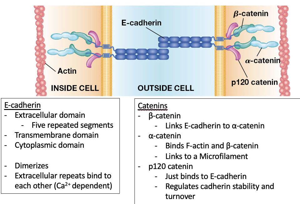
Adherens junctions: are cadherin-mediated junctions that interact with actin
Cadherins are characterized by:
“repeats” in their extracellular domain that are structurally similar
a transmembrane domain
widely varying cytosolic ends
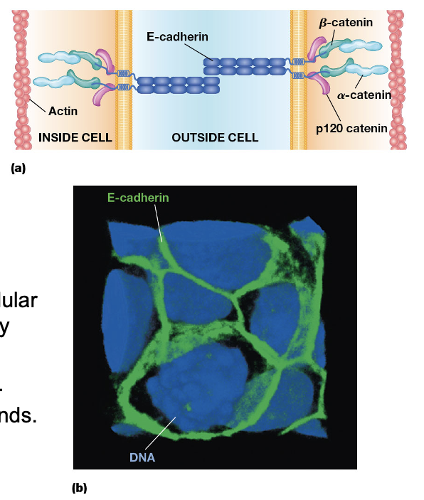
E-Cadherin
what are the 3 domains in E-Cadherin
characteristics
Extracellular domain: 5 repeated segments
Transmembrane domain
cytoplasmic domain
dimerizes
extracellular repeats bind to each other (Ca2+ dependent)
Catenins
name all 3 catenins, what do they do?
B-catenin:
Links E-cadherin to a-catenin
A-catenin:
Binds F-actin and B-catenin
Links to a microfilament
p120 Catenin:
just binds to E-cadherin
regulates cadherin stability and turnover
Tissue Specificity of Cadherins
where are E-cadherins found?
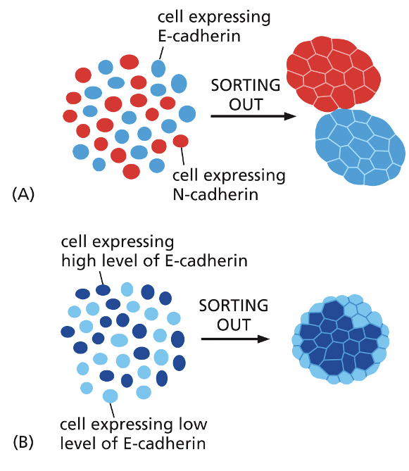
the amount and type of cadherins on cell surfaces help segregrate cells into specific tissues
Different types of cadherins are expressed in specific tissues
E-cadherins are found on epithelial cells, N-cadherins in neurons (anbd cardiac muscle)
The amount and type of cadherins on cell surfaces help segregate cells into specific tissues
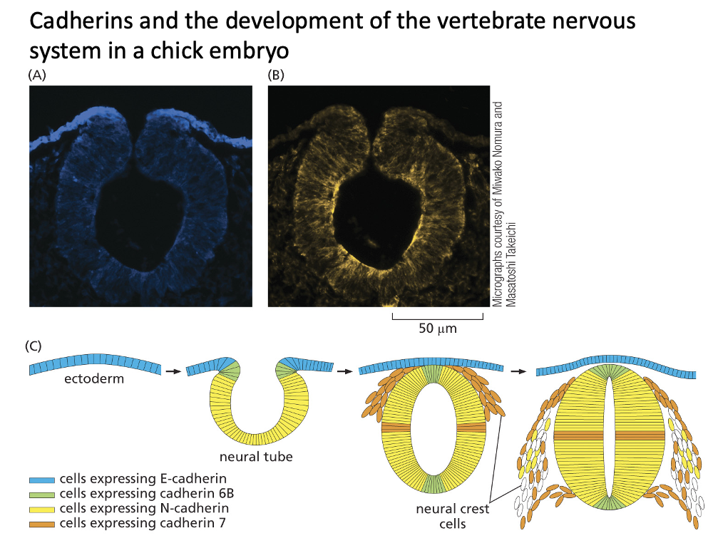
Cadherins and the development of the vertebrate nervous system in a chick embryo
Desmosomes
what are they?
what do they provide?
where are they abundant in?
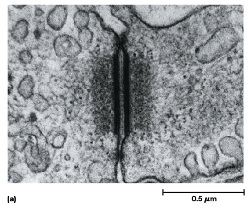
Desmosomes are button-like points of strong adhesion between adjacent cells in a tissue
they provide a tissue with structural integrity
they are especially abundant in cells that are under mechanical stress, like the skin, heart muscle, and the uterus
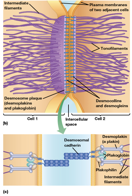
Desmosomes
name all the different types of desmosomes and their subtype
Transmembrane Proteins
Desmocollin (cadherin)
Desmoglein
Adaptor Protein
Plakoglobin (B-catenin)
Desmoplakin (a-catenin)
Plakophilin (p120 catenin)
Cytoskeleton
Intermediate Filaments
Desmosomes
Consider the Differences between adherens junction and desmosome
what are they?
what do they link?
Adherens Junction:
Continuous band of cell-cell connection
links the MF network in one cell to the MF network in a neighboring cell
Desmosome:
Localized disc of cell-cell connection
links the IF network in one cell to the IF network in a neighboring cell
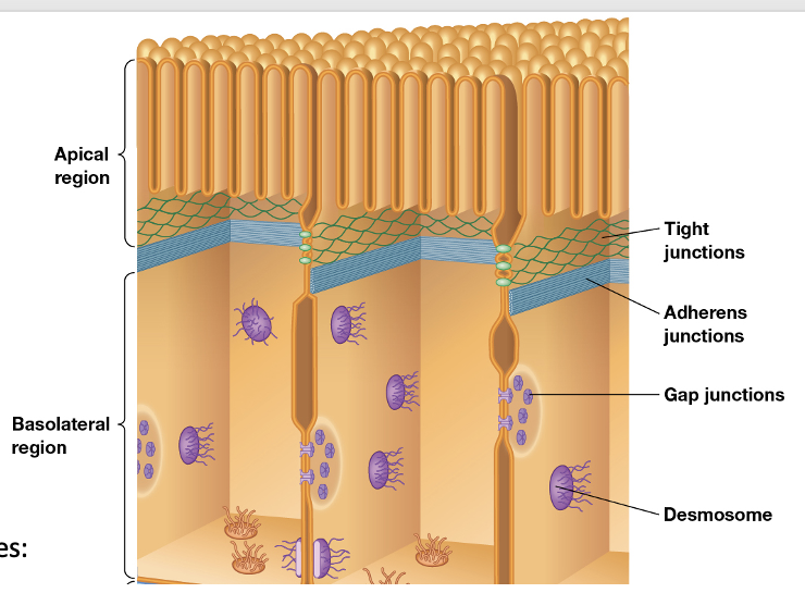
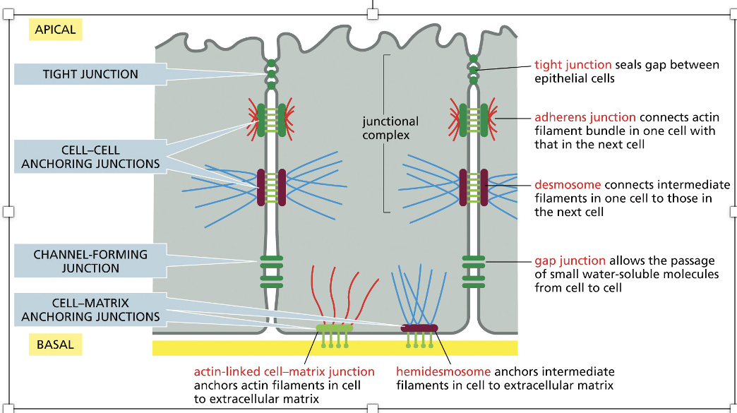
Tight Junctions, Adherens Junction, desmosome, gap junction, actin-linked cell-matrix junction, hemidesmosome
Tight junctions: seals gap between epithelial cells
Adherens Junction: connects actin filament bundle in one cell with that in the next cell
Desmosome: connects intermediate filaments in one cell to those in the next cell
Gap Junctions: allows the passage of small water-soluble molecules from ell to cell
actin-linked cell-matrix junction: anchors actin filaments in cell to extracellular matrix
Junction Images
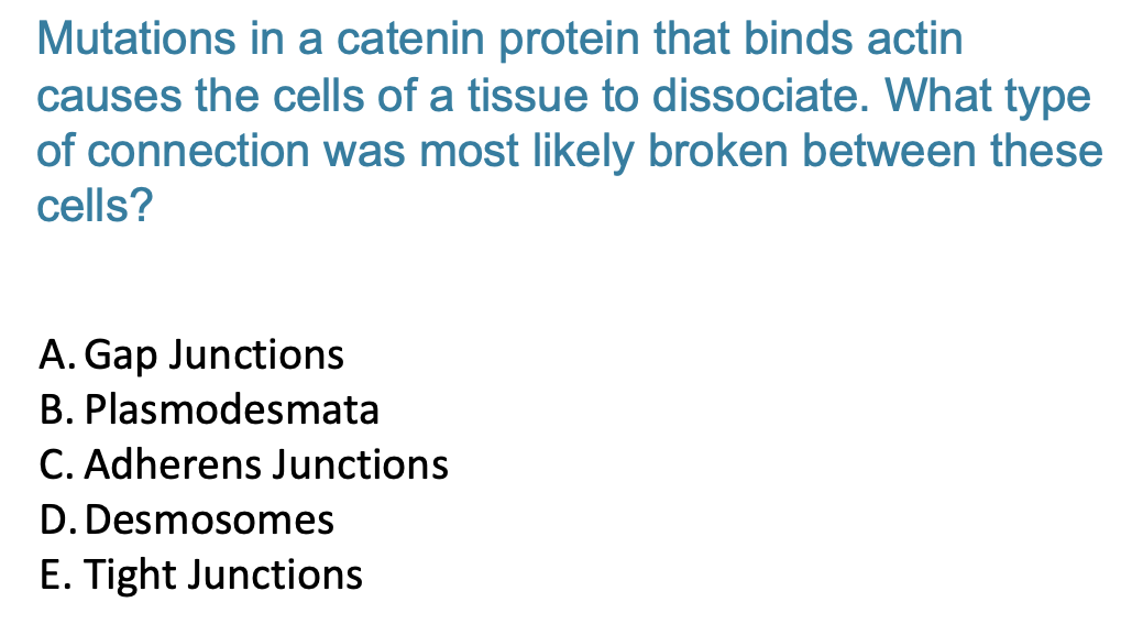
C. Adherens Junctions
Tight Junctions
what does it prevent?
what do epithelial cells need?
Prevent movement of molecules across cell layers
epithelial cells need specialized structures to seal them tightly together to form a barrier between the internal cells and the outside world
tight junctions serve this function, leaving no space between the plasma membranes of adjacent cells
they form a continuous belt around the apical ends of lateral surfaces of each cell; molecules cross the cell layer by passing through the cells
no connection to the cytoskeleton
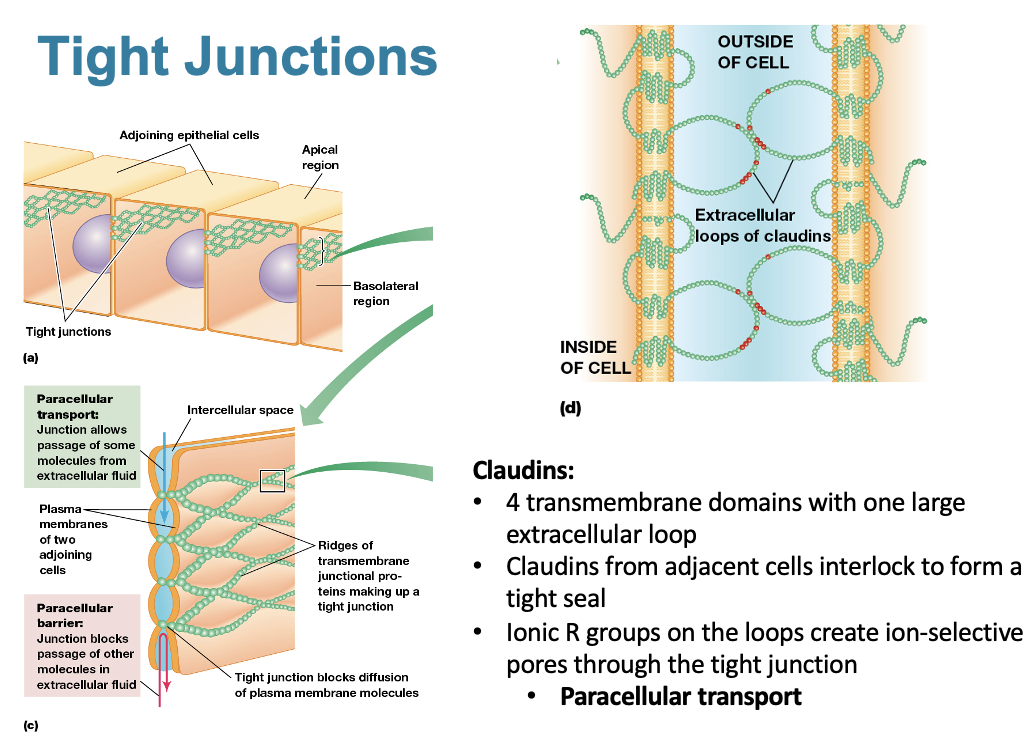
Tight Junctions
what are the functions of claudins
Claudins:
4 transmembrane domains with one large extracellular loop
claudins from adjacent cells interlock to form a tight seal
Ionic R group on the loops create ion-selective pores through the tight junction
Paracellular Transport
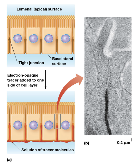
Tight Junctions create what?
what type of molecules can be seen by electron microscopy? where are they introduced to in the cell? what does it do?
Tight Junctions create a permeability barrier
Transmission electron microscopy
“Tracer” molecules that can be seen by electron microscopy are introduced in solution on one side of an epithelial cell layer
the tracer moves between the cells until it encounters the tight junctions and is stopped
Tight Junctions Permeability Barrier
Gap Junctions
what is it?
what is the gap lined by?
what passes through one cell to another?
Direct electrical and chemical communication between the cytoplasm of adjacent cells
A gap junction is a region where the plasma membranes of cells are aligned and brought into contact, with a very small gap between
The gap is spanned by small pipelines or passages between the cells
small molecules and ions can pass directly from one cell to another
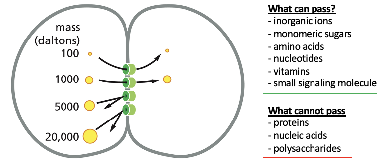
What can and cannot pass through Gap Junctions?
CAN
inorganic ions
monomeric sugars
amino acids
nucleotides
vitamins
small signaling molecules
CANNOT
proteins
nucleic acids
polysaccharides
Gap Junctions
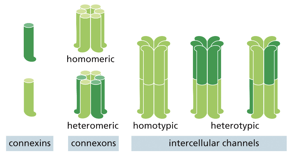
what transmembrane proteins are involved?
how many connexin subunits join to for a connexon?
what is the connexon?
transmembrane proteins involved: connexin
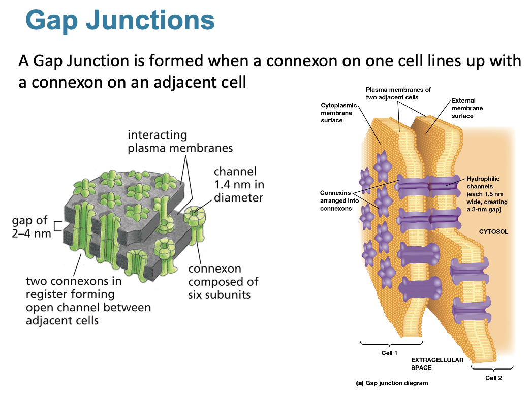
6 connexin subunits join to form a connexon
the connexon is a hollow protein cylinder with a 1.4 nm channel
Gap Junctions
when are gap junctions formed?
A Gap Junction is formed when a connexon on one cell lines up with a connexon on an adjacent cell
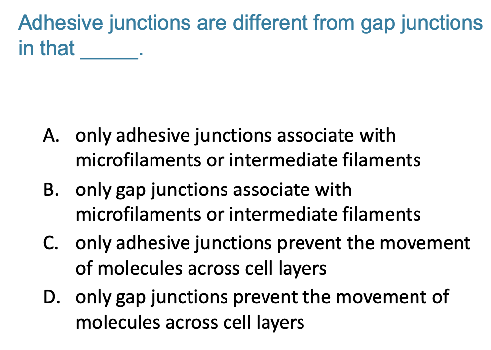
A. only adhesive junctions associate with microfilaments or intermediate filaments
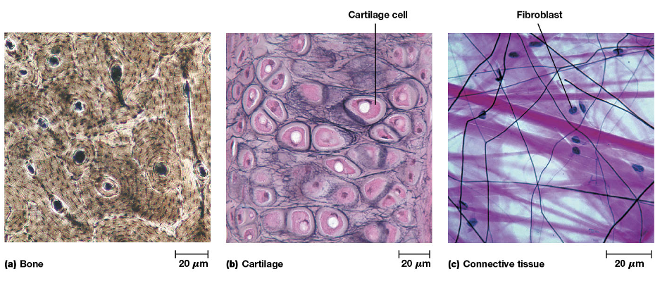
3 Types of ECM
Bone: Bone consists mainly of a rigid extracellular matrix that contains a small number of interspersed cells
Cartilage: is a tissue constructed mostly of matrix materials that is more flexible than bones
Connective Tissue: surrounding glands and blood vessels is relatively gelatinous and contains interspersed fibroblast cells
What is the ECM made of?
3 Classes of Molecules with different functions
A gel-like matrix in which the other molecules are embedded: Made of Proteoglycans (combinations of proteins and sugar polymers)
Structural proteins embedded in the matrix
Collagens: provide strength
Elastins
Adhesive glycoproteins that attach cells to the matrix. They must bind to molecules in the matrix (above) as well as transmembrane proteins on cells
Fibronectin: for most cell-ECM connections
Laminins: for epithelial cell connection to the Basal Lamina (other specialized cells with basal lamina- muscle, fat, Schwann cells)
Proteoglycans and the ECM
what are proteoglycans?
what are glycosaminoglycans?
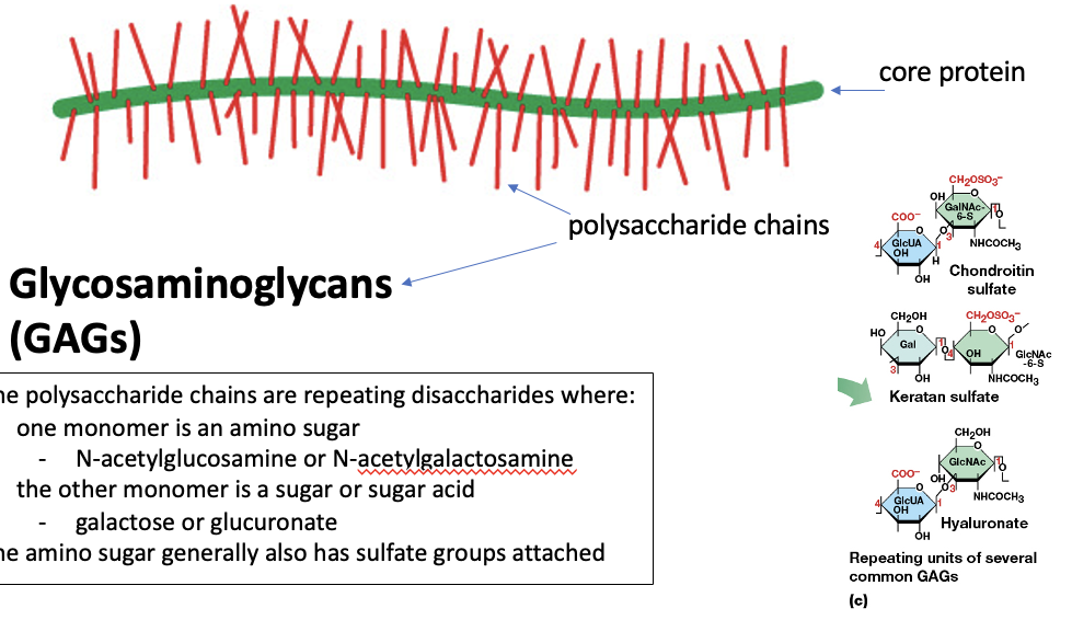
All Proteoglycans are combinations of core proteins and long polysaccharide chains
Glycosaminoglycans (GAGs)
The polysaccharide chains are repeating disaccharides where: 1 monomer is an amino sugar (N-acetylglucosamine or N-acetylgalactosamine
the other monomer is a sugar or sugar acid (galactose or glucuronate)
the amino sugar generally also has sulfate groups attached
GAGs: Glycosaminoglycans
GAG characteristics
GAG and Proteoglycan Diversity (tissues vs cartilage)
GAGs are hydrophilic and attract water and cations, forming a gelatinous matrix where collagen and elastin are embedded
GAG and Proteoglycan Diversity:
In many tissues proteoglycans are present as individual molecules
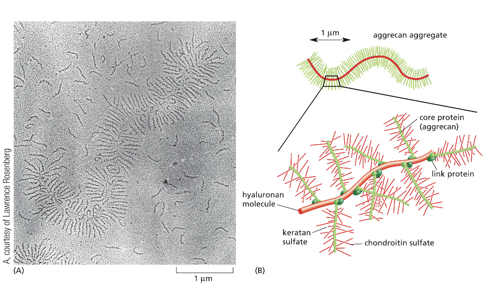
In cartilage, numerous proteoglycans become attached to long molecules of hyaluronate
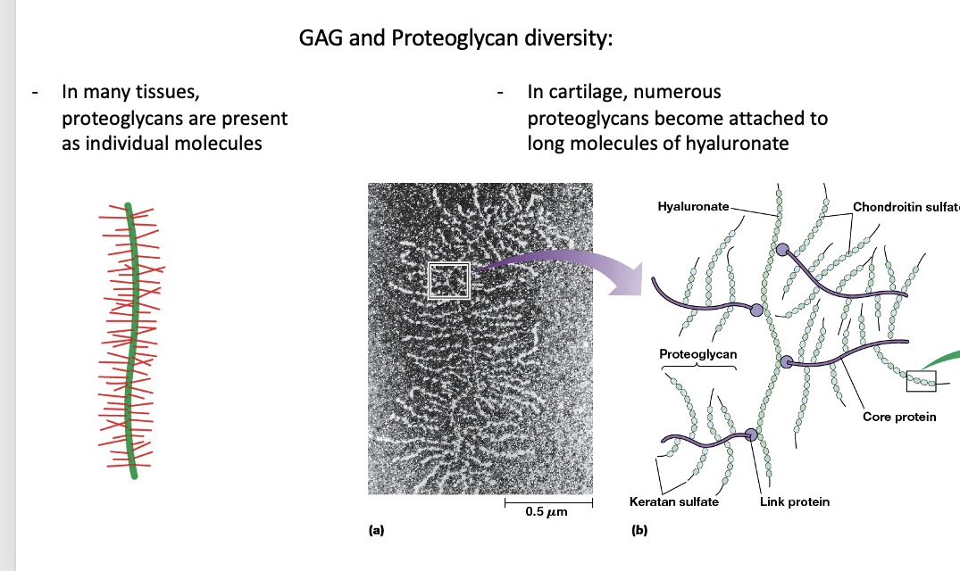
Individual Proteoglycans and a proteoglycan aggregate along what
Individual Proteoglycans and a proteoglycan aggregate along a single hyaluronate molecule
Free Hyaluronate
function, properties, where is it most abundant?
how do GAGs in the ECM exists, what is the exception?
Free hyaluronate LUBRICATES joints and FACILITATES cell migration
Most GAGs in the ECM exist only as components of proteoglycans
Hyaluronate is an exception that occurs both as a backbone of cartilage proteoglycans and as a free molecule
It has lubricating properties and is most abundant where friction needs to be reduced
Collagens are responsible for what?
what is the most abundant ECM component in animals?
what are collagen fibers visible in?
what is each fibril made out of?
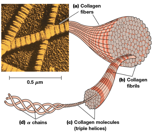
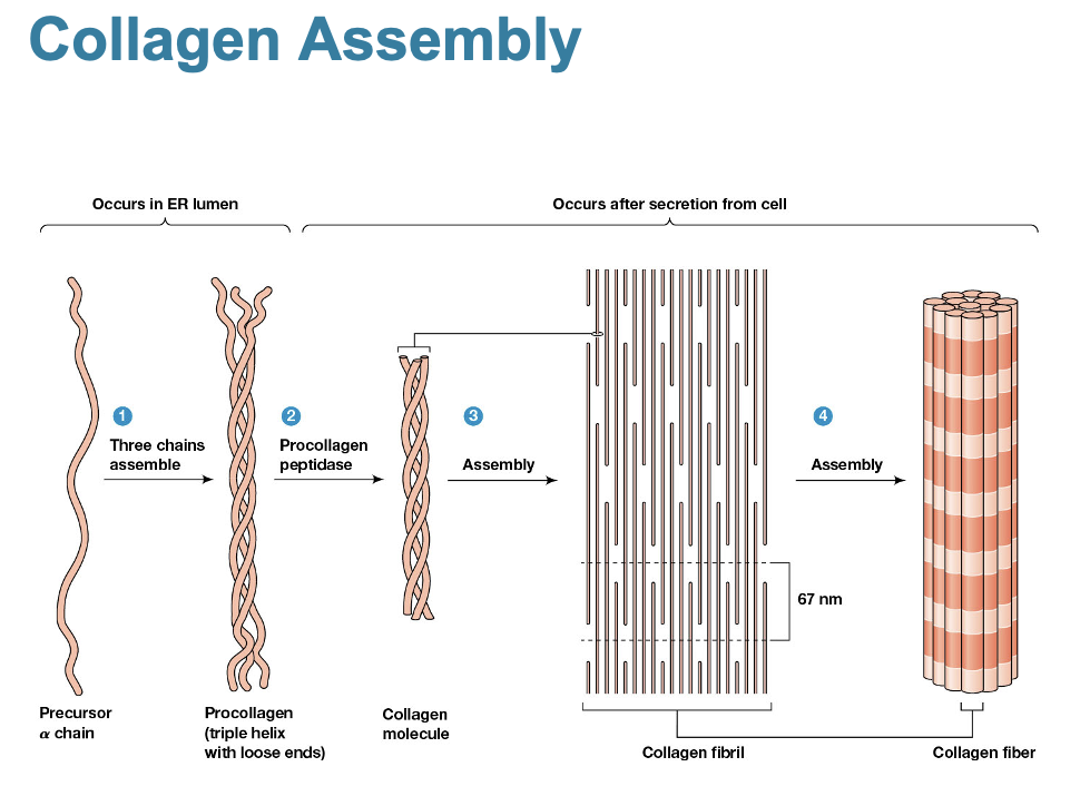
Collagens are responsible for the strength of the extracellular matrix
The most abundant ECM component in animals is a family of closely related collagens, which form fibers with high tensile strength
Collage fibers are visible in the ECM under scanning electron microscopy (EM)
The fibers are enormously strong and are composed of numerous fibrils
Each fibril is made of many collagen molecules, each composed of a 3 (a) chains twisted into a helix
Collagen Assembly Image
Elastins Impart Elasticity and Flexibility to the Extracellular Matrix
what provides elasticity to the ECM
what are they rich in?
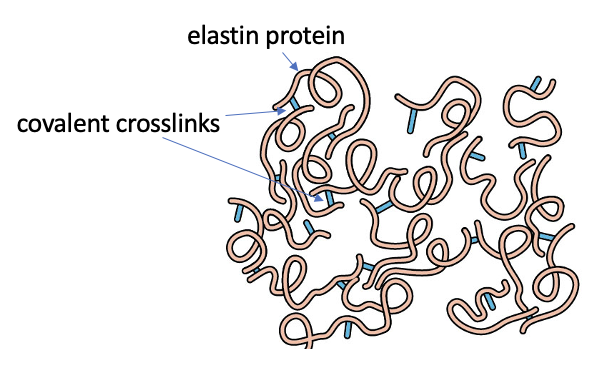
Elasticity is provided to the ECM by stretchable elastic fibers principally composed of elastins
these are rich in glycine and proline, and the molecules are crosslinked by bonds between lysine residues
Elastin Flexibility
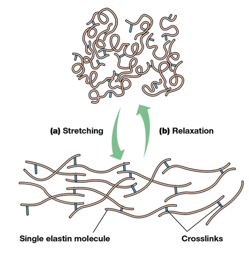
what does tension cause?
Tension causes the overall network of elastin to stretch, and release of tension causes individual molecules to relax
Adhesive Glycoproteins Anchor Cells to the Extracellular Matrix
what are the most common types of domains?
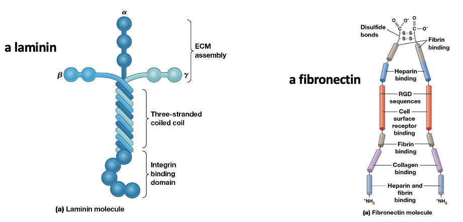
These glycoproteins have multiple domains to bind molecules in the ECM and receptors on membranes
Laminins and fibronectins are the most common types
Fibronectin
structure?
domains?
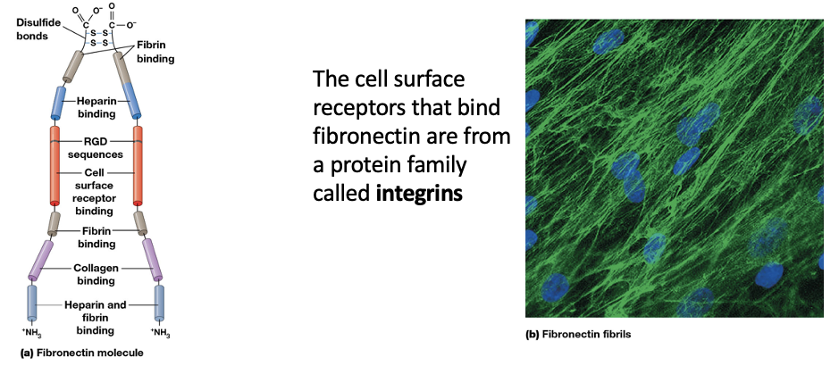
A fibronectin molecule has 2 large subunits linked near the C-terminals by two disulfide bonds
each fibronectin subunit is folded into a series of rodlike domains
several domains bind one or more ECM macromolecules, including several types of collagen
other domains recognize and bind cells surface receptors via the RGD (arg-gly-asp) sequence
Fibronectin
what are they?
what happens to the RNA transcribed from the fibronectin gene?
fibronectins are a family of closely related glycoproteins in the ECM
RNA transcribed from the fibronectin gene is processed to produce many different mRNAs and thus many different variants of the protein
Two main fibronectin variants
Insoluble Fibrils of fiibronectin in the ECM
Soluble fibronectin in blood and body fluids
Insoluble Fibrils of fibronectin in the ECM
what does fibronectin act as?
what requires fibronectin?
what over or under produces fibronectin? what does this effect?
fibronectin acts as a bridging molecule between cells and the ECM
cell migration requires fibronectin
many kinds of cancer cells over- or underproduce fibronectin compared to their tissue type
in cancer cells, this affects uncontrolled migration
Soluble fibronectin in blood and body fluids
what is the soluble form of fibronectin in blood?
what does it promote?
what can it attach to?
The soluble form of fibronectin in blood is called plasma fibronectin.
It promotes blood clotting because it has domains for binding fibrin, the blood-clotting protein.
It can attach blood platelets to fibrin as the clot forms
Laminins Bind Cells to the Basal Lamina
what is the basal lamina
function? and location?
what do basal laminae surround?
what are laminins?
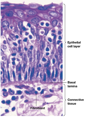
•The basal lamina is a thin sheet of specialized extracellular material.
•It underlies epithelial cells, separating them from connective tissues.
•Basal laminae also surround muscle cells, fat cells, and Schwann cells.
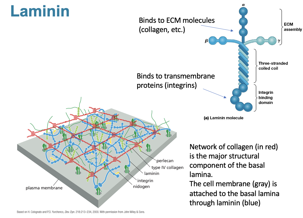
•The major adhesive glycoprotein in basal laminae are laminins.
Laminins Characteristics
what do all forms of basal lamina contain?
what do cells do here?
what do MMPs do? why is this important?
•All forms of basal lamina contain type Ⅳ collagen, proteoglycans, laminins, and another glycoprotein called nidogen.
•Cells can alter the properties of the basal lamina by secreting enzymes that catalyze changes in the lamina
•Matrix metalloproteinases (M M Ps) degrade the E C M locally, allowing cells to pass through.
•This is important for leukocytes to invade injured tissues and may be a factor in cancer cell invasiveness
Laminin Image
what is the major structural component of the basal lamina?
where is the cell membrane located at?
Network of collagen is the major structural component of the basal lamina
the cell membrane is attached to the basal lamina through laminin
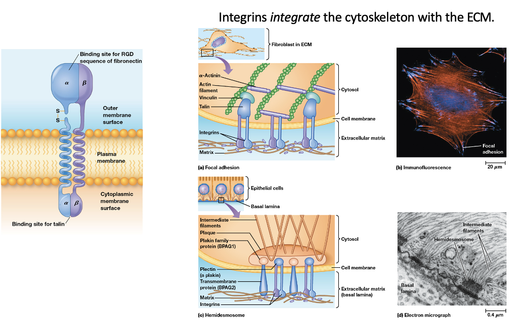
What are Integrins? Function?
Integrins are cell surface receptors that bind fibronectin and laminin
integrins integrate the cytoskelton with the ECM