CMNB Final Study Set
0.0(0)
0.0(0)
New
Card Sorting
1/100
Earn XP
Description and Tags
Study Analytics
Name | Mastery | Learn | Test | Matching | Spaced |
|---|
No study sessions yet.
101 Terms
1
New cards
Antagonist for nAChRs
α-bungarotoxin
2
New cards
Antagonist for muscarinic AChRs
atropine and scopolamine
3
New cards
NMDA competitive antagonists
APV
4
New cards
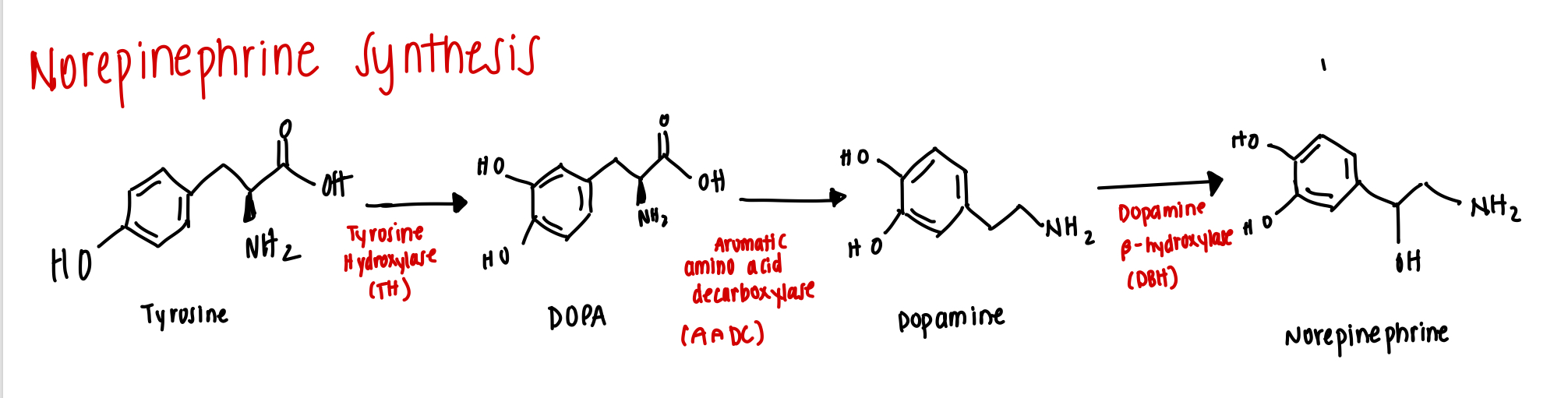
Enzyme in Norepinephrine Synthesis
Tyrosine --> DOPA
Tyrosine --> DOPA
tyrosine hydroxylase (TH)
5
New cards
Enzyme in Norepinephrine Synthesis
DOPA --> Dopamine
DOPA --> Dopamine
Aromatic amino acid decarboxylase
6
New cards
Enzyme in Norepinephrine Synthesis
Dopamine -> NE
Dopamine -> NE
dopamine β-hydroxylase
7
New cards
Major source of noradrenergic system
Locus Coeruleus
8
New cards
Locus Coeruleus Role
- Norepinephrine source
- Wakefulness and vigilance
- Wakefulness and vigilance
9
New cards
Cyclic AMP (cAMP) PK
Protein Kinase A
10
New cards
Cyclic GMP (cGMP) PK
Protein Kinase G
11
New cards
Phosphoinositide PK
Protein Kinase C
12
New cards
NE Terminating Enzymes
Phosphodiasterase and Protein phosphatase
13
New cards
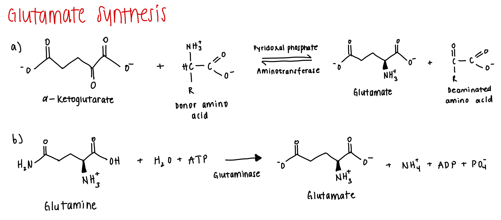
Enzyme to conver gln to glu
Glutaminase
14
New cards
Where is glutaminase ennriched?
Neurons
15
New cards
Enzymes for a-keto to glu
pydridoxal phosphate and aminotrasnferase
16
New cards
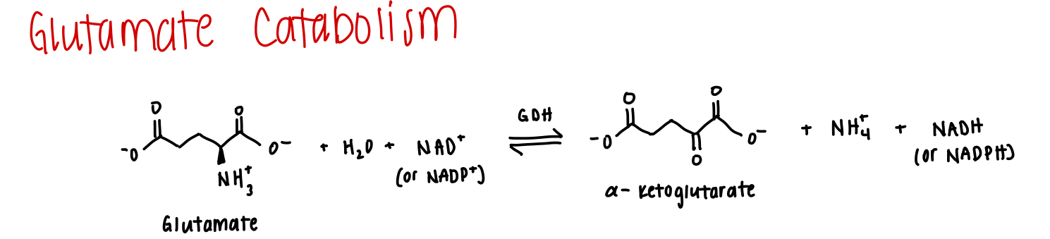
enzyme for glutamate catabolism
GDH (glutamate dehydrogenase)
17
New cards
Where is GDH enriched?
Astrocytes
18
New cards
Enzyme for GABA synthesis
glu -> gaba
glu -> gaba
GAD (glutamic acid decarboxylase)
19
New cards
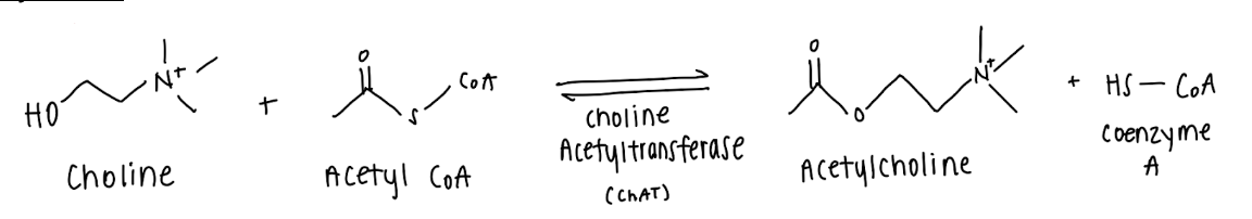
Enzyme for ACh synthesis
choline + acetyl coA -> ACh + coenzyme A
choline + acetyl coA -> ACh + coenzyme A
ChAT (choline acetyltransferase)
20
New cards
Draw ACh synthesis
\
(if you draw it correctly respond “right”)
\
(if you draw it correctly respond “right”)
Right
21
New cards
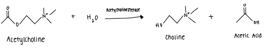
Draw ACh breakdown
\
(if you draw it correctly respond “right”)
\
(if you draw it correctly respond “right”)
Right
22
New cards
Where is ChAT found?
cytosol
Reminder: ChAT = choline acetyltransferase
Reminder: ChAT = choline acetyltransferase
23
New cards
Enzyme for ACh breakdown
ACh + h2o --> choline + acetic acid
ACh + h2o --> choline + acetic acid
acetylcholinesterase
24
New cards
where is acetylcholinesterase found
post and presynaptic membrane
25
New cards
NMDA Comp Antagonists
D-APV
26
New cards
NMDA Noncomp Antagonists
PCP, ketamine, CPP, MK-801
\
\*Important: Ketamine - it is noncompetitive meaning it does not compete with glutamate for NMDA receptor binding; however, the binding site for ketamine appears only after the conformational change caused by glutamate. When it binds, it blocks Ca2+ influx
\
\*Important: Ketamine - it is noncompetitive meaning it does not compete with glutamate for NMDA receptor binding; however, the binding site for ketamine appears only after the conformational change caused by glutamate. When it binds, it blocks Ca2+ influx
27
New cards
AMPA Competitive Anatgonist
NBQX
* Used by Zanos-Zarate to establish the necessity of AMPARs for Ketamine’s function
* Used by Zanos-Zarate to establish the necessity of AMPARs for Ketamine’s function
28
New cards
GABAA Agonist
Muscimol
29
New cards
GABAB Agonist
baclofen
30
New cards
GABAA Competitive Antagonists
Bicuculline and Benzodiazepines
31
New cards
GABAB Competitive Antagonists
Phaclofen
32
New cards
GABAA Noncomp Antagonists
Picrotoxin
33
New cards
Enzyme to go from glutamate to glutamine
glutamine synthetase
34
New cards
Loewi’s 1921 Experiment
Otto Loewi placed a beating frog’s heart into a saline bath chamber with its vagus nerve still attached. The saline in the first chamber was allowed to flow into a second saline bath chamber containing a second beating heart without the vagus nerve attached. Loewi found that stimulating the vagus nerve of the first heart caused the heart rate of the first heart to lower, but more shockingly it also caused the heart rate of the second heart to decrease. The hearts exhibited a physiological response to some diffusible chemical which was later found out to be acetylcholine.
35
New cards
Brain region with many cholinergic synapses
Basal forebrain area
36
New cards
What is Optogenetics
Method in which you introduce a foreign gene by using a virus as a vector. The virus is injected into a brain region
37
New cards
How was the role of acetylcholine examined using optogenetics?
Optogenetics was used to inject ACh into the prefrontal cortex of rats and use light to either stimulate or inhibit neurons to control ACh release. It was found that ACh played a role in improved detection of sensory stimuli.
Stimulating the neurons improved the detection of a very short tone by rats and when the tone was made easier to hear, stimulation did not further improve detection.
Inhibiting the neurons in the hard condition had no effect but drastically decreased detection in the easy condition.
Stimulating the neurons improved the detection of a very short tone by rats and when the tone was made easier to hear, stimulation did not further improve detection.
Inhibiting the neurons in the hard condition had no effect but drastically decreased detection in the easy condition.
38
New cards
Sillito and Kemp
Showed that ACh could enhance the receptive field property selectivity of a neuron, causing the neuron to respond more to a stimulus in its preferred orientation and direction; however, they also found that the effects can be inhibitory in some cells
39
New cards
What causes the loss of direction selectivity, in contrast to acetylcholine
Bicuculline, which we know to be a competitive antagonist of GABAA receptors
40
New cards
How was the role of acetylcholine examined through lesioning experiments?
Morris Water Maze Experiment
Found that lesioning the nucleus basalis, a brain region with many cholinergic pathways, resulted in the impairment of rats' ability to recall where a hidden platform was in opaque water.
Their ability to remember was even more impaired when researchers killed cholinergic neurons by the infusion of cholinergic neurotoxin into the cerebral ventricles.
41
New cards
What evidences supports that serotonin is a *bona fide* neurotransmitter?
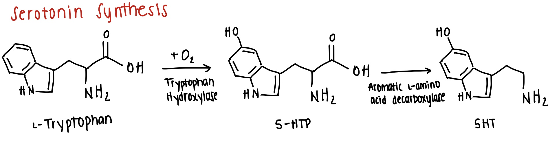
1. There’s enzymes (tryptophan hydroxylase) that synthesize it in axon terminals
2. There’s enzymes that break it down (monoamine oxidase -- MAO)
3. There’s vesicular transporters to allow 5HT to be packaged into vesicles
4. Presynaptic transporters on the plasma membrane to allow for uptake, indicating that the intracellular and extracellular concentrations of this molecule are important
5. Multiple membranous proteins (receptors) it binds to and evokes physiological responses
42
New cards
Where is serotonin synthesized?
In the raphe nuclei, located in the brainstem
43
New cards
How is serotonin synthesized? Draw it.
\
(if you draw it correctly respond “right”)
\
(if you draw it correctly respond “right”)
right
44
New cards
What G protein(s) coupled to serotonin receptors lead to adenylyl cyclase ***inhibition***? why is it important?
Gi and Go. It is important because adenylyl cyclase is needed for cAMP which regulates excitatory/inhibitory processes.
\
\*Gi and Go are also activated by D2 receptors
\
\*Gi and Go are also activated by D2 receptors
45
New cards
How is cAMP produced and how could it lead to cellular excitation?
cAMP could be produced by activating adenylyl cyclase through norepinephrine binding to β-adrenergic receptors or by serotonin binding to receptors coupled to Gs proteins. This could lead to excitation as increased cAMP concentrations could activate cAMP-dependent protein kinase which converts ATP to ADP, phosphorylating threonine and serine amino acid residues of proteins to create available VG Ca2+ channels.
46
New cards
What G protein(s) coupled to serotonin receptors lead to adenylyl cyclase ***activation***? why is it important?
Gs. It is important because adenylyl cyclase is needed for cAMP which regulates excitatory/inhibitory processes.
\
\*Gs also activated by D1 receptors
\
\*Gs also activated by D1 receptors
47
New cards
What G protein(s) coupled to serotonin receptors lead to phospholipase C (PLC) ***activation***? why is it important?
Gq and G11
48
New cards
Are all 5HT receptors G couple?
No. 5-HT3 receptors are excitatory ligand-gated ion channels
49
New cards
How is serotonin uptaken by presynaptic terminals?
Through 5-HTT, the serotonin transporter
50
New cards
What behavioral states is the ***locus coeruleus*** associated with? Is it activated or inhibited during this behavior
1. Awake, alerted by stimuli - activated
2. Grooming - inhibited
\
\*locus coeruleus is main source o norepinephrine
51
New cards
What behavioral states is the ***Dorsal Raphe*** associated with? Is it activated or inhibited during this behavior
1. Awake. alerted by stimuli - activated
2. Grooming - activated
3. Slow Wave Sleep - inhibited
4. REM Sleep - completely shut down
\
\*Raphe nuclei - serotonergic
52
New cards
What behavioral states is the ***Dorsal Tegmental Nucleus*** associated with? Is it activated or inhibited during this behavior
1. Awake, alerted by stimuli - activated
2. REM sleep - activated
\
\*These neurons are important for the regulation of wakefulness. The DTN is serotonergic
53
New cards
What behavioral states is the ***Substantia Nigra and Ventral Tegmental Area*** associated with? Is it activated or inhibited during this behavior
1. Awake, alerted by stimuli - activated
\
\*Both of these regions are critical for their role in the production of dopamine
54
New cards
What functions does serotonin play?
1. Sleep-wake regulation
2. Feeding
1. increased serotonin release suppresses appetite
3. Migraine relief
4. Vomiting (blocking 5H3 treats nausea)
5. Visual hallucination
6. Depression (blocking reuptake of 5HT treats depression)
7. Sense of well-being
55
New cards
What behavioral method is used to study depression in rodents and how is it induced?
Learned helplessness is induced in rats by placing them into operant chambers and delivering shocks that they are not able to stop (a second group is given a lever to stop shocks and they do not obtain learned helplessness).
\
The animals were then underwent a forced swim test and fond than those who ‘obtained’ learned helplessness gave up and became immobile quicker. Anti-depressants were given to the animals to examine any reduction in their mobility.
\
Another way to test immobility is through the tail-suspension test which is suitable for mice (not rats).
\
The animals were then underwent a forced swim test and fond than those who ‘obtained’ learned helplessness gave up and became immobile quicker. Anti-depressants were given to the animals to examine any reduction in their mobility.
\
Another way to test immobility is through the tail-suspension test which is suitable for mice (not rats).
56
New cards
Describe the acute effects of antidepressants
They block catecholamine and NT uptake and AutoRs inhibit release
57
New cards
Describe the chronic effects of antidepressants
After chronic presence of an antidepressant, post-synaptic receptor activation strengthens due to the down-regulation of AutoRs which would typically inhibit release
58
New cards
How would MAO inhibitors work as anti-depressants?
Monoamine oxidase functions to catabolize serotonin and remove other NT from terminals, like norepinephrine and dopamine. Inhibiting this from occurring would result in an increase in NT release and an increased postsynaptic response, despite a reduction in β-Adrenergic and 5-HT2 receptors
59
New cards
Outline the up-regulation of second messengers in response to chronic anti-depressant treatments
An increase in NE and 5-HT caused by treatment produces a down-regulation of their receptors, up-regulating the cAMP pathway - first adenylyl cyclase is activated which produces more cAMP, activating cAMP-dependent protein kinase (PKA), increasing CREB activity, producing more proteins like BDNF.
60
New cards
What is BDNF and why is it important? How is it linked to depression?
BDNF (also known as brain-derived neurotrophic factor) are endogenous proteins essential for the development and survival of neurons and synapses.
\
Low BDNF expression has been linked to stress and depression-like symptoms. This occurs as a result of chronic defeat stress inducing histone methylation, creating a more closed chromatin state that results in less BDNF gene transcription. Chronic antidepressant treatment increasing the expression in BDNF, not by reversing methylation but by increasing histone acetylation which induces a more open chromatin state.
61
New cards
What are glucocorticoids and what’s their relationship to stress and depression?
Glucocorticoids are hormones released in response to stress and have been found to been found be in abnormally elevated levels in depressed individuals.
\
High glucocorticoid levels have also been linked to decreased BDNF transcription.
\
High glucocorticoid levels have also been linked to decreased BDNF transcription.
62
New cards
MDMA
A neurotoxin that induces euphoria by enhancing the release of 5-HT and DA. It also inhibits the reuptake of 5-HT.
63
New cards
How does ketamine treatment work?
Ketamine de-phosphorylates eukaryotic elongation factor 2 (eEF2), allowing for the de-suppression of protein synthesis caused by excess ambient glutamate causing chronic GluR activation. This de-suppression promotes the transcription of BDNF, which in turn increases AMPARs
64
New cards
What were Miller et al’s procedures and findings?
\*Note: Miller et al. did not focus on metabolite but how ketamine and depression function. He thinks ketamine acts on pyramidal neurons.
\
Finding 1: Ketamine blockade of NR2B-containing NMDARs in pyramidal neurons is critical for anti-depressive action
* Knocking out NR2B had the same effect on immobility as ketamine. Injecting ketamine into a NR2B-KO did not further decrease immobility time
* NR2b-KO occludes Ketamine’s effect
\
Finding 2: NR2B-NMDARs are activating by ambient glutamate and ketamine blocks the current activated by ambient glutamate
* Mg2+ blocks removed, NR2A blocked by Zn2+ and AP blocked with TTX to look at the current flow through NR2B-NMDARs solely caused by ambient glutamate. Found that the ambient glutamate caused current through NR2B Rs that could be blocked by ketamine, but also found that there was no current flow through NR2B knockouts (ketamine wasn’t needed).
\
Finding 3: GLT-1 (glutamate transporter) works as an antidepressant like ketamine, reducing NMDAR activation by ambient glutamate
* Removing ambient glutamate by up-regulating glutamate transporter expression with ceftriaxone has antidepressant effects, reducing immobility in a tail suspension test
\
Finding 2: Ketamine promotes synaptogenesis, especially in the prefrontal cortex
\
Finding 1: Ketamine blockade of NR2B-containing NMDARs in pyramidal neurons is critical for anti-depressive action
* Knocking out NR2B had the same effect on immobility as ketamine. Injecting ketamine into a NR2B-KO did not further decrease immobility time
* NR2b-KO occludes Ketamine’s effect
\
Finding 2: NR2B-NMDARs are activating by ambient glutamate and ketamine blocks the current activated by ambient glutamate
* Mg2+ blocks removed, NR2A blocked by Zn2+ and AP blocked with TTX to look at the current flow through NR2B-NMDARs solely caused by ambient glutamate. Found that the ambient glutamate caused current through NR2B Rs that could be blocked by ketamine, but also found that there was no current flow through NR2B knockouts (ketamine wasn’t needed).
\
Finding 3: GLT-1 (glutamate transporter) works as an antidepressant like ketamine, reducing NMDAR activation by ambient glutamate
* Removing ambient glutamate by up-regulating glutamate transporter expression with ceftriaxone has antidepressant effects, reducing immobility in a tail suspension test
\
Finding 2: Ketamine promotes synaptogenesis, especially in the prefrontal cortex
65
New cards
What were Monteggia et al’s procedures and findings?
Finding 1: Ketamine has a unique property in comparison to other non-competitive NMDAR antagonists: it’s long-lasting effect.
* They measured the reduction of immobility in rats undergoing a forced-swim test after being treated with three different NMDA antagonists: ketamine, CPP, and MK-801. They compared the effects various times after injection: 30 mins, 3 hrs, 24 hrs, and a week.
\
Finding 2: BDNF KO occlude’s ketamine’s effect, meaning BDNF is essential for Ketamine’s action
* When *BDNF* was knocked out, there was not a strong reduced immobility response to ketamine 30 ins or 24 hours after injection (but there was in the control group).
\
Finding 3: Ketamine works as an antidepressant by prompting eEF2 de-phosphorylation which promotes BDMF synthesis
* injected cells with **rottlerin** which prompts eEF2 de-phosphorylation and noted increased BDNF expression, decreased phosphorylation and decreased immobility
* Everyone agrees with this
\
__Against Zanos-Zarate et al.)__
\*Finding 4: Ketamine’s primary action is via Ketamine at NMDARs
* Miller would agree
\
\*Finding 5: Like ketamine, HNK works as an antagonist of NMDARs
* He recorded spontaneous excitations (current flow) through NMDARs and compared the effect NMDAR antagonists have on them -- including ketamine. He found that at high enough concentrations, HNK greatly reduced NMDA current flow just like the other NMDAR antagonists.
\
\
Finding ?: AMPAR changes in the hippocampus, not the prefrontal cortex (supported by Zarate)
66
New cards
What were Zarate et al’s procedures and findings?
Finding 1: Like Monteggia, noted Ketamine’s long lasting effect but compared to imipramine.
* Also conducted a forced swim test and measured their immobility at two points after injection: 30 mins and 2 weeks
\
Finding ?: AMPAR changes in the hippocampus, not the prefrontal cortex
\
__Zanos-Zarate et al.__
\*Finding 1: The antidepressant effects of ketamine are actually the result of the ketamine metabolite HNK acting on AMPARs (hydroxynorketamine)
* According to Zanos-Zarate, instead of functioning as an antidepressant by ketamine binding to NMDARs and preventing excitation, HNK binds to AMPARs, increases the level of AMPARs on the membrane, which in hand may increase BDNF expression.
* They measured the current flowing through AMPARs in 4 different situations (in hippocampal synapse):
1. DNQX present (no current)
2. Baseline
3. Saline (same as baseline)
4. HNK (enhanced current)
\
Finding 2: Ketamine and HNK require AMPAR activity to work as an antidepressant
* Used NBQX AMPAR antagonist and measured immobility. There was no reduced immobility in response to ketamine or HNK when NBQX was also administered
\
Finding 3: HNK has very different binding site from MK-801 and ketamine
* Used a receptor binding assay and found that at high enough doses ketamine displaced the binding of MK-801 but HNK never displaces it.
67
New cards
What happens to synapses when a CS and US are repeatedly paired?
Well, neurons receiving inputs from US start with strong synaptic connections to their postsynaptic partner while neurons receiving inputs from CS start with weak synaptic connections to the US postsynaptic cells. Repeatedly pairing them causes the strengthening of the synaptic connection between the CS and postsynaptic cells. This strengthening is LTP.
68
New cards
What is a tetanus and how does it relate to synaptogenesis?
Tetanus refers to the rapid delivery of stimulating pulses to a neuron. A tetanus can convert a weak synapse to a strong synapse, leading to LTP
69
New cards
Where is LTP mostly studied?
Pyramidal neurons in the CA1 region of the hippocampus which receives excitatory glutamatergic inputs from CA3 neurons.
70
New cards
What retrograde messengers play a role in LTP induction? Describe the pathway in which these messengers are synthesized.
Nictric oxide and arachidonic acid.
\
LTP induction requires the influx a Ca2+ through NMDARs which are activated by the release of presynaptic glutamate. Ca2+ further increases intracellular Ca2+ released from stores in response to Ca2+ activating mGluRs that activate phospholipase C, liberating IP3 which released Ca2+ stored in the endoplasmic reticulum. This greatly enhanced Ca2+ presence is linked to the activation of protein kinases and other biochemical process that produce nictric oxide and arachidonic acid which permeate the cell membrane and travel from the post- to presynaptic axon, stimulating further glutamate release. NO enhances vesicular release by binding to and activating guanylyl cyclase, raising cGMP in presynaptic terminals which enhances Ca2+ release from stores, further enhancing NT vesicular release. This release increases postsynaptic excitation and LTP.
71
New cards
What experiment do you suppose scientists did to come to the conclusion that NMDARs are needed for LTP induction but NOT needed for LTP expression?
Scientists could have conducted an experiment in which an NMDA receptor antagonist was inserted into the cell before and after the tetanus stimulation. If NMDARs are needed for LTP induction, then an antagonist would prevent LTP induction from the tetanic stimulus. If NMDARs are need for LTP expression, then inserting the antagonists after LTP induction would block an enhanced EPSP from a single test pulse
72
New cards
Difference between early and late LTP
E-LTP lasts a few hours and L-LTP lasts a few days
73
New cards
Outline the underlying mechanism of LTP
Ca2+ current flows in through NMDARs, activating protein kinases including CaMKII (calcium/calmodulin kinase II) which increase the insertion of additional AMPARs to the dendritic spine membrane, enhancing glutamate sensitive and creating a stronger synapse → LTP. This process is a modulation of receptor trafficking.
74
New cards
LTP vs LTD
LTP involves an increased rate of AMPAR insertion through exocytosis, enhancing the cells sensitivity to glutamate. LTD, on the other hand, involves the withdrawal of AMPARs from the membrane through enhanced endocytosis, reducing the cells sensitivity to presynaptic outputs.
75
New cards
What type of AMPARs are added during LTP?
AMPARs containing the GluR1 subunit. They are eventually substituted with GluR2/3 subunits which form the new steady state of receptors on the membrane.
76
New cards
LTD follows from activity of……
Group 1 mGluRs (mGluR1 and mGluR5). These receptors induce LTD in the postsynaptic cell and LTP in the presynaptic neuron, increasing Ca2+ uptake and, therefore, promoting NT release
77
New cards
Mechanism of Action of Group I mGluRs
Glu activated mGluR which is coupled to Gq type of g-protein, which we know activates phospholipase C which liberates two second messengers by hydrolyzing PIP2: IP3 and DAG. DAG activates protein kinase C and IP3 releases stores Ca2+
78
New cards
Fragile X Syndrome
Fragile X syndrome is an important example of abnormalities in Group 1 mGluR expression. Fragile X Syndrome is a single gene mutation in FMR1 which inhibits the translation of FMRP. FMRP typically binds to group 1 mGluRs to inhibit local protein translation that inhibit LTD. The mutation prevents this inhibition of mRNA translation, resulting in the over-expression of proteins that increase LTD, resulting in spine abnormalities and autism-like behaviors.
79
New cards
What structures provide evidence of synaptic plasticity capabilites in adult human brains?
Filopodia -- they are immature, actin-filled dendritic protrusions that account for the large portion of silent synapses in our brains which only contain NMDARs
80
New cards
What evidence shows that filopodia has only NMDARs and can become a functional synapse?
GFP used to visualize filopodia and immunocytochemistry used to visualize antibodies for synaptic proteins. ICC revealed that filopodia have NMDARs (GluN1) but not AMPARs (GluR1), and that they have bassoon which is an essential presynaptic protein that orchestrates vesicular release, indicating the filopodia are capable of NT release if receptors were activated.
81
New cards
Malinow et. al (goal, methods, and findings)
Goal: Study the role of GluR1-receptor trafficking at thalamo-amygdala synapses in associative fear conditioning
\
Methods:
1. Exogenous GluR1 introduced into amygdala with GluR1-GFP gene that were tagged with markers: GFP and increased AMPAR rectification (so, it can be seen visually with electrophysiology)
2. GluR1-C-tail-GFP acts as dominant-negative, preventing synaptic incorporation of endogenous GluR1 receptors
3. GFP as a control, should do nothing to cells
4. Mice are trained to associate tone (CS) with shock (US)
\
Behavioral Control and Measurement:
* Control: unpaid auditory fear conditioning
* Measured: % freezing
\
Findings:
1. There is little basal level trafficking of GluR1 into synapses when animals have not undergone fear conditioning.
1. This was noted because cells transfected by the virus had the same rectification as non-infected cells which indicate that the GFP-GluR1 receptors had not been incorporated into synapses
2. GluR1 is incorporated into the synapse in response to threat conditioning
1. When associated, tone alone began evoking a freezing response. On a molecular level, this occurred when GluR1 was incorporated into the synapse, indicated by a high rectification index
3. Unpaired conditions produced less membrane incorporation of exogenous GluR1
4. LTP requires GluR1 insertion into synapses
1. Using a GluR1-c-tail-GFP tag, which overfloods neuron with C-terminals, inhibits the incorporation of endogenous GluR1 to the membrane. This was shown to inhibit LTP from tetanus but short-term plasticity/potentiation still occurred.
5. GluR1-trafficking into synapses is necessary for retention of associative learning but not for conditioning.
1. Conditioning still occurred when amygdala was transfected with GluR1-C-tail-GFP but testing freezing response 3 hrs and 24hrs later showed there was no retention of the association learned.
\
Methods:
1. Exogenous GluR1 introduced into amygdala with GluR1-GFP gene that were tagged with markers: GFP and increased AMPAR rectification (so, it can be seen visually with electrophysiology)
2. GluR1-C-tail-GFP acts as dominant-negative, preventing synaptic incorporation of endogenous GluR1 receptors
3. GFP as a control, should do nothing to cells
4. Mice are trained to associate tone (CS) with shock (US)
\
Behavioral Control and Measurement:
* Control: unpaid auditory fear conditioning
* Measured: % freezing
\
Findings:
1. There is little basal level trafficking of GluR1 into synapses when animals have not undergone fear conditioning.
1. This was noted because cells transfected by the virus had the same rectification as non-infected cells which indicate that the GFP-GluR1 receptors had not been incorporated into synapses
2. GluR1 is incorporated into the synapse in response to threat conditioning
1. When associated, tone alone began evoking a freezing response. On a molecular level, this occurred when GluR1 was incorporated into the synapse, indicated by a high rectification index
3. Unpaired conditions produced less membrane incorporation of exogenous GluR1
4. LTP requires GluR1 insertion into synapses
1. Using a GluR1-c-tail-GFP tag, which overfloods neuron with C-terminals, inhibits the incorporation of endogenous GluR1 to the membrane. This was shown to inhibit LTP from tetanus but short-term plasticity/potentiation still occurred.
5. GluR1-trafficking into synapses is necessary for retention of associative learning but not for conditioning.
1. Conditioning still occurred when amygdala was transfected with GluR1-C-tail-GFP but testing freezing response 3 hrs and 24hrs later showed there was no retention of the association learned.
82
New cards
Context vs Cued Threat Conditioning
1. In cued threat conditioning a subject is made to associate a cue with an unconditioned stimulus and response. However, in context threat conditioning subjects are left to learn about the context/environment in which they are in before they are exposed to an unconditioned stimulus. If removed and placed again into the environment without the unconditioned stimulus, it is expected that the subject will display the fear response they’d have to the unconditioned stimulus.
2. Contextual fear conditioning does not happen in the amygdala like cued threat conditioning, but instead is found to occur through LTP in the hippocampus
83
New cards
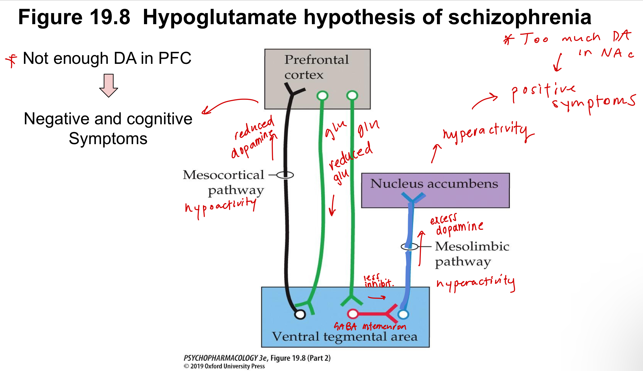
What are the three dopaminergic pathways? What anomalies in these pathways are linked to diseases?
1. Mesostriatal pathway
1. Midbrain/SN → Striatum
2. Loss of this pathway → Parkinson’s
2. Mesolimbic pathway
1. Midbrain → Limbic system (nucleus accumbens, hippocampus, amygdala)
2. Hyperactivity of this pathway causes positive symptoms of schizophrenia
3. Mesocortical pathway
1. Midbrain → Cortex
2. Hypoactivity of this pathway causes negative symptoms of schizophrenia
84
New cards
Outline the pathways that cause positive and negative symptoms of schizophrenia
\
(write '“right” if you draw it correctly)
\
(write '“right” if you draw it correctly)
right
85
New cards
How do you create an animal model for parkinson’s disease?
Lesioning cells in the substantia nigra, inhibiting dopaminergic release to striatum
86
New cards
What is the effect of ***amphetamine*** in an animal who’s striatum is lesioned in one hemisphere?
Amphetamine increases dopamine release, so it will increase the release of dopamine on the intact hemisphere of the striatum. This will cause the animal to rotate towards the lesioned side since the intact side controls the other side of the body.
87
New cards
What is the effect of ***apomorphine*** in an animal who’s striatum is lesioned in one hemisphere?
Apomorphine causes the animal to rotate towards the intact side of the brain because, instead of increasing dopamine release, it works to activate postsynaptic D1 receptors which have become supersensitive on the denervated side due to up-regulation of the receptors, causing them to be more responsive in that side than in the intact side.
88
New cards
How does nicotine affect the dopaminergic pathway?
nAChRs are excitatory so those on dopaminergic neurons will induce the release of dopamine, increasing extracellular dopamine levels
89
New cards
How does met-amphetamine affect the dopaminergic pathway?
It reduces dopamine transporter activity, reducing reuptake and increasing extracellular dopamine
90
New cards
How does amphetamine affect the dopaminergic pathway?
Enters the presynaptic terminal though the dopamine transporter and induces dopamine release
91
New cards
How does cocaine affect the dopaminergic pathway?
By blocking dopamine transporters, increasing dopamine levels in the extracellular space
92
New cards
Ahmari et al. (presynaptic)
Main points:
1. Transport of active zone proteins along developing axon in discrete ***packets***
2. Physical contact between neurons stabilizes transport packets
\
Methods:
1. Tagged VAMP2-GFP and watched movement over 5 DIV
1. found that formed puncta
2. These puncta oscillated between stationary and saltatory (spontaneously jumped)
2. Monitored endocytosis with red dye to establish that puncta were located in regions capable of vesicular release, making it presynaptic
1. Cells bathed in FM4-64 which underwent reuptake through endocytosis. External dye was rinsed off and internal visualized, along with VAMP-GFP. Colocalization of these proteins were observed
3. Dendritic contact initiates clustering of VAMP puncta → synaptogenesis
1. Observed area where dendrite made contact with axon and noted how over time there was an accumulation of VAMP at the contact point. Within 1 hour, FM4-64 began appearing indicating the gained ability of vesicular release
4. Axons can also initiate synaptogenesis
5. VAMP and many other presynaptic proteins present in the same mobile transport packets
1. Colocalization of VAMP with synapsin 1a, amphiphysin i, and voltage gate calcium channel subunits
\
93
New cards
Components of PSD-95
PDZ domain, SH3 domain, GuK doman.
\
\*PDZ domains bind to c-terminus of NMDARs, nNOS (neuronal nitric oxid synthase, stargazin
\
\*PDZ domains bind to c-terminus of NMDARs, nNOS (neuronal nitric oxid synthase, stargazin
94
New cards
Bresler et al. (postynaptic)
Main point:
1. While the active zone of presynaptic terminals assembles in mobile transport packets, this does not happen for PSD proteins in the postsynaptic cell
1. Instead, there is a gradual recruitment of individual proteins with similar kinetics
\
Findings:
1. NR1 puncta are synaptic
1. NR1:GFP juxtaposed with anti-synapsin I (a presynaptic protein)
2. Gradual accumulation of postynaptic Nr1-GFP, scaffolding proteins and PSD-95 that moved slowly
1. this differs from the presynaptic packets that oscillated between stationary and saltatory
3. Again, no evidence of mobile packets but of gradual accumulation of GFP proteins visualized through photobleaching
1. photobleached GFP:ProSAP1 which, in response to an intense light source, would cause fluorescent clusters in an axon to disappear. It was observed that the fluorescence would gradually reappear through the clustering of pre-existing GFP puncta
95
New cards
Washbourne et al. (postsynaptic)
Main point: Postsynaptic proteins, including GluR1 and NR1, DO form transport packets that travel to and from the membrane surface
\
Findings:
1. PSD-95 has a mechanism for anchoring AND not anchoring
1. GFP tags reveal that in a live cell, NR1 and PSD-95 (an NMDA anchoring protein) may come really close together but NR1 can continue moving
2. NR1 cluster can be on surface and intracellular
1. Immunolabel surface GFP:NR1 and use antibody to marker extracellular NR1.
3. NMDAR clusters undergo cycling
1. Biotin - which cannot pass the bilayer - is placed on cells, associating with surface NR1 Rs. After some time, extracellular biotin is stripped and what remains is considered to have been internalzied.
96
New cards
__Washbourne vs Bresler__
Washbourne’s et al. conclusion differed from Breslers in that they determined that NR1 subunits traveled in packets of post synaptic proteins, not individually accumulating gradually. However, Washboourne looks at younger (3-4 DIV) cortical neurons while Bresler looks at older (4-9 DIV) hippocampal neurons
97
New cards
Gerrow et al. (postsynaptic)
Main point: postsynaptic proteins can pre-form as a complex and still be motile
\
Findings:
1. Packets containing multiple layers o postsynaptic proteins can be motile
1. colocalization and comigration of PSD-95 and shank proteins
1. although most PSD-95 clusters are stationary
2. \
\
Findings:
1. Packets containing multiple layers o postsynaptic proteins can be motile
1. colocalization and comigration of PSD-95 and shank proteins
1. although most PSD-95 clusters are stationary
2. \
98
New cards
Glutamate synthesis
right
99
New cards
Glutamate breakdown
right
100
New cards
Norepinephrine synthesis
right