Psychology - Biopsychology
1/47
Earn XP
Description and Tags
Name | Mastery | Learn | Test | Matching | Spaced | Call with Kai | Chat |
|---|
No analytics yet
Send a link to your students to track their progress
48 Terms
What are the divisions of the nervous system
Nervous system
CNS
Peripheral nervous system
Autonomic and somatic nervous system
sympathetic and parasympathetic nervous system
What are the functions of the nervous system
Consists of CNS and PNS and communicates via electrical signals.
fast acting and short term effects
Two roles:
Processes and responds to information from the environment
Coordinates the working of different glands and organs
What is the CNS
involves complex processing
consists of the brain (responsible for conscious and most unconscious processing)
consists of spinal cord (responsible for reflex actions + passes messages to and from brain + connects nerves to PNS)
What is the PNS
neurones transmit impulses to and from CNS
sensory - to CNS
motor - away from CNS
What is autonomic nervous system
unconscious, involuntary system
governs vital functions in the body such as breathing and heart rate
controls internal organs
What is somatic nervous system
conscious, voluntary system
governs muscle movement and receives information from sensory receptors
controls skeletal muscles
What is sympathetic and parasympathetic nervous system?
S → increases bodily functions, releases adrenaline to prepare body for fight or flight, increases heart rate and breathing rate, dilates pupils and inhibits digestion and saliva production
PS → decreases bodily functions, decreases heart and breathing rate, contracts pupils and stimulates digestion and saliva production
What is the endocrine system
It instructs glands to release hormones which act on target cells
Communicates via chemicals
It acts slowly but has widespread and long-term effects
Outline the fight or flight response
the body senses a stressor and the amygdala reacts
the amygdala sends signals to the hypothalamus which activates the sympathetic nervous system, and the adrenal medulla releases adrenaline into bloodstream
adrenaline induces sympathetic state which causes changes such as: increased heart and breathing rate, dilated pupils and inhibited digestion and saliva production
parasympathetic system takes over once threat has passed and returns body to resting state
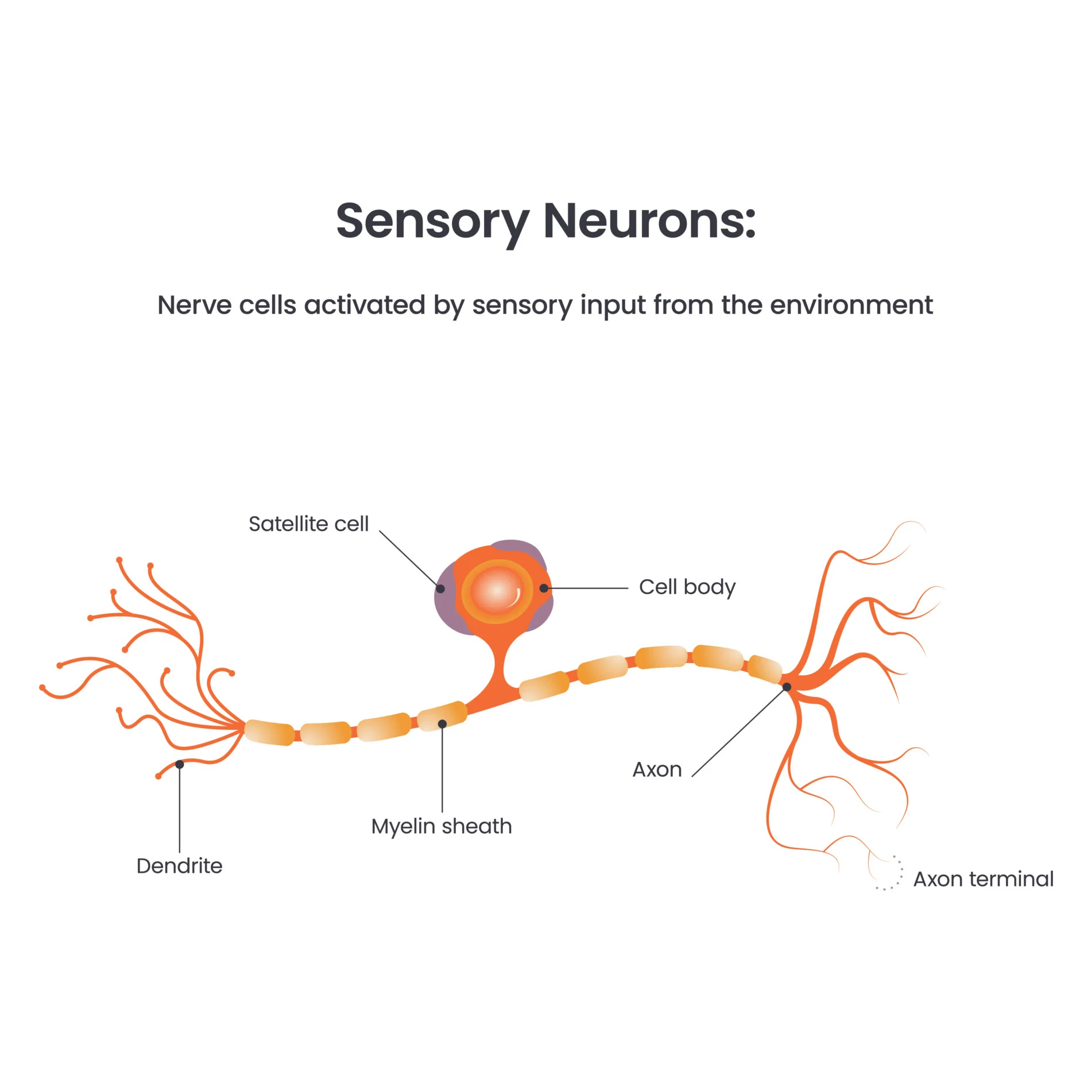
Structure and function of sensory neurone
long dendrites and short axon
unipolar → only one process extends from cell body
detects sensations from sensory site and sends action potential to CNS via PNS
found in various locations around body + PNS
Structure and function of relay neurone
short dendrites + axon and no myelinated sheath
found in CNS
communicator between sensory and motor neurone
Structure and function of motor neurone
short dendrites and long axon
begins in CNS and projects to muscles
detects signal from relay neurone in CNS via synaptic transmission and sends to effector along myelinated axon to contract
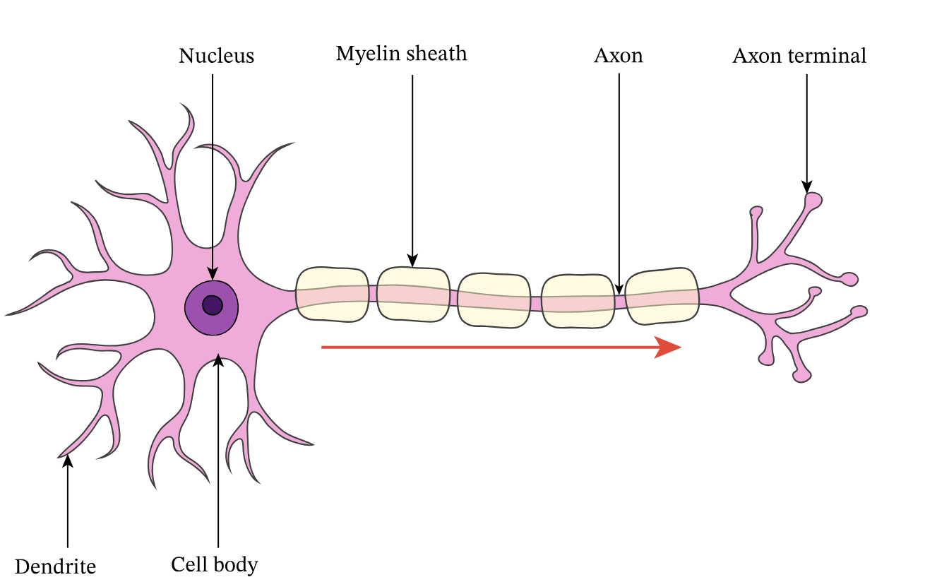
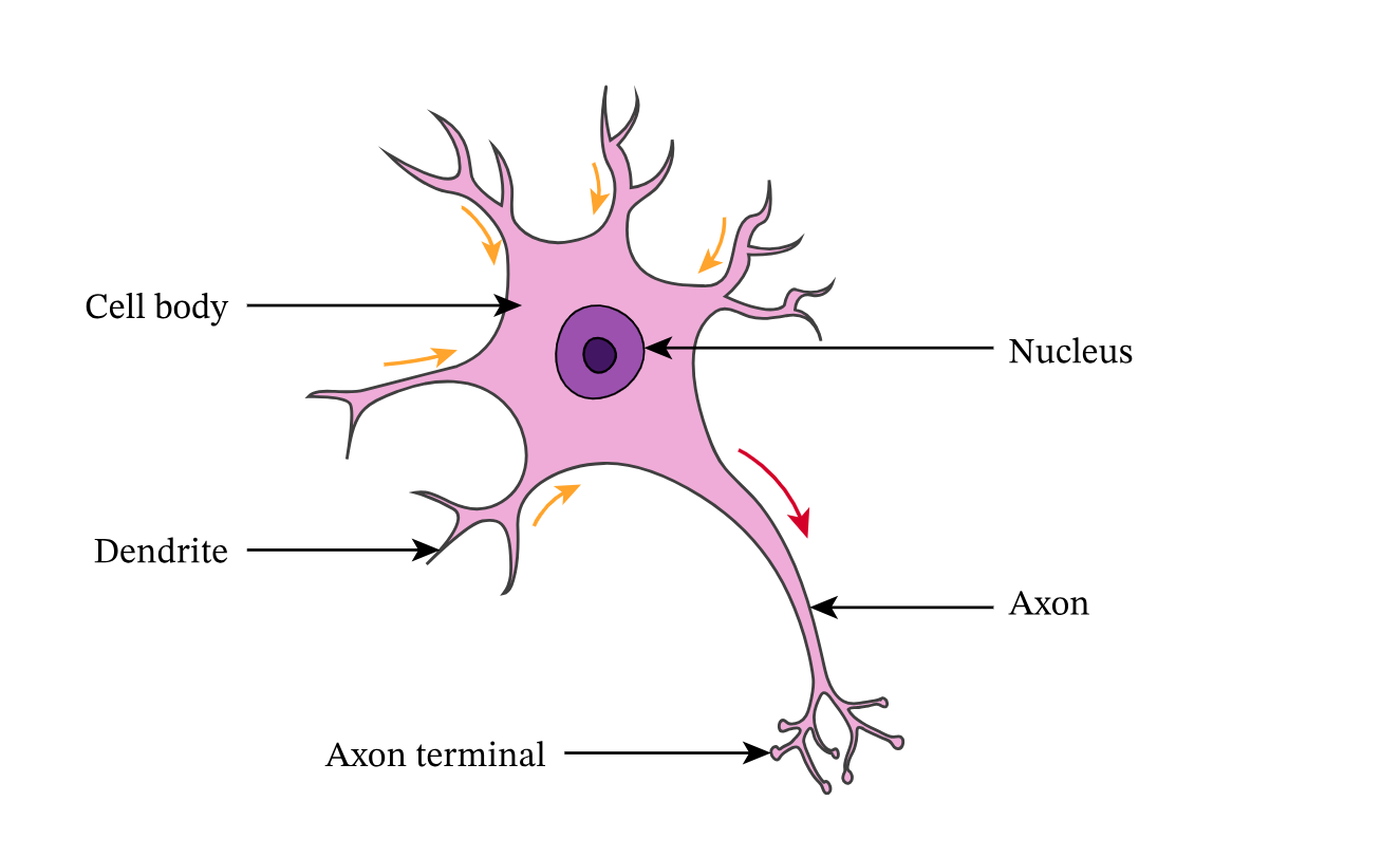
Describe the structure and function of a neuron (6)
neurons enable communication within the nervous system
the soma (cell body) contains the genetic material
dendrites extend from cell body and receive information from other neurons
axons, which can be myelinated to increase speed of transmissions, carry messages away from cell body
axon terminals contain neurotransmitters
What is a synapse
junction where 2 neurons meet
What is a neurotransmitter + types
chemical messages released by neurones that diffuse across synapse
excitatory → increases likelihood of neurone firing action potential
inhibitory → decreases likelihood of neurone firing action potential
what is summation
excitatory and inhibitory influences are summed and certain threshold needs to be reached so action potential of postsynaptic neurone is triggered
if net effect if excitatory, it increases likelihood of neurone firing action potential
if net effect is inhibitory, it decreases likelihood of neurone firing action potential
Outline synaptic transmission
Action potential reaches presynaptic neurone, causing it to release vesicles containing neurotransmitters to synaptic gap
Neurotransmitters diffuse across synaptic gap and bind to receptors on postsynaptic membrane
Neurotransmitters either excite (depolarise) or inhibit (hyperpolarise) postsynaptic neurone
Neurotransmitters return to presynaptic neurone via transport proteins via reuptake
What is localisation
theory that specific areas of the brain are associated with certain physical and psychological functions
What lateralisation
theory that certain hemispheres control certain physical and psychological functions
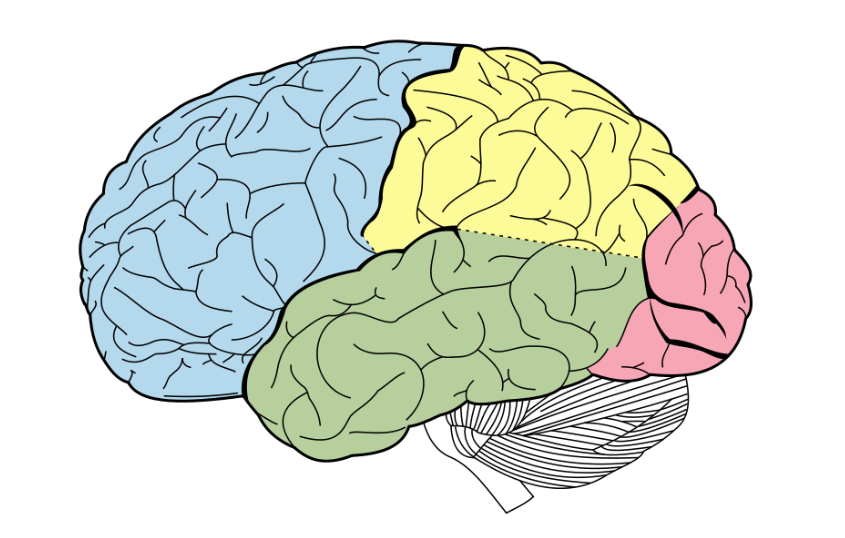
label ts
blue → frontal lobe
yellow → parietal lobe
green → temporal lobe
red → occipital lobe
Function of frontal lobe & area & cortex
controls cognitive activity
Broca’s area (in left frontal lobe) → language production, allows speech to be fluent
damage leads to Broca’s aphasia → difficulty producing speech e.g. slow speech, cant name objects & struggle with conjunctions and prepositions
Motor cortex → controls voluntary movement
damage leads to loss of movement
Function of parietal lobe and cortex
processes sensory information and directs movement
Somatosensory cortex → processes sensory information from skin
Function of temporal lobe and area
processes auditory information and understanding speech
Wernicke’s area → language understanding, allows speech to be meaningful
damage leads to Wernicke’s aphasia → difficulty understanding language, but can produce it (often fluent, meaningless speech w/ nonsense words (neologicisms))
auditory area → analyses speech based info, damage leads to hearing loss
Function of occipital lobe and cortex
processes visual information
Visual cortex → Each eye sends information from right visual field to left visual cortex and vice versa
AO3 for Localisation of function in the brain
✅Evidence from neurosurgery → cingulotomy isolates part of brain responsible for OCD, (Dougherty et al.) of 44 people, 30% that underwent cingulotomy had successful response, suggesting mental disorders are localised, ↑ construct validity / ❌ only 30%, mental disorders may involve multiple brain regions
✅Evidence from brain scans → Peterson et al. dual tasks show Broca and Wernicke’s areas are responsible for language during reading task and listening task → objective methods to measure brain activity increase validity + provide scientific evidence, ↑credibility
❌Language may not be localised to just Wernicke and Broca’s areas → e.g. advanced tech like fMRI shows language processing includes more areas, holistic theory may be more accurate
✅Localisation is supported by case studies such as Phineas Gage / ❌ case study, lacks generalisability, subjective, lacks validity, oversimplifies brain processes, undermining theory
What is split brain research
the study of individuals whose corpus callosum have been severed to reduce effect of epilepsy
Outline Sperry’s procedure
11 people whose corpus callosum was severed (to treat their epilepsy) were studied
Sperry projected information to each visual field, controlling info each hemisphere had access to
Ppt were required to either say what they saw or draw it
Outline Sperry’s findings
Information presented to RVF controlled by LH meant ppt could say what they saw, but not spoken if showed to LVF controlled by RH
Ppts could draw object or associate it if presented to LVF controlled by RH
∴ Brain is lateralized, language centre is in LH and spatial judgement is in RH
AO3 strengths of Lateralisation of function
✅Hemispheres process information differently→ PET scans show RH is more active when looking at global elements of an image and LH is more active when looking at finer details → can help stroke patients recover by encouraging RH to compensate for lost functions, ↑ practical usefulness
✅ Split brain research support → Luck et al. found split brain ppt were faster at identifying odd object than normal ppl as their superior LH isn’t watered down by RH. Supports Sperry’s findings that hemispheres are distinct, ↑ reliability + construct validity
AO3 weaknesses of Lateralisation of function
❌Plasticity is more important than lateralisation as after damage functions can switch sides but lateralisation doesn’t deal with loss of function e.g. lateralised chickens are adaptive as they can perform 2 tasks at once but plasticity is more adaptive / ❌ can’t be generalised to humans
❌Ethical implications → ppt couldn’t fully consent due to severed corpus callosum. So, the socially sensitive research may lead to a change in the way ppl w/ severed corpus callosums are treated
What is plasticity
the brain’s tendency to change and adapt as a result of new learning and experiences
synaptic pruning → frequently used synapses become stronger and unused synapse connections get lost, making brain’s communication system more efficient
What is functional recovery
a form of brain plasticity
after trauma, the brain redistributes or transfers functions from damaged areas to undamaged areas also known as neural reorganization
neuroscientists suggests it occurs quickly after trauma (spontaneous recovery)
the brain rewires itself, forming new synaptic connections near damage + activates secondary neural pathways
What happens to brain during functional recovery?
Axonal sprouting → Existing neurons grow new axons & connect with adjacent neurons. Neural regeneration is the growth of new neurons
Denervation super sensitivity → To compensate for loss of axons, remaining axons become more sensitive (more likely to fire). Can result in side effects like pain
Recruitment of homologous areas → Specific tasks can still be performed by opposite side of brain e.g. if Broca’s area was damaged, language production would be carried out in RH
AO3 strengths of Brain Plasticity and Functional Recovery
✅scientific research support for plasticity → Draganski et al. used a control group & MRI scans to measure posterior hippocampus to establish casual relationship + objective + reduces researcher bias + increases reliability
✅functional recovery has RWA → led to neurorehabilitation & axonal sprouting led to new therapies, e.g. constraint-induced therapy where patient practices moving affected arm while unaffected arm is constrained, ↑ ecological validity + positive social + economic implications
AO3 weaknesses of Brain Plasticity and Functional Recovery
❌Draganski’s research - correlation ≠ causation → cant be sure studying alone caused changes to posterior hippocampus + cant establish casual relationship due to inability to control potential participant variables e.g. diet ↓ internal validity
❌Plasticity may lead to negative behavioural consequences → e.g. the brain’s adaptation to constant drug use leads to poorer cognitive functioning & an increased risk in dementia + 60-80% of amputees have phantom limb syndrome as reorganisation leads to maladaptive consequences due to changes in somatosensory cortex → adaptation to damage isn’t always beneficial
Outline fMRI for studying the brain
detects changes in blood flow & oxygenation to the brain that occur due to neural activity in certain brain area
when a brain area is more active, it consumes more oxygen and more blood flow is directed to area (haemodynamic response)
AO3 for fMRIs
✅high spatial resolution ≈ 1mm so images are very detailed
✅safe as it doesn’t use radiation so can safely provide a clear picture of how brain activity is localized → economic implications - ppl can return to normal activities like work immediately after scanning + no radiation-related side effects, ↓ strain on NHS from as no follow up treatment needed
❌expensive, poor temporal resolution - 5s lag between neural activity and image so fMRIs may not truly represent brain activity as its too fast to measure
Outline EEGs for studying the brain
a method of recording electrical activity in the brain using electrodes placed on scalp
electrodes detect brain wave patterns generated by firing of neurons in the brain
by measuring characteristics of brain wave patterns, EEGs can help diagnose certain brain conditions
AO3 for EEGs
✅high temporal resolution → detects brain activity within the millisecond, accurately represents brain activity
❌low spatial resolution → information is generalised & source isn’t pinpointed so can’t distinguish activity of different neurones
Outline Event Related Potentials (ERPs) in studying the brain
measuring small changes in brain activity that happen when a person responds to a specific stimulus or event
removes background electrical noise unrelated to stimulus
waveform’s peaks and dips show cognitive processes after stimulus is presented
AO3 for Event Related Potentials (ERPs)
✅high temporal resolution → detects brain activity quickly, valid representation of brain activity
❌lack standardisation → many researchers use different methods for recording & analysing brain data, harder to compare + replicate, ↓ reliability
Outline Post Mortem Examinations for studying the brain
technique of analysing person’s brain after death
dissects unusual brains to examine areas that caused deficit or disorder, then compared to neurotypical brain
AO3 for Post Mortem Examinations
✅high spatial resolution → allows study of microscopic brain structures down to neuronal level
❌unethical → invasive procedure, no informed consent provided after death, challenges whether benefits outweigh costs, damaging psychological research and its reputation
AO3 strengths for Circadian rhythm
✅RWA for shift workers → e.g. night shift creates desynchronisation in biological rhythms + cause period of reduced concentration at 6am, economic implications, workers less productive during circadian trough so not able to contribute to economy + employers can provide days off to recalibrate sleep schedule / ❌ research is correlational so desynchronisation not cause of lower productivity, may be divorce
✅RWA for medical treatments → knowledge of circadian rhythm can be used to time medication to maximise effects since C.R. coordinates basic processes e.g. heart rate, researchers found taking aspirin is most effective at night as it reduces heart attacks which are most common in morning
AO3 weaknesses for Circadian rhythm
❌shifting the school day may not be effective → e.g. starting at 10 benefits teenagers mental & physical health & academic achievements, but is disruptive for parents and teachers and teenagers may still stay up later and still be exhausted - not practical
❌generalisations are difficult to make → sleep/wake study cycles use small sample (e.g. Siffre) ↓ population validity + variation within people’s sleep cycles, idiographic approach may be more suitable than nomothetic
AO3 for infradian rhythms
✅menstrual cycle synchronisation is evolutionary → may have been adaptive for ancestors to menstruate together so babies who lost their mothers could access breastmilk to increase survival chance, so synchronisation is an adaptive strategy
❌menstrual synchronisation is not always present in females → e.g. Stern & McClintok’s findings are not reliable as they cant be replicated due to confounding variables that may affect menstrual cycles like stress, so menstrual synchrony studies may be flawed + raises doubt of pheromones as exogenous zeitgebers
AO3 for Ultradian rhythms
✅lab experiment → sleep studies occur in labs using machinery like EEGs where extraneous variables can be controlled, ↑ internal validity + reliability + objective machinery (feature of science), ↑ scientific credibility
❌individual differences between peoples sleep patterns → stages 3 and 4 differ, influences are biologically determined, hard to generalise sleep patterns, idiographic approach more suitable than nomothetic