A&P 2: TopHat Lab 15
1/50
There's no tags or description
Looks like no tags are added yet.
Name | Mastery | Learn | Test | Matching | Spaced | Call with Kai |
|---|
No analytics yet
Send a link to your students to track their progress
51 Terms
Respiratory system
System of tubes and ducts facilitated by a pumping system; primary function is exchange of gases between the atmosphere and the body; protects the body from rapid changes in internal temperature and dehydration by warming and moistening the inhaled air; also used to produce sound for communication
External nares
Openings connecting the nasal cavity to the outside; air passes through these openings during respiration
Nasal cavity
Cavity within the nose and above the palate; divided by the nasal septum into two passageways by the nasal septum; contains the olfactory epithelium and warms, moistens, and filters the air passing through the nose
Paranasal sinuses
Cavities located within the skull that lighten the skull and serve as resonating chambers for speech
Internal nares
Openings at the rear of the nasal cavity that open into the nasopharynx; air passes though these openings into the nasopharynx
Pharynx
Passageway known as the throat; composed of three regions, the nasopharynx, oropharynx, and laryngopharynx
Nasopharynx
Uppermost portion of the pharynx superior to the soft palate; the openings of the Eustachian, or auditory tubes, are located in this region
Oropharynx
Portion of the pharynx between the soft palate and the epiglottis (a flap of skin); common passageway for the digestive and respiratory systems
Laryngopharynx
Lower portion of the pharynx lying between the hyoid bone and the larynx
Larynx
Connects the pharynx to the trachea; contains the paired vocal cords used for speech; also called the “voice box”
Epiglottis
Flap of tissue that covers the opening to the larynx, the glottis, during swallowing; prevents food and fluid from entering the lower portion of the respiratory system
Trachea
A cartilage-reinforced tube that is commonly called the “windpipe”; begins at the larynx and ends as it splits into the right and left bronchi, in the thorax; passageway for air entering the lungs
Lungs
Two large respiratory organs located in the thoracic cavity; site of external respiration, where oxygen diffuses into the bloodstream
Hilus
Commonly called the “root” of the lung; located on the medial edge of the lung, where the bronchi and blood vessels enter and leave the lung
Bronchi
Passageway for air in the lungs. Primary bronchi are the two tubes that split from the base of the trachea and enter the right and left lungs. Secondary bronchi split from the primary bronchi. Tertiary bronchi split from the secondary bronchi.
Bronchioles
The tertiary bronchi split into these smaller tubes within the lungs; eventually lead to the alveoli
Alveoli
Small sacs lined with simple squamous epithelium; form the functional unit of the lungs where gas exchange occurs
Diaphragm
A large, flat, skeletal muscle that separates the thoracic and abdominal cavities; acts to change air pressure within the lungs during inhalation and exhalation

Identify A
Paranasal sinus - picture

Identify B
Nasal cavity - picture

Identify C
Internal nares - picture
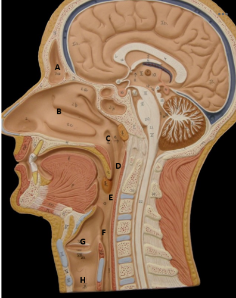
Identify D
Nasopharynx - picture

Identify E
Oropharynx - picture

Identify F
Laryngopharynx - picture

Identify G
Larynx - picture

Identify H
Trachea - picture

Identify B
Hilus - picture

Identify C
Primary Bronchi - picture

Identify D
Secondary Bronchi - picture
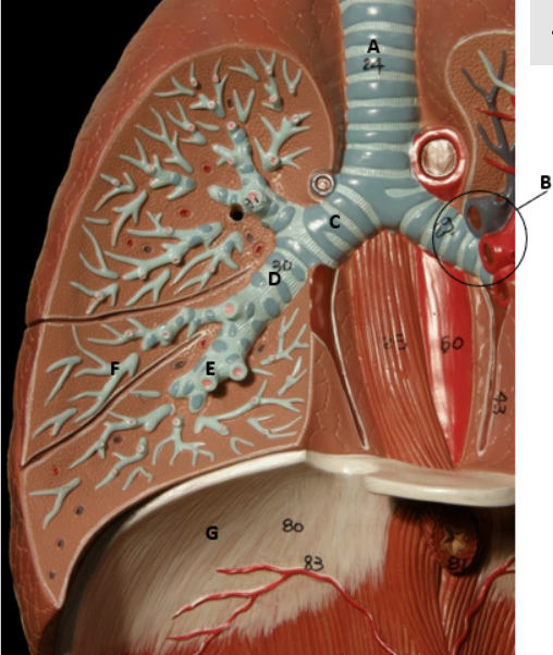
Identify E
Tertiary Bronchi - picture

Identify F
Bronchioles - picture

Identify G
Diaphragm - picture

Identify A
Inspiratory Reserve Volume - picture

Identify B
Tidal volume - picture

Identify C
Expiratory Reserve Volume - picture

Identify D
Residual Volume - picture

Identify E
Inspiratory Capacity
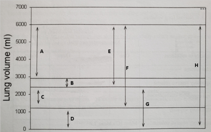
Identify F
Vital Capacity - picture

Identify G
Functional Residual Capacity

Identify H
Total Lung Volume - picture
Boyle’s Law
The pressure of a gas in a closed container is negatively related to the container's volume; aka when the volume of the lungs increases, the air pressure within the lungs decreases
Exhalation
When the volume of the lungs is decreased by the relaxation of the intercostals and the diaphragm muscles; passive
Inhalation
When the volume of the lungs is increased by the contraction of the intercostal muscles and diaphragm; active
Residual Volume
Amount of air remaining in the lungs after fully exhaling; remains in the alveoli, keeping them open for respiration; measuring this volume in a lab setting is effectively impossible, so we used an average of 1200ml
Tidal Volume
Volume of a single inhalation during normal, quiet breathing; mean value is approximately 500 ml for a healthy individual. Approximately 70% of this reaches the respiratory surfaces of the lungs; remaining 30% stays in the non-respiratory parts of the system, (like the trachea, the various bronchi, and the bronchioles)
Expiratory Reserve Volume (ERV)
Amount of air which can be forcibly exhaled after the tidal volume has been exhaled; average for healthy males is about 1100ml while the average for healthy females is about 700ml
Vital Capacity (VC)
Maximum amount of air that can be exhaled after a deep inhalation; on average, about 4800ml for males and 3100ml for females; the sum of inspiratory reserve volume, tidal volume and the expiratory reserve volumes
Inspiratory Capacity (IC)
Total volume of air that can be inhaled after the exhalation of the tidal volume; on average, it is about 3800ml for males and 2400ml for females; the sum of the tidal volume and inspiratory reserve volume
Minute Respiratory Volume (MRV)
The volume of air inhaled each minute; calculated by multiplying the tidal volume by normal breathing rate
Inspiratory Reserve Volume (IRV)
Amount of air inhaled when taking a very deep breath in. The average for healthy males is about 3300ml and 1900ml for healthy females
Total lung volume (TLV)
Total volume of air that can be inhaled; on average total volume is about 6000 ml for males and 4200ml for females; calculated by adding the average residual volume, expiratory reserve volume, tidal volume, and inspiratory reserve volume