8 - Junctions
1/165
There's no tags or description
Looks like no tags are added yet.
Name | Mastery | Learn | Test | Matching | Spaced | Call with Kai |
|---|
No analytics yet
Send a link to your students to track their progress
166 Terms
How do multicellular orgasnims hold their cells together? (3) --> these influence body architecture and cell-cell communication
1. They could be sticky - adhesion.
2. They could be physically connected - junctions.
3. They could be bound by a secreted matrix - ECM
cells are themselves small ____ yet multicellular organisms, through cell-cell adhesion, contacts, and ECM interactions, can be massive, strong, and ____
cells are themselves small malleable yet multicellular organisms, through cell-cell adhesion, contacts, and ECM interactions, can be massive, strong, and stable
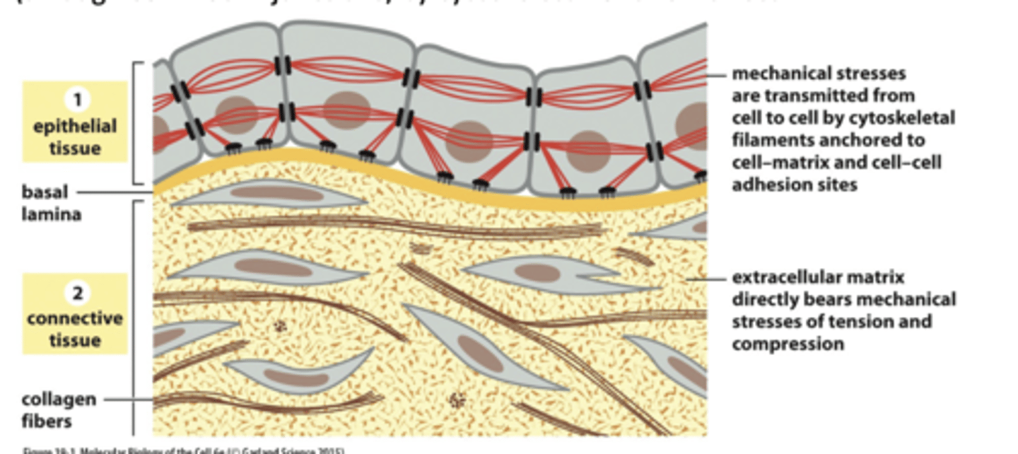
List 2 types of animal tissues cell category + where are they found
1. Connective - bone, tendon
2. Ephithelial - skin , lining of gut
What is connective tissues (3)
1. h as a lot of extracellular matrix (ECM), which is a secreted networkof proteins and polysaccharides
2. frequently rich in collagen; bears most of the mechanical stress,
3. providessupport; strong attachments with the ECM (through cell-matrix junctions) by cytoskeleton and few direct attachments with each other.
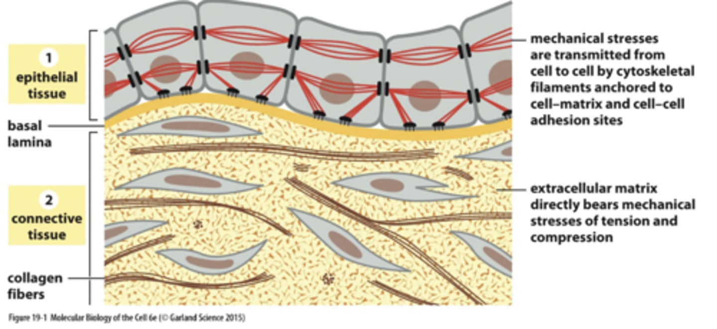
What is epithelial tissues
sheets of cells tightly boundtogether by cell-cell junctions (viacytoskeleton) --> creates ECM

What is ECM + where is it found
Extracellilar matrix is a thin mat on basal sidecalled the basal lamina (akabasement membrane)
--> linked to cytoskeleton via cell -matrix junctions
Draw and all the The junctions (schematic diagram )
.

Where are cell-cell adhesions mostly clear seen in?
Ephitlia (such as intestinal lining) --> where there is strong, direct anchorage of adjacent cells
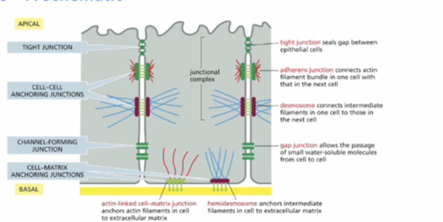
How many different categories of achnoring junctions are there --> draw them + what are the sub junction?
four:
1. tight junction
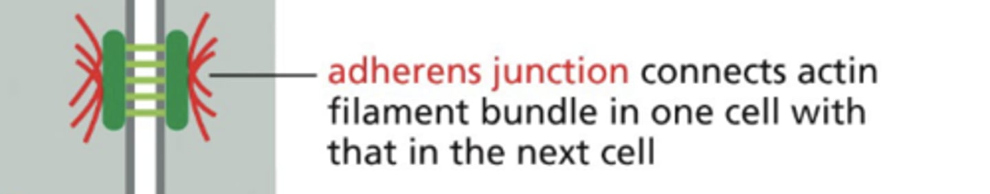
2. Cell-cell adhering junction
--> Adherenes junction
--> Desmosome
3. Channel forming junction
--> gap junction
4. Cell-matrix anchoring junction
--> Actin-linked cell matrix junction
--> Hemidesmosomes
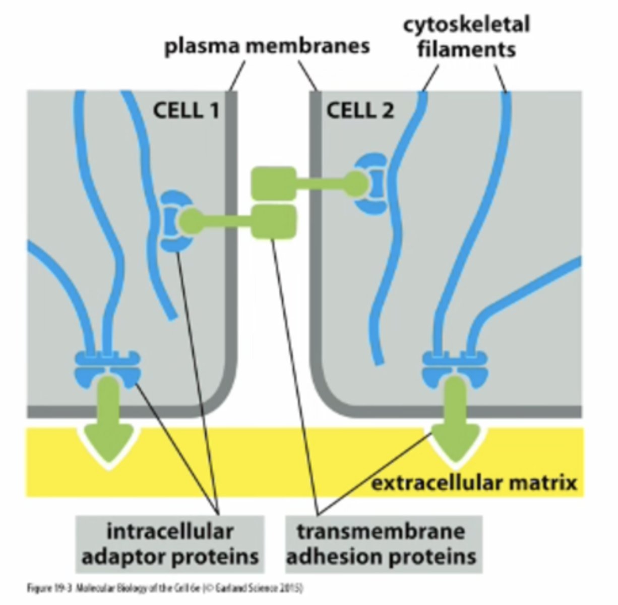
what are Transmembrane adhesion protiens
Protines that spans the PM that helps anchor cytoskeleton filaments --> helps cell-cell adhesions
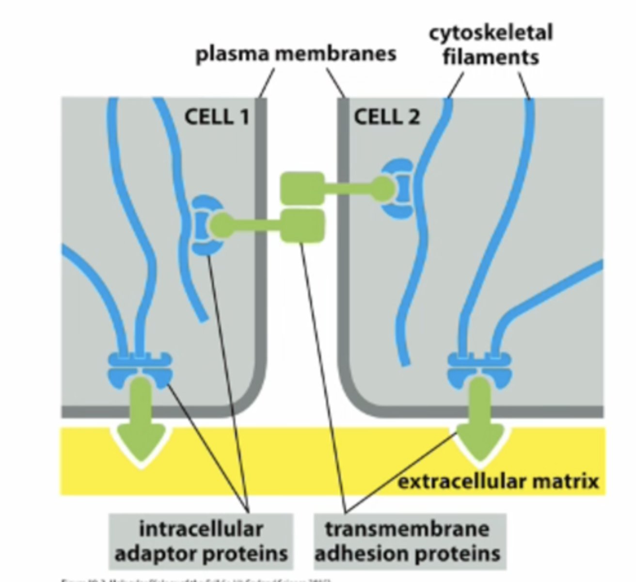
What are the 2 superfamilies of transmembrane adhesions proteins + where are they involve in?
cadherins - generally cell-to-cell attachment
integrins: generally cell-to-ECM attachment
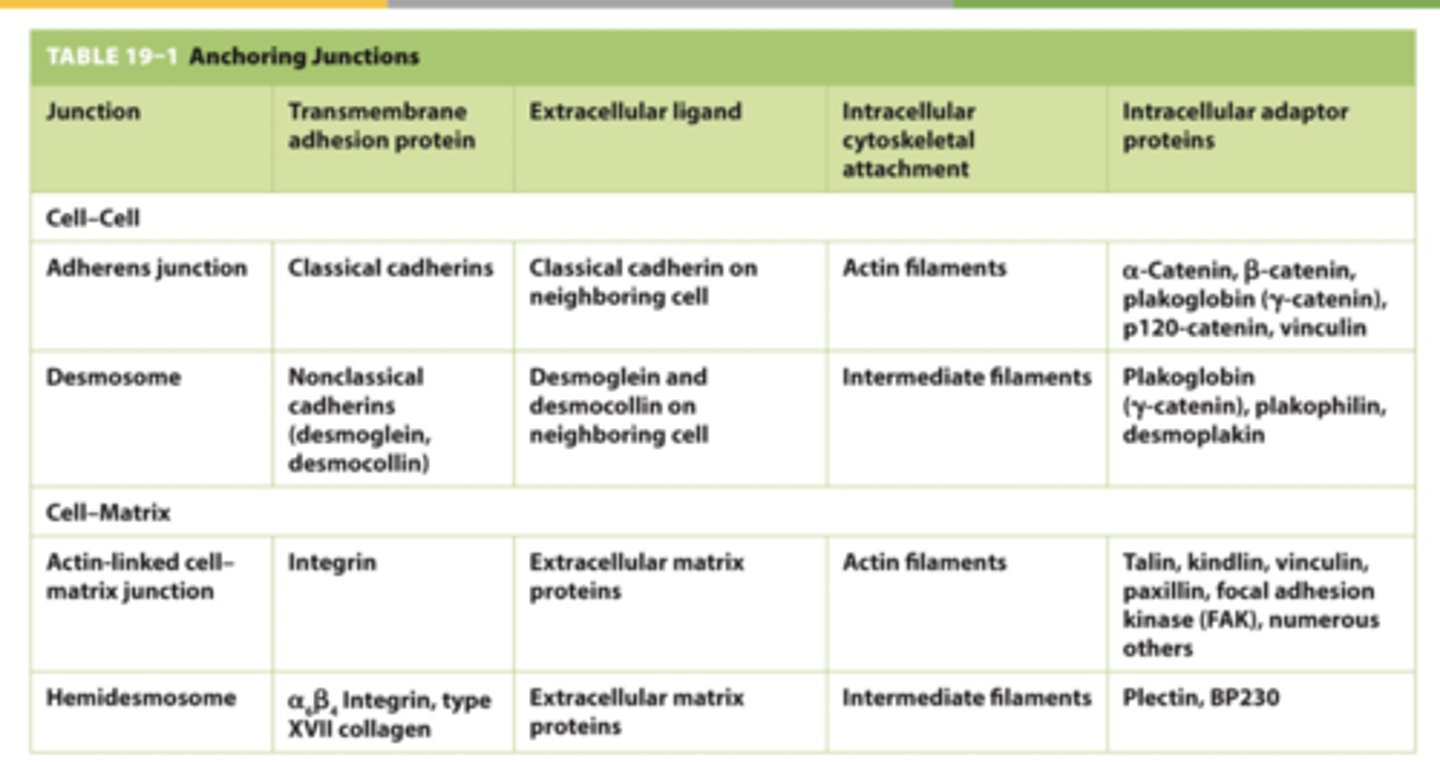
Adherenes junction vs Desmosome vs Actin-linked cell matrix junction vs . Hemidesmosomes (write the table)
.
Where are cadherins vs integrins found in?
Cadherins - found in cell-cell junctions, depend on interacting cytoskeletal protein;
Integrins - found in cell-matrix junctions;; depend on cytoskeletal interactions

where are cadherins vs integrates anchored by?
Cadherins - anchored by catenins
Integrins - anchored by diverse collection of protiens

Which organisms have cadherins?
Presnet in all multicellualr animsals.. absent from fungi and plants
How were cadherins discovered?
Some types of cells (especially embryonic) can be readily dissociated by removing calciumfrom the extracellular medium; sometimes the addition of a protease (e.g. trypsin) is alsorequired. If dissociated cells are placed back into normal medium with calcium, they canreassociate
What was the cadherin found in epithelial cell vs nerve vs placenta/epidermis --> what are these called
Epithelial cell = E-cadherin
Nerve = N-cadherin
Placenta/epidermis = P-cadherin
--> These are called "classical cadherins"
Describe 2 characteristics of classical cadherins
1. closely related throughout their sequence
2. perform well-defined adhesive functions.
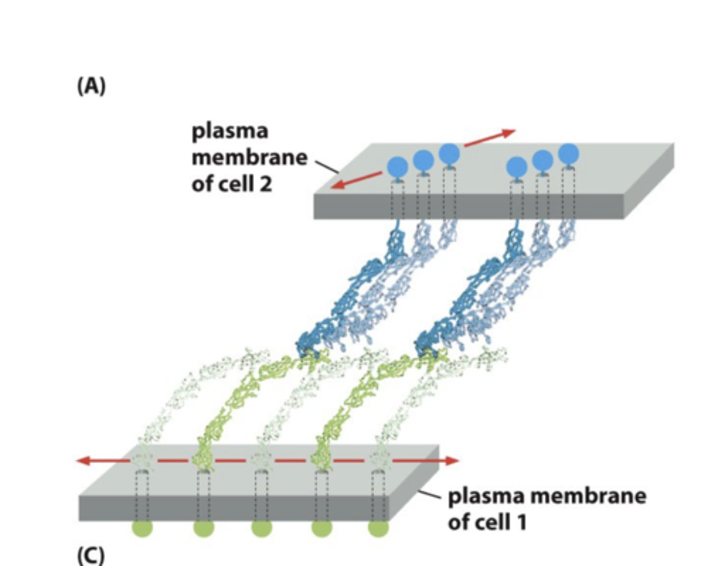
cadherins from one cell usually bind to cadherins in opposing cells through a _____ interaction ; the anchoring junction is _____
cadherins from one cell usually bind to cadherins in opposingcells through a homophilic interaction ; the anchoring junction is symmetrical.
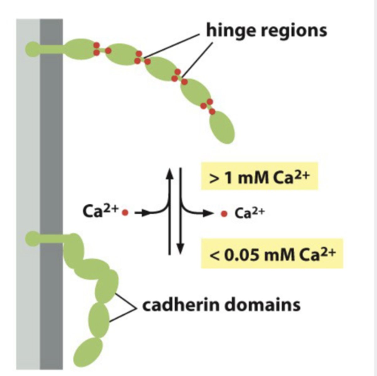
What does cadherin domains form?
a rigid unit and adjacent cadherin domains are separated by a hinge region that is stabilized and made rigid by Ca2+ ions
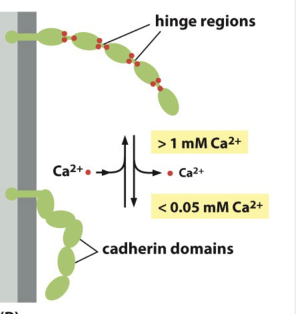
What makes cadherin rigid?
Ca2+ ions

What happens to the hinge of cadherin when Ca2+ ar remove>
hinges can flex and the structurebecomes floppy
How does cadherin weakens its affinity for marching cadherin on opposite cell
conformation change at N-terminus
When do we degrade cadherin + what degrades it?
Destabilized caderin --> degraded by protease
What is the function of cadherin
"Velcro fro cell" = cell-cell adhesion
--> bind at N-terminal tups
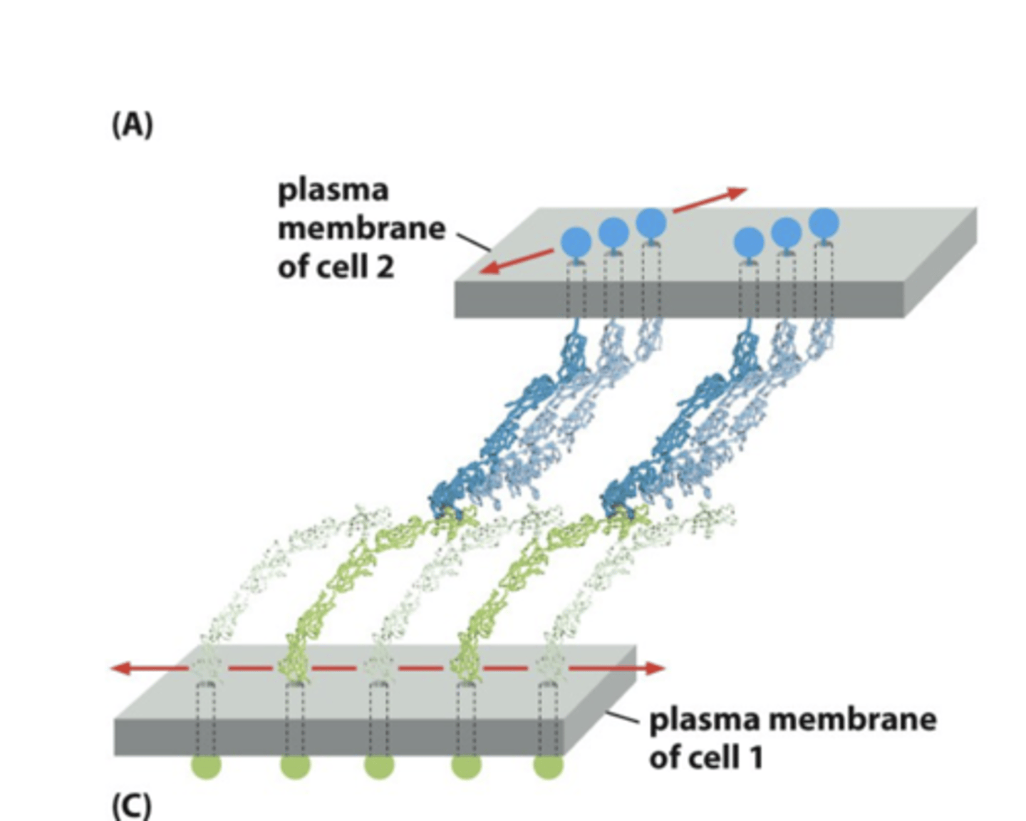
How are cadherin disassembled
By breaking molecules sequentially from the side like velcro
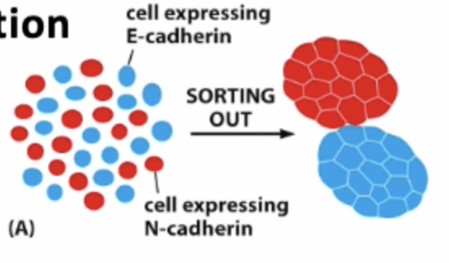
There are many different types of cadherin.... but they only bind with the _____ type
There are many different types of cadherin.... but they only bind with the _same ____ type = specificity
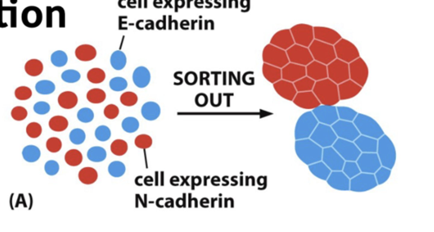
Explain how they showed that cadherin binding is specific with amphibian embryonic cells
1. disaggregated amphibian embryonic cells can be randomly reaggregate
2. tend to sort themselves out according to cell type, on the basis of the particular cadherins they express; example: amphibian embryo with epidermal, mesodermal, neural plate cells (below)

during ______ development, can see change in expression from one cadherin to another at specific times and stages
during embryonic development, can see change in expression from one cadherin to another atspecific times and stages
What happens to mouse embryos that lack E-cadherin?
fall to pieces; mutant phenotype is embryonic lethal
What controls Tissue segregation ?
Homophilic binding
What happens to fibroblast that normally dont express cadherins, transfected with E-cadherins?
Will become adherent
--> transfect with different cadherin = will sort according to type
When can sorting out occur?
occur among cells that are expressingdifferent levels of the same cadherin

What happens to cell expressing high level of E-cadherin vs low level --> how are they sorted out?
High level = would end internally
Low level = would end out externally
What are mesenchymal cells?
dispersed unattached cells
How can mesenchymal cells form epithelial cells + where does this occur
the expression of cadherin in dispersed unattached cells (mesenchymal cells) can cause them tocome together and form an epithelium
--> occur in tissue regneration, grafting
When does epithelial-to-mesenchyal transition occur in?
during development
Epithelial cells vs mesenchymal cells (4 characteristics of each)
Epithelial cel
1. form sheets (act as barrier) connected by junctionalcomplexes
2. move in harmony
3. have a clear polar character from one side to another
4. basal lamina is a foundation, contacts only onesurface of the cell
Mesenchymal cells
1. loosely organized and loosely attachedcells (more like bold individuals)
2. can migrate as individual cells
3. can also adhere in 3-dimensional clumps
4. basal lamina may surround the cells(muscle or fat cells
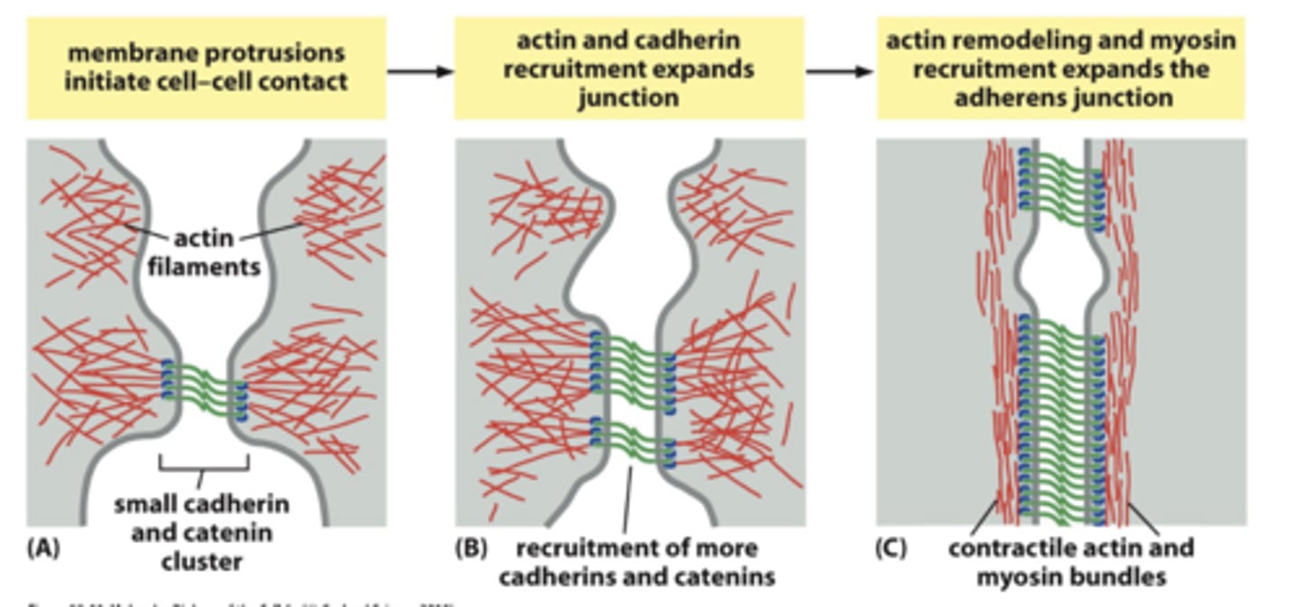
What is required for the assembly of Adherens junctions (2)
1.actin-associated proteins
2. non-muscle mycosin
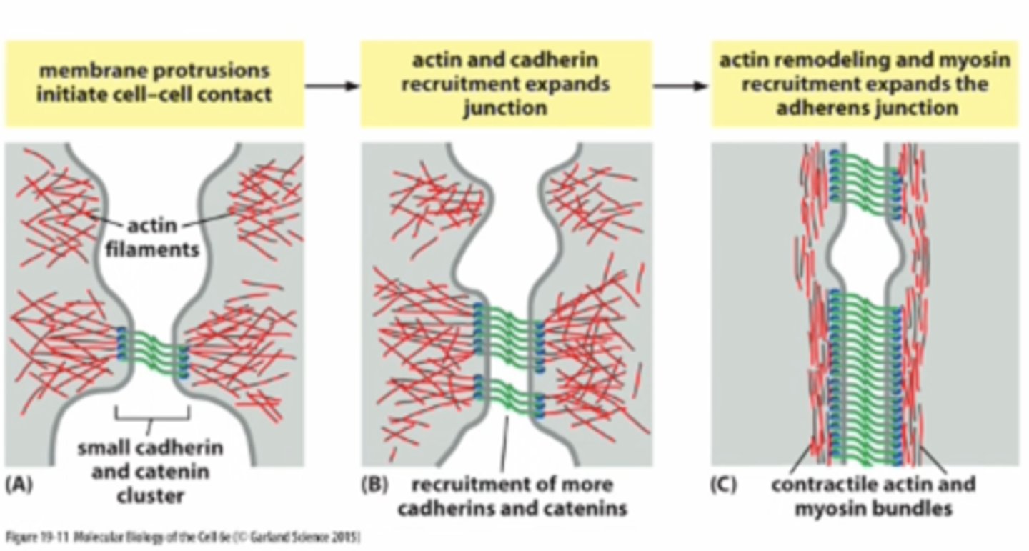
What is the function of adheren junction (2)
1. pulling forces important for junctionassembly and maintenance
2. site for mechanotransduction
What links cadherins to the cystoskeleon?
Catenins
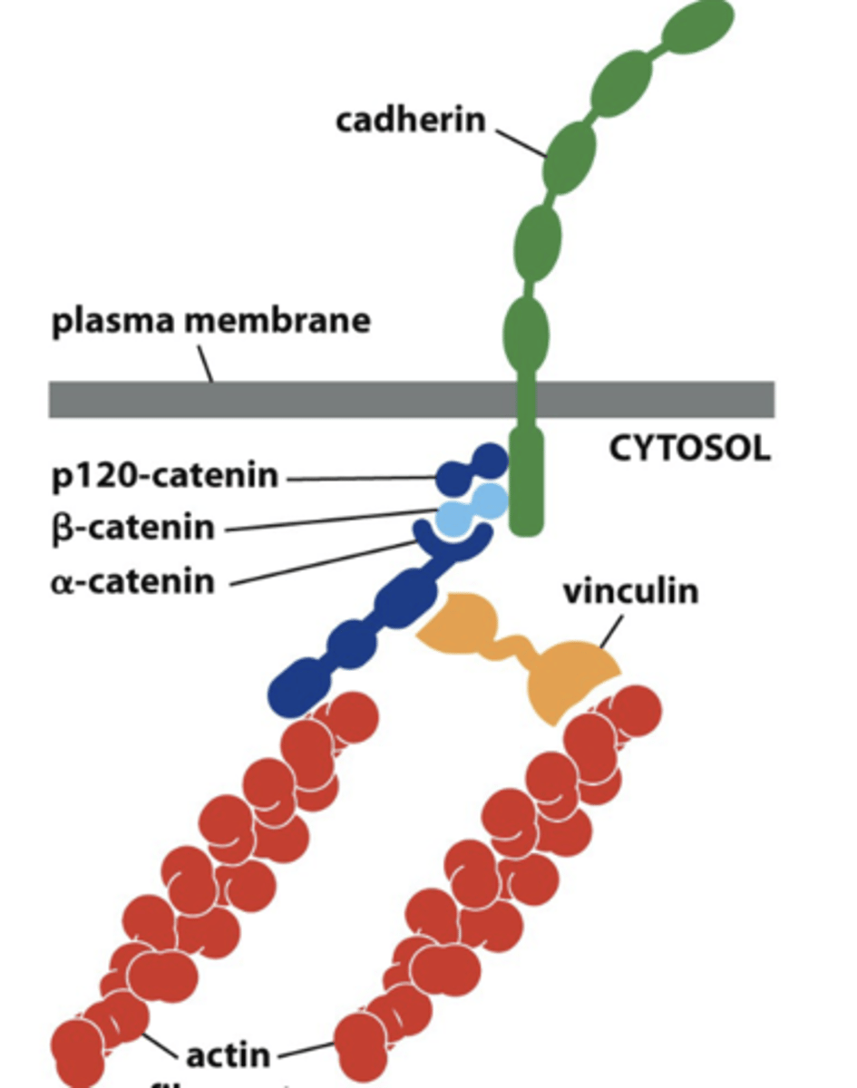
What kind of catenin is required to actin cytoskeleton (in adherens junctions) vs intermediate filaments (in desmosomes)
actin cytoskeleton (in adherens junctions) --> p120-catenin and B-catenin
intermediate filaments (in desmosomes) --> gamma-catenin (plakoglobin)

Loss of B-catenin and plakoglobin in the heart can lead to?
arrhythmogenesis

List the two cell-cell -anchoring junctions
1. adheren junction
2. desmosomes

Where do we see adherens junction
Epithelial cells
--> these junctions organized as continuous adhesion belt beneath apical surface of cells (zonula adherens)
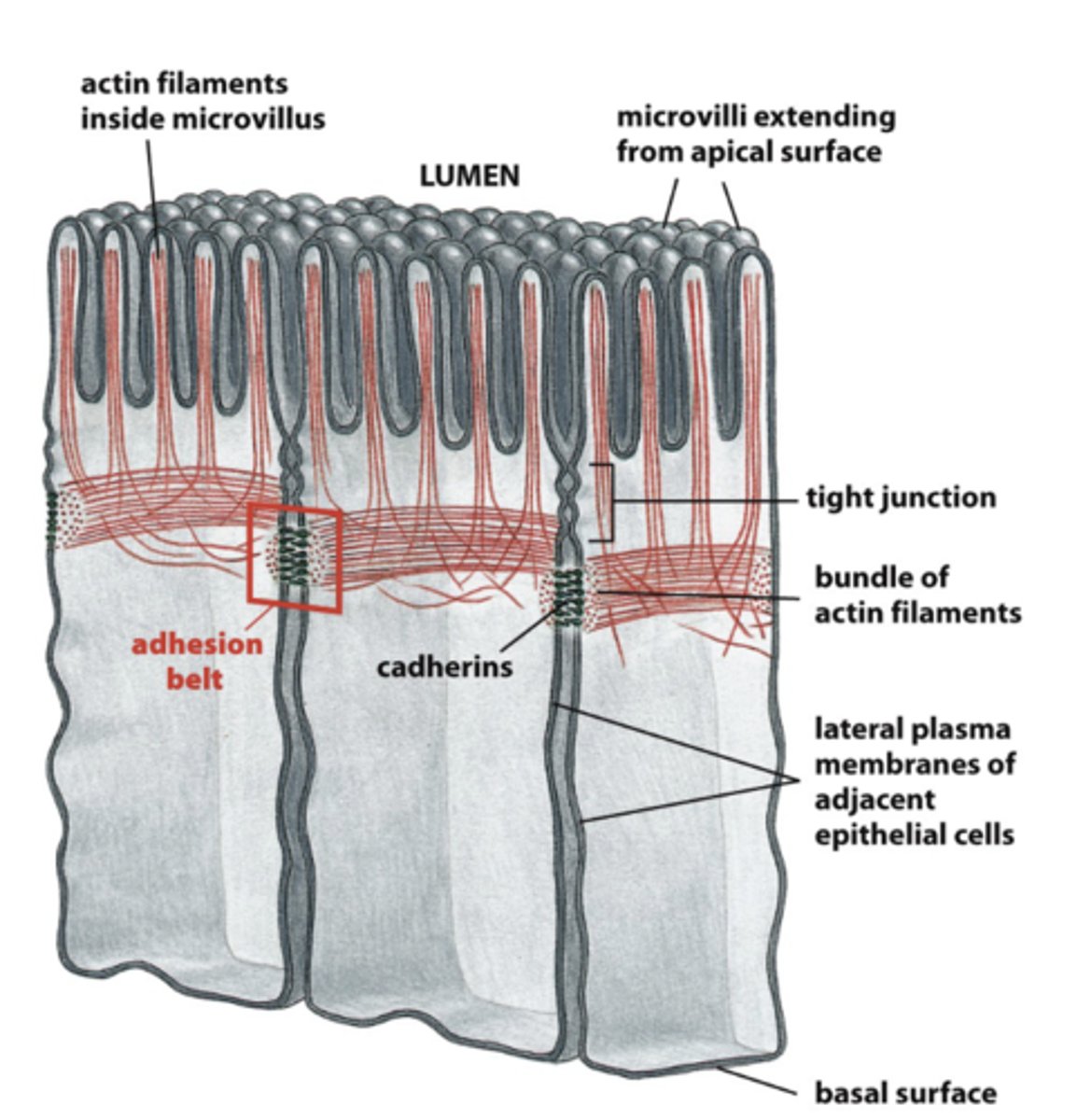
What is zonal adherens
continuous adhesion beltbeneath apical surface of cells

What are zone adherens junctions tethered to?
tethered to bundles of contractile actin, formstranscellular network, such that entire sheet of epidermalcells can act as coordinated unit; can contract throughaction of myosin and actin

What are desmosomes tethered to? + what do they link?
linkage of cadherins to intermediate filaments of cytoskeleton, organized in button-like spot

Where can we find desmosomes?
tissues subjected to high stress:
What are the intermedtae filaments that anchors demoes in epithelia vs heart muscle
epithelia - keratin
heart muscle - desmin
What is pemphigus (disease)
disorder resulting from disruption ofdesmosomes, autoimmune, blistering of skin & leakage offluids into loosened epithelium
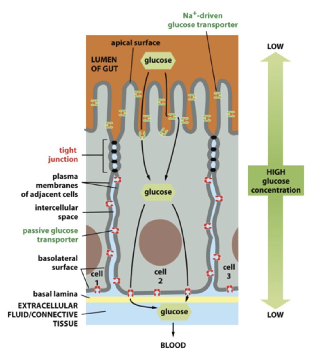
What are tight junctions function
seals gaps between epithelial cells = sheets of epithelial cells enclose
--> prevent apical and basolaterla proteins from diffusing into irong area

epithelia are structurally ________ attached to one tissue on basal side and to “extracellular fluid” on oth
epithelia are structurally polarized, attached to one tissue on basal side and to “extracellular fluid” on other
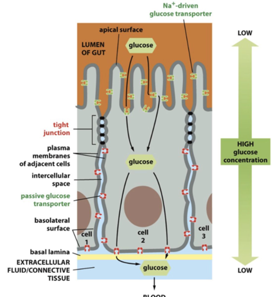
What does the tight junction cause? (2)
1. transcellular transport, need specific (and different)transporters on apical and basolateral surfaces
2. paracellular transport - movement of substances between epithelial cells
What is another name for tight junction
Occluding junction
Why is tight junction also called Occluding junction
environment (space and fluid) on apical side ofepithelium is separate from space and fluid onbasal side

What is paracellular transport
movement of substances between epithelial cells
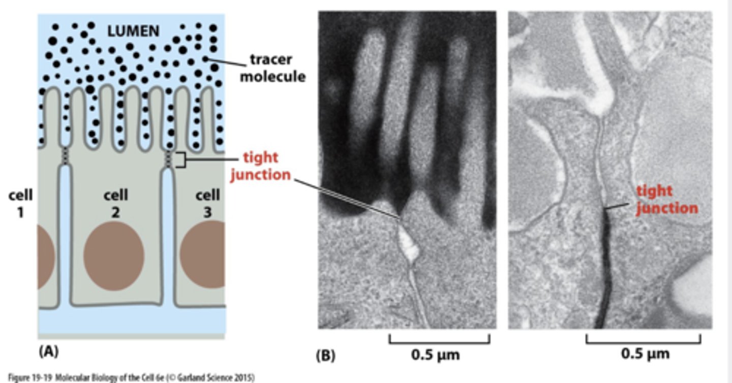
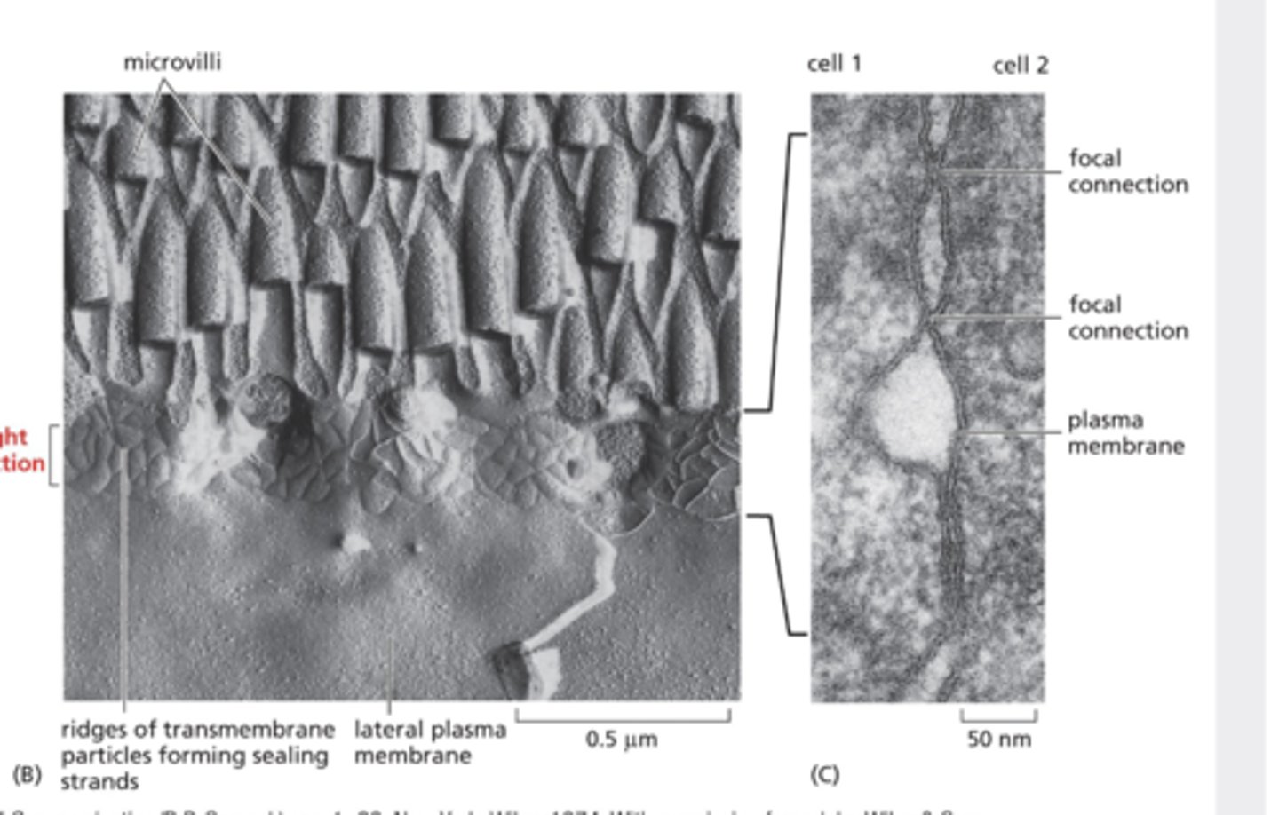
How were tight junctions observed?
freeze-fracture EM asbranching networks of sealing
--> each strand is composed of a longrow of transmembranehomophilic adhesion proteinsembedded in PM of adjacent cells- extracellular domains of theseadhere directly to one another toocclude intercellular space
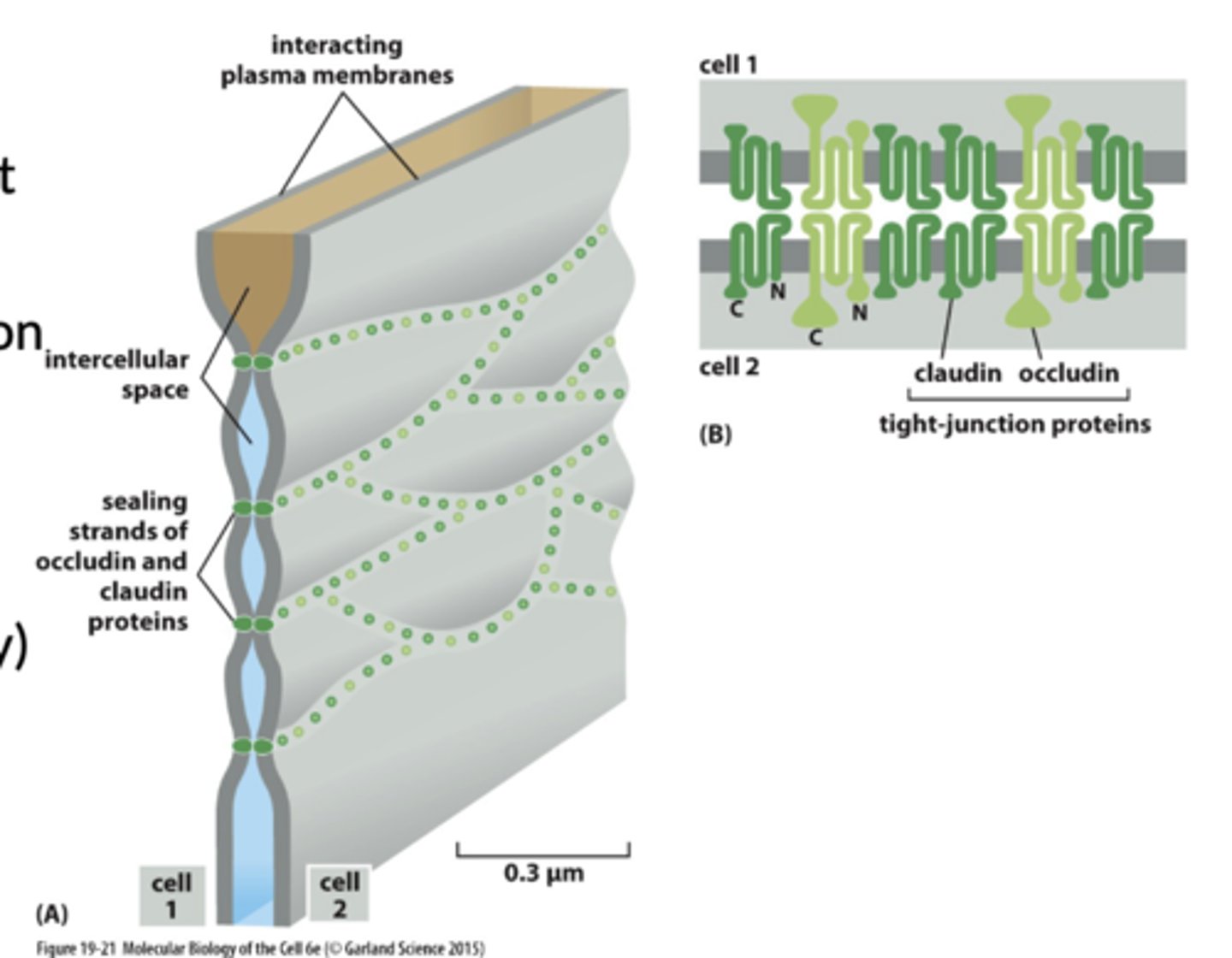
What protins composes (create the seal) in tight junctions (2(
1. Claudins
2. Occludins
What protein prevent leakage at tight junction
tricellulin

What are paracellular pores
associated withdifferent degrees of permeability and selectivity ofpermeability
--. ex selective retention of Mg2+ ions in kidney)
What is Clostridium perfringens enterotoxin (CPE) (disease)
binds to a subset of claudins
--> gastrointestinal symptoms of one of the most common foodborne illnesses
--> pore formation in PM of host mucosal cells is mediated by the N-terminal domains, leading to cell death
--> cCPE (C-terminal domain) is not cytotoxic
What is cCPE
cCPE (C-terminal domain) is not cytotoxic --> promising Claudia modulator
Explain how cCPE can be a Claudin modulator (2)
1. I ncreases paracellular permeability; could be used to improve drug delivery across tissue barriers
2. also target claudin-overexpressing tumors (deregulation of claudin expression/function associated withtumor proliferation/growth
claudins and occludins interact with each other through ______ _______, organization depends on binding to cytoplasmic domains by ________ _______ proteins
claudins and occludins interact with each other through extracellular domains, organization depends on binding to cytoplasmicdomains by intracellular scaffold proteins
What is an example of a intracellular scaffold protines
ZO proteins (zonula occludens
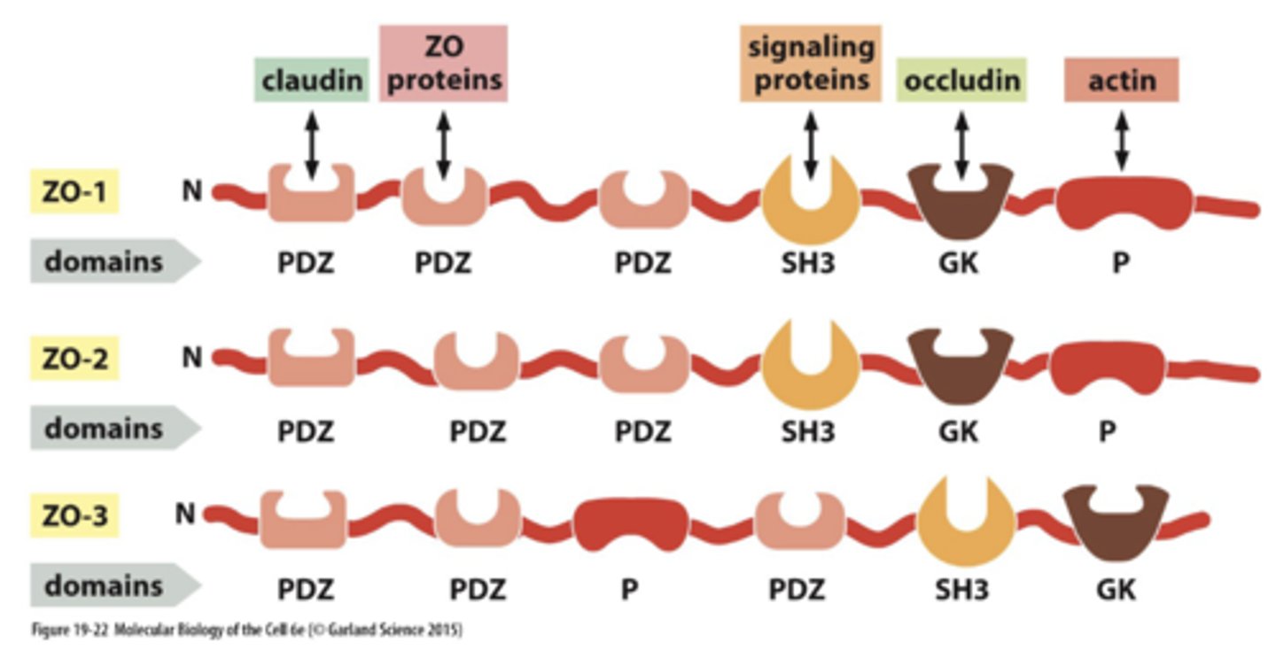
How can sealing strand form>
ZO protiens have to be in right position
--> sually apical to adherens and desmosome junctions - together = junctional complex

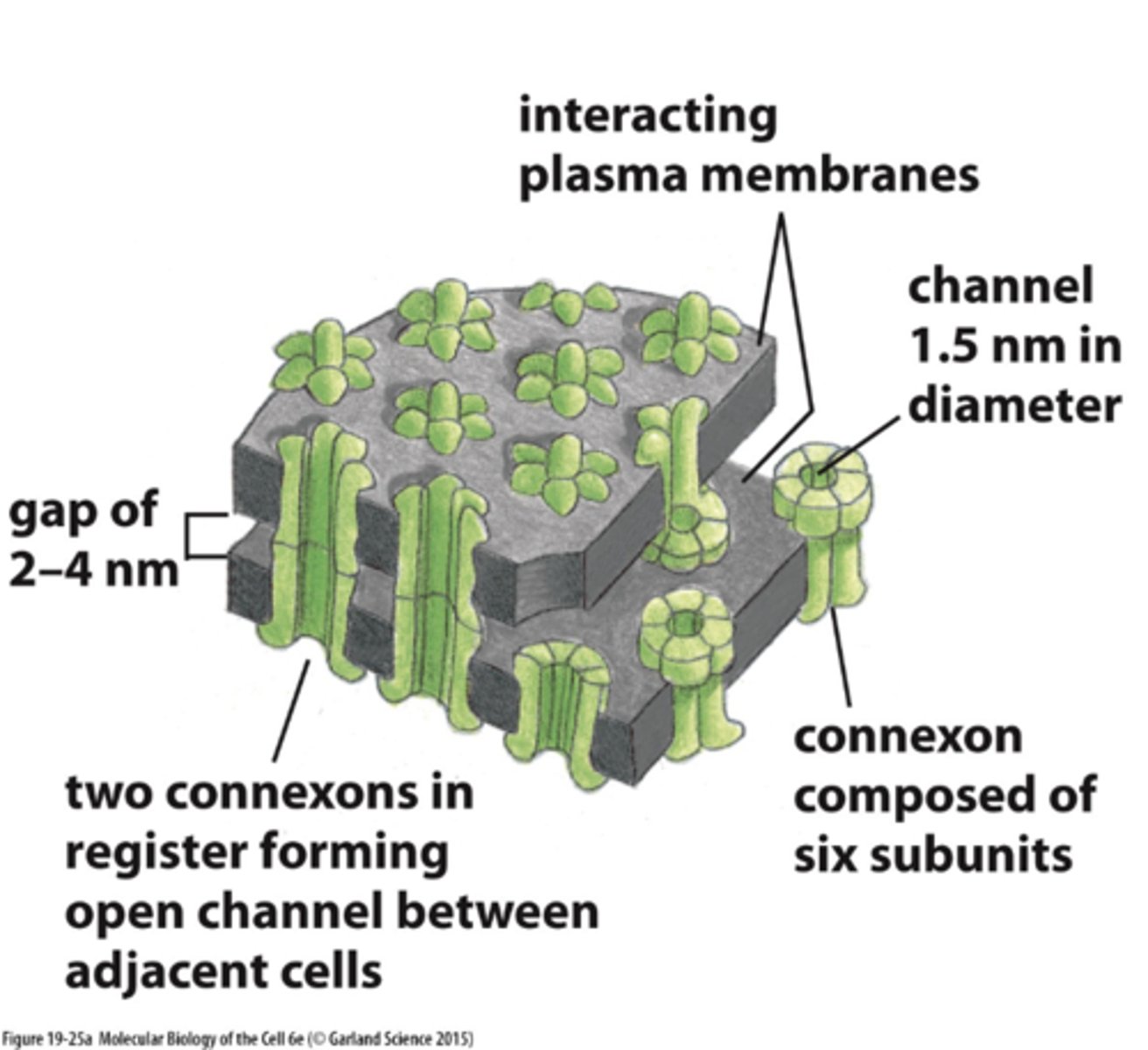
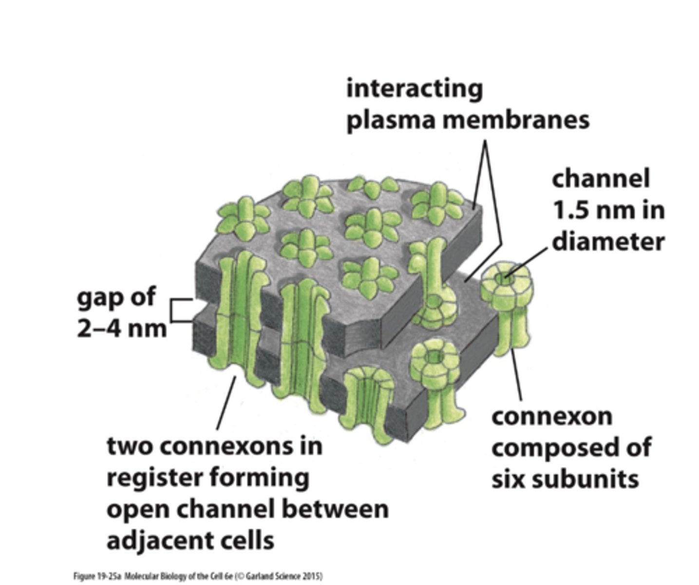
What are gap junctions function
bridges gaps between adjacent cells tocreate direct channels ---> separated by narrow, uniform gap (2 - 4 nm)(seen with TEM)

What electron microscopy help visual gap junction
TEM
Gap junction vs Plasmodesmata
Gap junction - in animals
Plasmodesmata
--> both are "channels"

What is the pores size of gap junction + what size of molecules can go through ?
1.4 nm
--> small molecules (<1000 Da) , can pass
List 6 molecules that can pass through gap junctions
--> small molecules (<1000 Da) , can pass
1. inorganic ions
2. sugars
3. amino acids
4. nucleotides
5. vitamins
6. signalling molecules (cAMP, IP3)
passage of ______ and ___________ molecules in gap junctions... means that epidermal cells are metabolically and electrically connected
passage of inorganic ions and small water-soluble molecules in gap junctions... means that epidermal cells are metabolically and electrically connected
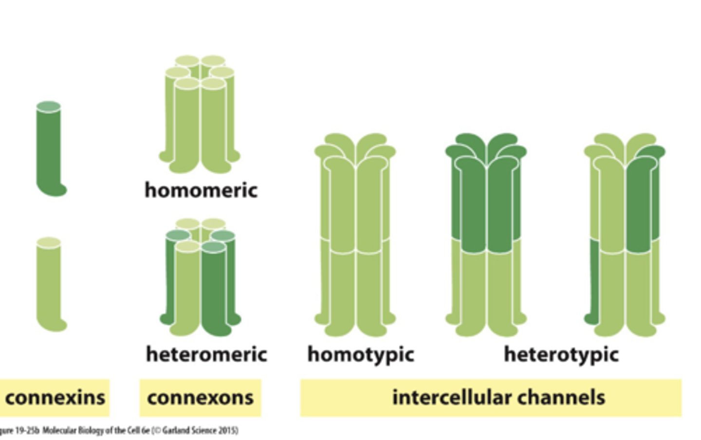
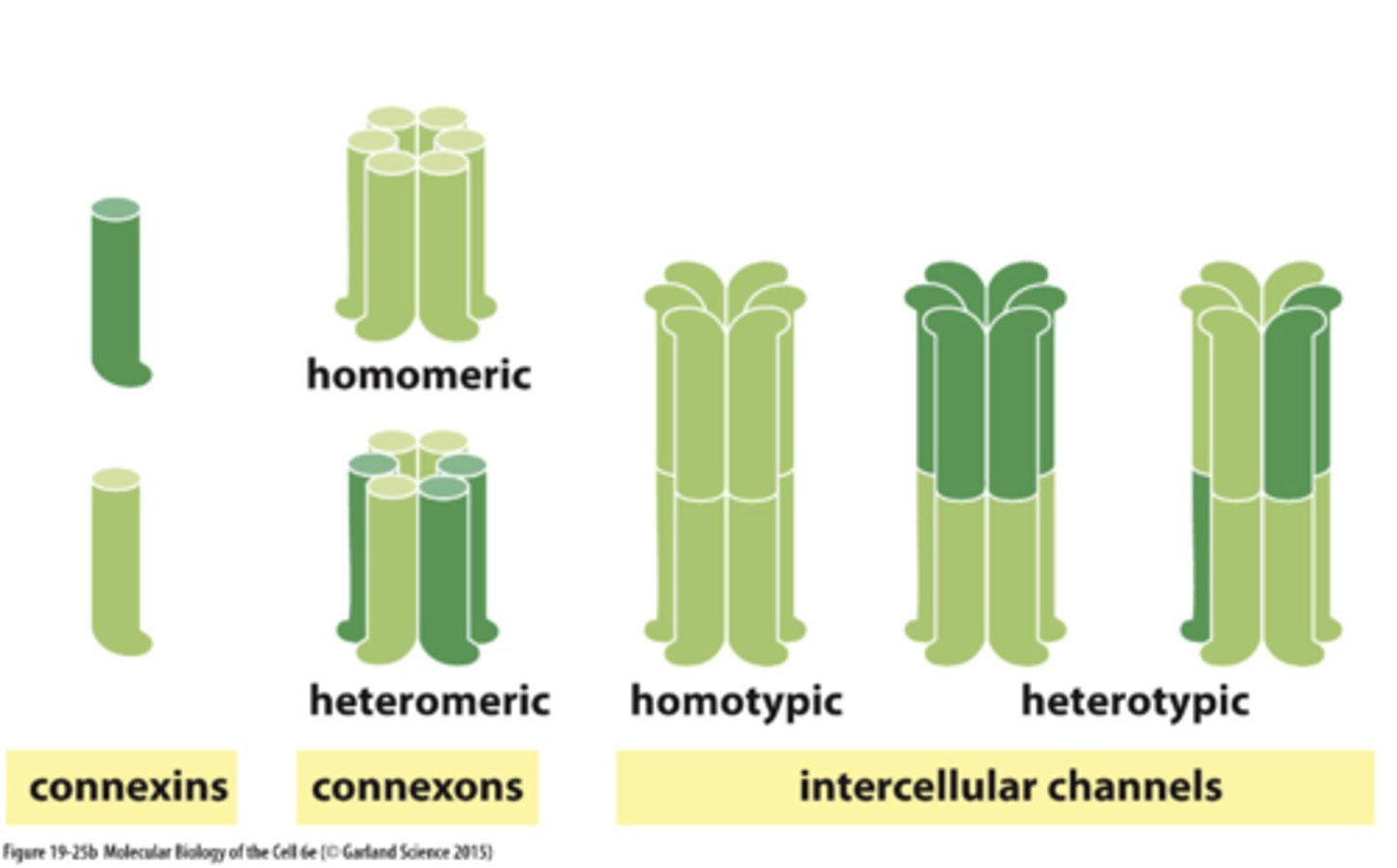
What are gap junctions composed of (proteins) (2)
1. connexins
2. innexins
How are connexins innexins similar and different to each other
unrelated in sequence but similar in shape andfunction
______ connexins assemble to form hemichannel =connexon
six connexins assemble to form hemichannel =connexon

How does 2 cells form a continuous aq channel between eachother
connexons in PM are in contact + aligned
gap junctions in different cells have different properties/permeabilities due to-________ connexins
gap junctions in different cells have differentproperties/permeabilities due to different connexins
gap junction channels switch between "open" and"closed" states, in response to specific ______ (as for ion channels)
gap junction channels switch between “open” and“closed” states, in response to specific __stimuli ____ (as for ion channels)
What confers gap junction function
types of connexis that bind together to from connexions
--> homotypic vs heterotypic vs homomeric vs hetermeric
Each gap junctional plaque is ______ - can be assembled,disassembled, remodelled;
Each gap junctional plaque is dynamic – can be assembled,disassembled, remodelled;
--> New connexons continually added around periphery; oldconnexons removed from core; half-life of a few hour
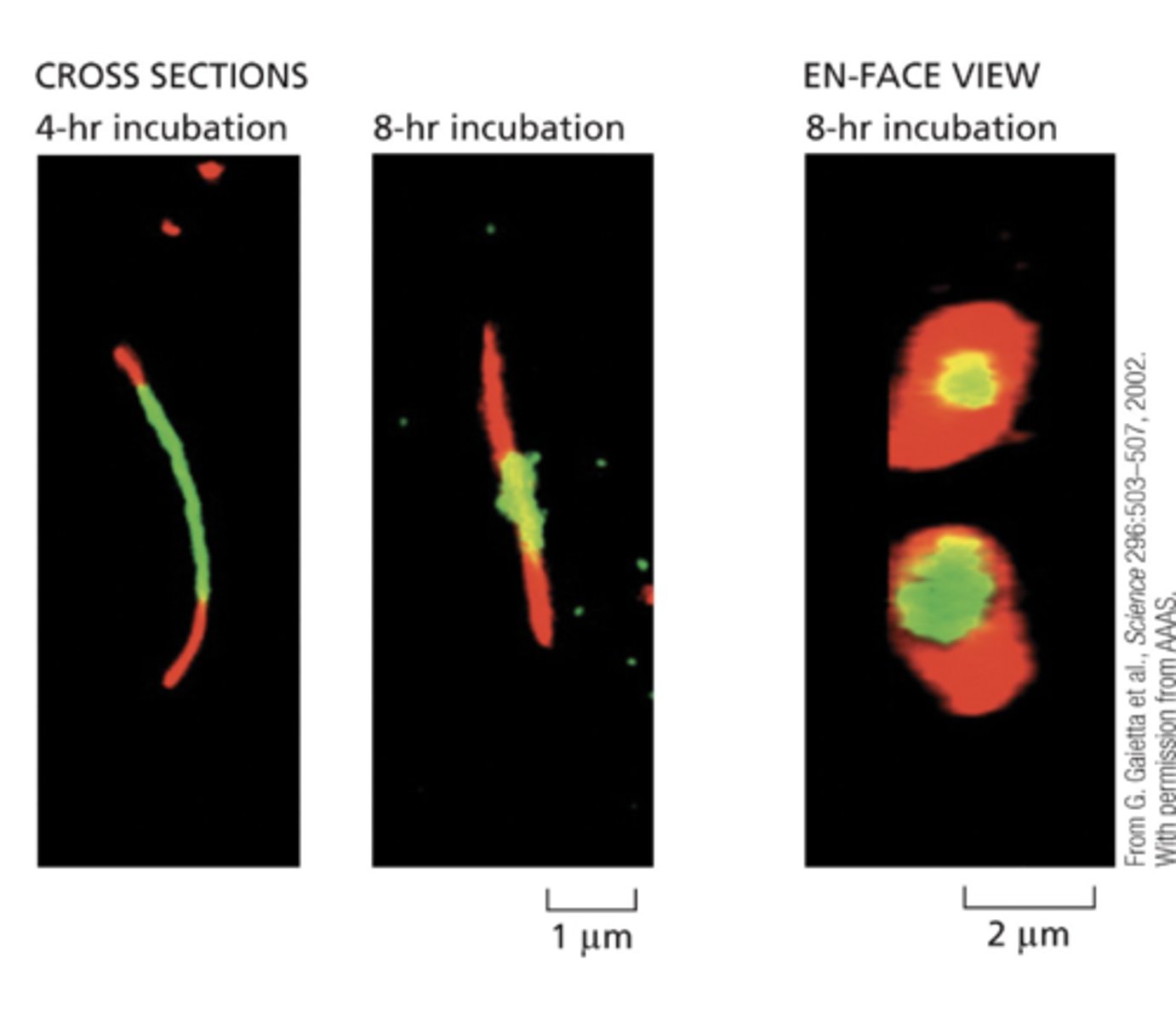
Route of delivery of new connexons is via ________; then diffusion in plane of membrane until encounter periphery of plaque and get trapped.
Route of delivery of new connexons is via exocytosis; the ndiffusion in plane of membrane until encounter periphery of plaque and get trapped.

What is the ECM
Extracellular Matrix (ECM) -Functions IN support, regulation, survival, development, migration, proliferation,shape, function
What produces the ECM in connective tissue vs cartilage vs bone
Connective - fibroblast
Cartilage - chondroblast
Bone - osteoblast
What is the basal laminate
s specialized ECM found in epithelial tissue, almost all multicellularanimals
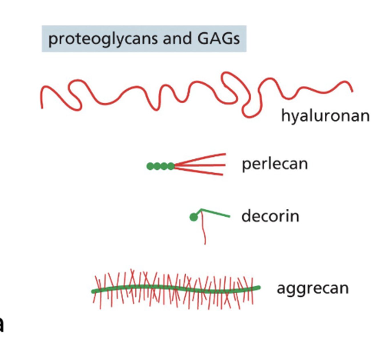
What does proteogylcvans linked with GAGs form?
form hydrated, gel-likesubstance (resist compression, allow diffusion)\
What are N-linked glycoprotines make
Non-collagen
What does the basal laminate compost of? (4)
All are glycoproteins
1. Laminin
2. Type IV collagen
3. Nidogen
4. Perlecan
What molecules are found in mesh like network of the ECM (5)
1. Collagen XV11
2. Fibronectin
3. Elastin
4. Decorin
5. Hyaluronan
The primary organizer of the basal lamina is (and also seen in early development)
laminin
What does GAGs stand for
Glycosaminoglycans (GAGs
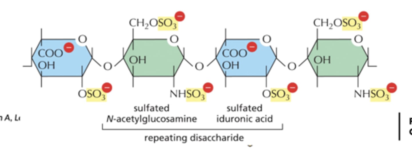
What are Glycosaminoglycans (GAGs) 3
1. unbranched polysaccharide chains composed of repeating disaccharide
2. 1 of sugars always either GlcNAc or GalNAc, usually sulfated
3. 2nd sugar usually uronic acid (glucuronic or iduronic)- highly negatively charged thus very hydrophilic

What are the 4 main groups of Glycosaminoglycans (GAGs)
1. hyaluronan
2. chondroitin sulfate
3. dermatan sulfate
4. heparansulfate; keratan sulfate

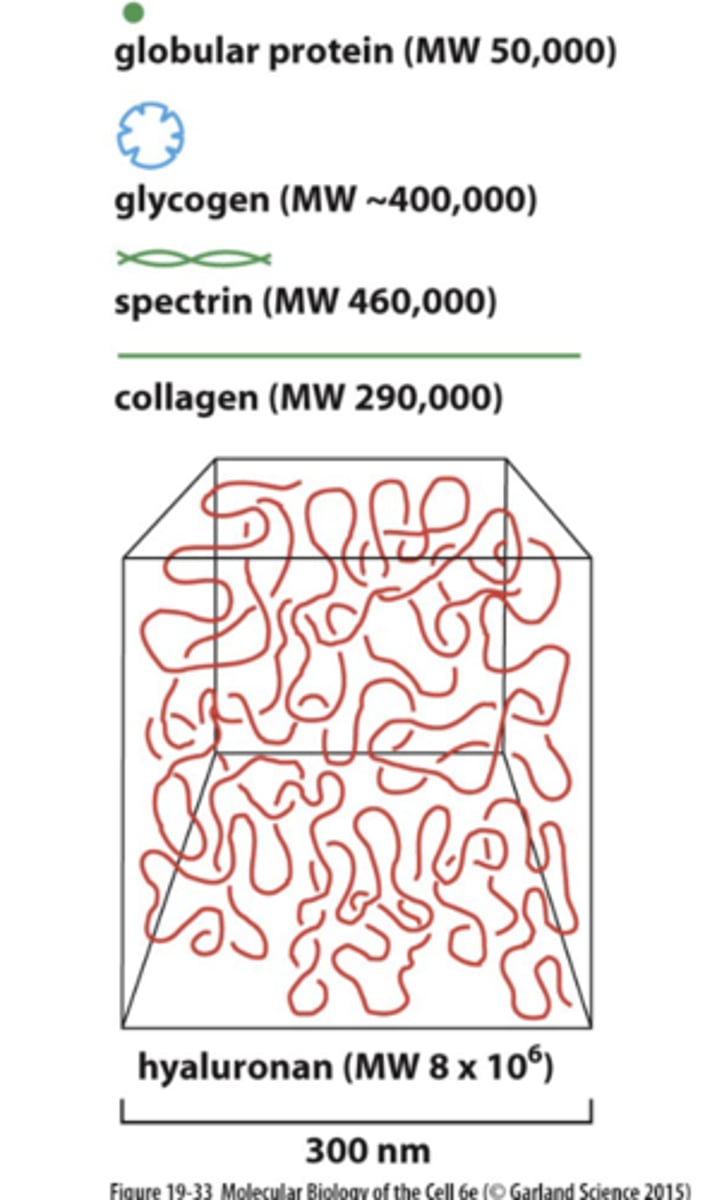
GAGs occupy huge _____ relative to mass, tend to adopt extended conformations, form hydrated gels at low concentrations
GAGs occupy huge _volume __ relative to mass, tend to adopt extended conformations, form hydrated gels at low concentrations
--> weight of GAGs about 10% of that of proteins
What does the GAGs do?
attracts “cloud” of cations, esp Na+, that are osmotically active, which causes large amounts of water to be sucked into matrix. ---> creates turgor, enables matrix to resist compression
What is the most simplest GAGs
hyaluronan (repeat, up to 25,000 disaccharide units)
Where are hyaluronan found in (4)
1. all tissues and fluids
2. especially abundant in embryo
3. required for compression resistance; high amounts in wound healing
4. lubricant in joints
How is hyaluronan different from other GAGs (2)
1. no sulfation, disaccharides identical, chain length verylong, not generally linked to protein core
2. spun out directly from complex (embedded in PM) at cell surfac

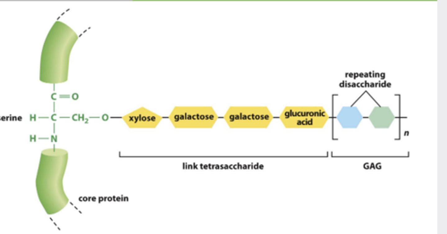
GAGs that are not hyaluronan ... how are they linked + synthesized + and secreted?
ll other GAGs linked to protein core, synthesized inside cell & exocytose
What are core protein
protein that attaches to GAG
(directly or indirectly )
