6. afferent processes
1/75
There's no tags or description
Looks like no tags are added yet.
Name | Mastery | Learn | Test | Matching | Spaced | Call with Kai |
|---|
No analytics yet
Send a link to your students to track their progress
76 Terms
afferent vs efferent neurons
afferent neurons carry sensory msgs to brain
efferent neurons carry motor msgs to the muscles
stimulus
any energy capable of exciting a receptor
3 kinds of stimulus-dependent(?) receptors
mechanoreceptors - touch, pressure, vestibular, audition
chemicoreceptors - taste, olfaction
photoreceptors - light
4 things required to perceive a sensation
a stimulus sufficient to initiate a response in the NS must be present
a receptor must convert the stimulus to a nerve impulse
the conduction of the nerve impulse must be transmitted from the receptor to the brain
interpretation of the impulse must occur in a specific portion of the brain
2 things all receptors do
absorb physical energy from the env
convert (transduce) the energy into a neural impulse
rate law
the lvl of response is determined by the intensity of the stimulation
2 types of receptors
tonic receptors
phasic receptors
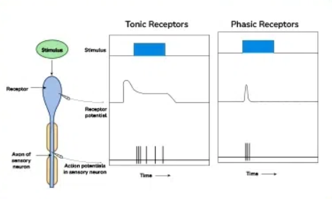
tonic receptors
continue to fire at a relatively constant rate as long as the stimulus is maintained, slow to adapt
phasic receptors
respond w a burst of APs when the stimulus is first applied, but quickly reduce their rate of firing when the stimulus is maintained
sensory receptors + what it does
specialized nerve cells that transduce energy into neural signals, lack axons but form synapses w dendrites of other sensory neurons
CANNOT PRODUCE APs — only inc or dec NT released in response to detection of stimunli
law of specific nerve energies
sensory messages are carried via separate pathways to different areas of the brain, but all use the same neural impulse
the range of energy lvls which eyes, ears, and taste buds detect
eye: 380-760 nM (wavelength)
ear: 20-20,000 Hz (frequency)
taste buds: specific chemicals
function of visual system + 3 functions of vision
detect electromagnetic radiation (EMR) emitted by objects
discriminate figure from background (food or rock?), detect mvmt (predator/prey?), detect color (is fruit ripe?)
wavelength
measured in nM, related to perceived color/hue
intensity
amplitude of radiation, related to brightness
an eye consists of 3 things:
aperture (pupil) to admit light
lens that focuses light
photoreceptive elements (retina) that transduce the light stimulus
describe the path of light in the eye
cornea → aqueous humor → pupil (opening in iris) → lens (rounded for nearby objects; flattened for distant objects) → vitreous humor → retina (image is upside-down and reversed)
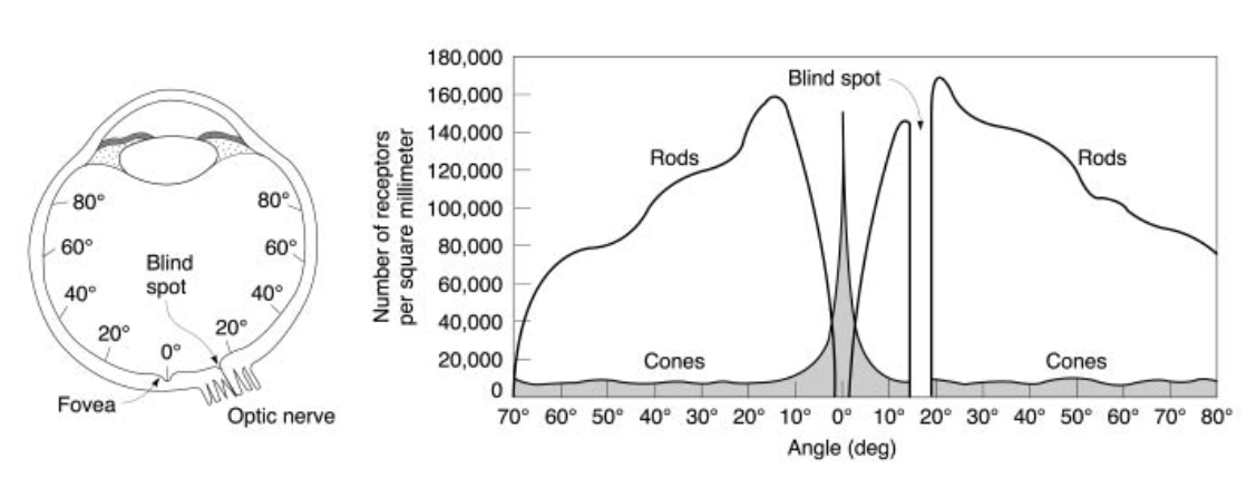
blind spot + where is it closest to
bundle of axons running back to the brain, has no photoreceptors, closest to the nose
how is color vision in peripheral vision?
poor
floaters
dead photoreceptors
3 types of eye mvmts
vergence mvmts
pursuit mvmts
saccadic mvmts
vergence mvmts
the eyes rotate to keep an object on corresponding parts of the retina
pursuit mvmts
following an object to keep it on the same part of the retina (the fovea, to maximize detail and color appreciation)
saccadic mvmts
eyes fixate on objects during “smooth pursuit”
eyes also “jump back and forth” — they are not stationary?
light passes through the … and is focused by the … onto the … at the back of the eye
pupil, lens, retina
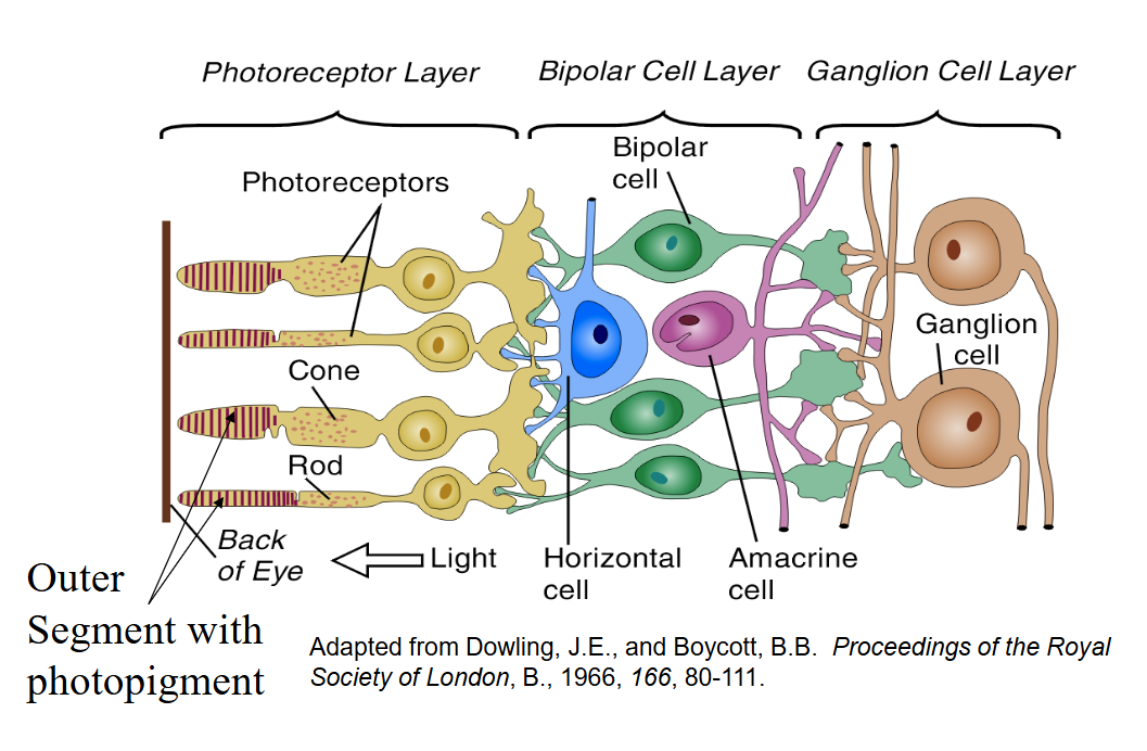
3 layers of cells of the retina
photoreceptor layer (back of eye) - transduce light into electrochemical impulses (eventually, APs), FARTHEST FROM LIGHT
bipolar layer
ganglion cell layer, CLOSEST TO LIGHT
2 types of photoreceptors + traits
rods - light sensitive (not color), found in periphery of retina, low activation threshold
cones - color sensitive, found mostly in fovea (center of retina)
the outer segments of a rod or cone contain different … that react to light
photopigments
what does each photopigment consist of? what do they do?
an opsin (a protein) and a retinal (a lipid)
in the dark, membrane Na+ and Ca2+ channels are kept open by cGMP → NT (glutamate) is released which hyperpolarizes the bipolar cell membrane
light splits the photopigment — opsin and retinal — apart
activates G protein (transducin)
activates phosphodiesterase → destroys cGMP
reduced cGMP → reduced NT release (closes presynaptic Na+ and Ca2+ channels)
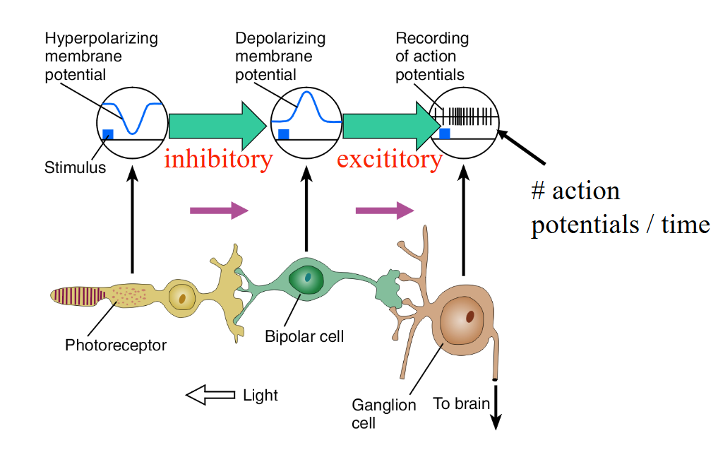
net effect of light is to … the photoreceptors and … the release of NT
hyperpolarize, reduce
“on” vs “off” cells/pathways
on - detects a stimulus and ganglia cells turn on (this is what we’re focusing on)
off - detects a stimulus and turns off
“bleached” photoreceptor
occurs when light splits the photopigment, causing it to become white, inhibits the bipolar cells less
signals from the ganglion cells of the retina are sent to the … via the …
thalamus, optic nerve/tract
6 layers of the lateral geniculate nucleus (LGN) + functions
magnocellular layers - the inner 2 layers that contain large cells
form, mvmt, depth, brightness
parvocellular layers - the outer 4 layers that contain small cells
color and fine detail discrimination
neurons of the LGN project thru the optic radiations to a region of occipital cortex termed …
primary visual cortex (striate)
vision researchers say we have 4 retina. how?
we have 2 on each eye
2 retinas on the outside of the eyeball are TEMPORAL RETINAS
2 retinas close to the inside are NASAL retinas
light from LVF and RVF strike what side of both eyes?
light from LVF strikes RIGHT side of the eyes
light from RVF strikes LEFT side of the eyes
what part of the LGN do nasal and temporal retinas go?
nasal retinas transmit info to the CONTRALATERAL LGN of the thalamus
temporal retinas transmit info to the IPSILATERAL LGN of the thalamus
optic radiation
transfers visual info back to the brain
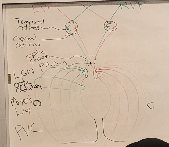
Meyer’s loop
outer areas of the optic radiation, located LOW going into the temporal lobes and processes info that’s HIGH (superior vision)
medial areas of the optic radiation
location is HIGH and goes up to the parietal lobes but processes info that’s LOW (inferior vision)
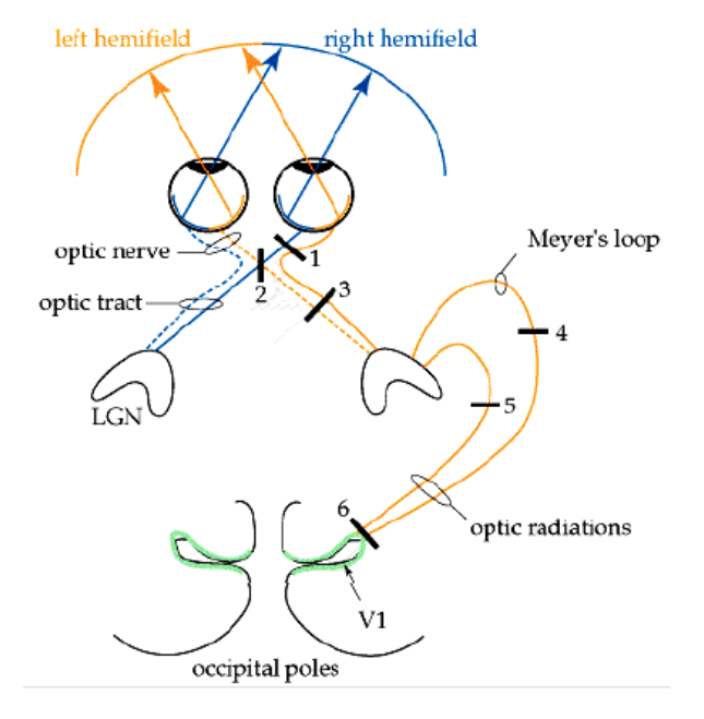
lesion to right eye
everything being transduced in right eye is lost → CAN’T SEE FROM RIGHT EYE

lesion to optic chiasm + potential cause
inputs from the nasal retina are cut, so you’d lose PERIPHERAL VISION on both sides. can be caused by a pituitary tumor

lesion to Region 3
loss of left hemifield (left nasal retina, right temporal retina). both eyes will be blind to anything on the left side of the world
anopsia
inability to see
left hemianopsia
lost the left side of the world

lesion to region 4
right Meyer’s loop has been cut, so vision will be lost in the upper visual world but only in the left hemifield (so upper left)
left superior quadrantanopsia

lesion to region 5
parietal portion of the optic radiations were cut, so the lower visual world would be affected on one side
left inferior quadrantanopsia

lesion in region 6 (primary visual cortex)
at first, appears to be a straightforward loss of one hemifield, but vision at the fovea is spared (foveal sparing), perhaps bc there’s such a large representation of fovea in the cortex
what happens if we lesion the parvocellular layers of the LGN in the right hemisphere?
we lose color vision in the LVF
ganglion cells in the retinal periphery vs ganglion cells in the fovea
ganglion cells in the retinal periphery receive input from many photoreceptors
ganglion cells in the fovea receive input from one photoreceptor → much higher resolution at fovea, higher sensitivity
why are cones better at detecting small details?
they’re highly dense in the fovea and ganglion cells hook up 1:1 for every high resolution (tho there’s a higher threshold necessary compared to a rod for a cone to activate)
why are rods better for night vision?
they’re extremely sensitive (only need 1 rod to activate the ganglion cell bc many of them are attached to one ganglion cell)
to see better in the dark, use your periphery vision instead of the fovea
horizontal cells functions
perform “lateral inhibition” — inhibit cells directly adjacent to them
at the edge of light activation, bipolar cells at the edge that are in darkness are still inhibited
allow us to see the edges of light very clearly (Mach Bands)
2 color vision theories
trichromatic theory
opponent theory
trichromatic theory
argued there are 3 different receptors in the eye, w each sensitive to a single hue
blue, red, green
any color could be accounted for by mixing 3 lights in various proportions
opponent theory
notes that ppl perceive 4 primary colors: yellow, green, blue, red
negative color afterimages suggest that red and green are complementary colors as are blue and yellow
photoreceptors in primate retina and how it supports trichromatic theory
3 types, and each cone uses a different opsin which is sensitive to a particular wavelength (blue, red, green), supporting trichromatic theory
at the ganglion cell lvl, how does the system respond?
in an opponent-process fashion (yellow on, blue off. blue on, yellow off. red on, green off. green on, red off.)
firing rate increases to green/yellow, decreases for red/blue
rebound effect + what does it cause?
when ganglion cells fire faster or slower than normal due to being excited or inhibited for a prolonged period of time
negative afterimages
protonopia
an inherited form of defective color vision in which red and green hues are confused; “red” cones are filled w “green” cone opsin (can’t see the color red)
see the world in shades of yellow and blue; both red and green look yellowish to them
deuteranopia
an inherited form of defective color vision in which red and green hues are confused; “green” cones are filled with “red” cone opsin (can’t see the color green)
see the world in shades of yellow and blue; both red and green look yellowish to them
tritanopia
an inherited form of defective color vision in which hues w short wavelengths are confused; “blue” cones are either lacking or faulty
have trouble seeing yellows and blues
see the world in greens and reds
blue looks green and yellow looks pink
rare
microelectrodes
used to record the firing activity of a single sensory neuron (impulses/sec)
primary visual cortex aka … aka …
striate cortex, V1
orientation sensitivity
some cells fire best to a stimulus of a particular orientation and fire less when orientation is shifted (striate cortex)
spatial frequency (low vs high)
cells vary firing rate according to the sine wave frequency of the stimulus (V2)
low frequency = large areas of light/dark
high frequency = fine details
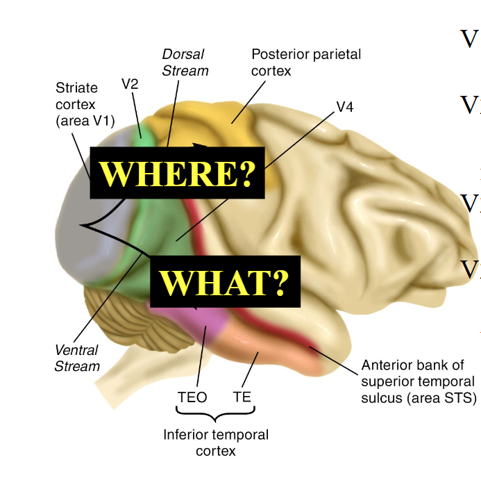
V3 of brain is involved in
gestalt processing
V4 of brain
only in humans, part of learning process
have categories of neurons (e.g., 9, dogs, horses, etc.)
for symbolic, visual association, learning
list out the basic functions of V1 to V4
V1 - orientation
V2 - shape/object
V2-V4 temporal: what
V2-V4 parietal: where
fusiform face area (FFA) (location, what determines the degree to which we respond, its role in autism)
at base of brain, grouping of neurons that responds to faces of all kinds
degree to which we respond to activation related to lvl of expertise
ASD (autism) tends to have reduced activation of FFA; they avoid faces, can’t understand facial expressions well
but their FFA lights up for their fixations → indicates expertise
dorsal stream
dorsal → parietal lobe: where/how stream; where is an object in space and how to interact with it?
ventral stream
ventral → temporal lobe: what stream; what is an object? shape, object recognition
2 clinical symptoms of lesions to temporal and parietal lobes
temporal
trouble identifying objects by sight (touch should help)
trouble w “boston naming test”
parietal
trouble w location/mvmt of objects
grasping problems
anomia
may be indicated by failure in boston naming test (BNT), is the inability to name objects due to a lesion in the Broca’s Area (indicated by if giving a phoneme cue helps them name it)
prosopagnosia
inability to recognize faces, even close friends and family
area just below FFA affected: anterior inferior temporal region (Alzheimer’s degradation)