The Peripheral Nervous System
1/96
There's no tags or description
Looks like no tags are added yet.
Name | Mastery | Learn | Test | Matching | Spaced | Call with Kai |
|---|
No analytics yet
Send a link to your students to track their progress
97 Terms
What is the central nervous system composed of?
The brain and spinal cord
What is the peripheral nervous system composed of?
The cranial and spinal nerves
What is a plexus?
Branching network where nerve fibres from different spinal segments cross over and combine, so that all fibres going to a specific body part are together in one nerve.
What is a ganglion?
A collection of nerve cell bodies
What is the parasympathetic nervous system responsible for?
Autonomic, rest and digest
What is the sympathetic nervous system responsible for?
Autonomic, fight or flight
What does decussate mean?
Where nerves cross over onto the opposite side of the body.

What cranial nerve is labeled A?
Olfactory (I)

What cranial nerve is labeled B?
Oculomotor (III)

What cranial nerve is labeled C?
Trochlear (IV)

What cranial nerve is labeled D?
Abducens (VI)

What cranial nerve is labeled E?
Vestibulocochlear (VIII)

What cranial nerve is labeled F?
Hypoglossal (XII)

What cranial nerve is labeled G?
Accessory (XI)

What cranial nerve is labeled H?
Optic (II)

What cranial nerves is labeled I?
Trigeminal (V)

What cranial never is labeled J?
Facial (VII)

What cranial nerve is labeled K?
Glossopharyngeal (IX)
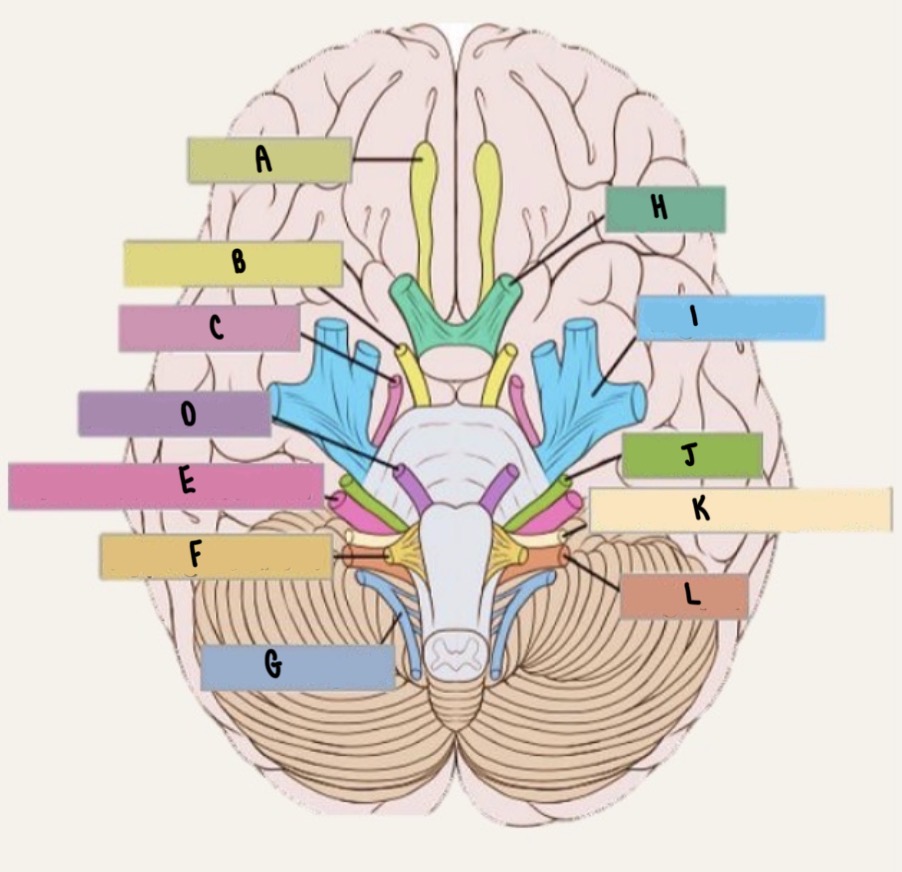
What cranial nerve is labeled L?
Vagus (X)
What are the three possible compositions of cranial nerves?
Purely sensory, purely motor or mixed
Briefly describe the action of the olfactory nerve (I):
Chemical signals stimulate sensory cells in the nasal epithelium which generate an action potential. Sensory afferent fibres attach to the olfactory bulb via the cribiform plate.
Where is the olfactory bulb found?
In the rostral forebrain
What are the three neurons involved in the sequence of sensing light in the optic nerve (III)?
Bipolar cell - receives information from the neuroepithelium of the retina.
The ganglion cell - its axon continues through the optic chiasm and along the optic tract on the opposite side.
Has its cell body in the lateral geniculate nucleus - its axons project through the optic cortex in the occipital cortex in the optic radiation.
Briefly describe the pupillary light reflex:
a light is shone into one eye
this is detected by the retina and conveyed by the optic nerve through the optic chiasm and along the optic tract.
Parasympathetic fibres of the oculomotor nerve are stimulated to signal to the pupil via the ciliary ganglion and ciliary nerves to the muscles of the pupil.
What are the three primary branches of the trigeminal nerve?
Opthalmic (V1) Maxillary (V2) and Mandibular (V3)

What nerve innervates the area shaded in light blue?
Opthalmic branch of the trigeminal nerve
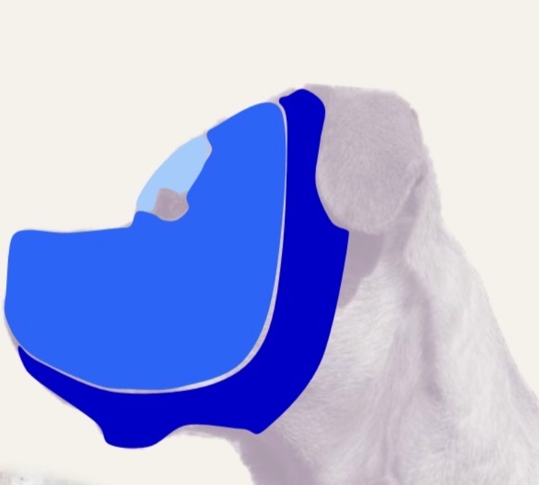
What nerves innervates the area shaded in royal blue?
Maxillary branch of the trigeminal nerve

What nerve innervates the area shaded in dark blue?
The Mandibular branch of the trigeminal nerve
What is the function of the opthalmic branch of the trigeminal nerve?
Sensory to the orbit
What is the function of the maxillary branch of the trigeminal nerve?
Sensory to the upper eyelid, nasal mucosa, upper teeth, lips and nose.
What is the function of the mandibular branch of the trigeminal nerve?
Sensory to the cheek, lower lip, lower teeth, tongue and some skin of the head. Motor to muscles of mastication, ventral and throat palate.

What branch of the facial nerve is labeled A?
The Auriculpaplpebral

What branch of the facial nerve is labeled B?
The Caudal auricular

What branch of the facial nerve is labeled C?
The Ventral buccal
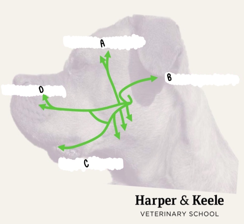
What branch of the facial nerve is labeled D?
Dorsal buccal
What is the motor function of the facial nerve (VIII)?
Control of the muscles of facial expression and superficial muscles of the head, external ear, caudal digastricus, stapedius, stylohoideus and playsma.
What is the parasympathetic function of the facial nerve?
Innervates the lacrimal, salivary and mucosal glands
What is the facial nerves roles in special taste?
The Chordae tympani branch is responsible for taste to the palate and rostral 2/3 of the tongue.
What are some of the clinical signs of paralysis of the facial nerve?
Inability to blink, drooping of the face and dry eye. All of which can be unilateral. Can be caused by an ear infection.
Briefly describe the action of the vestibular root of the vestibulocochlear nerve:
Innervates the hair cells of labyrinth, cell bodies are in the vestibular ganglion, sends afferent signals to the brain on head position, plays an important role in balance.
Briefly describe the cochlear root of the vestibulocochlear nerve:
Cell bodies are in the spiral ganglion, innervates hair cells in the cochlear duct, plays a key role in the special sense of hearing.
Where are the cell bodies of the glossopharyngeal nerve found?
In the nucleus ambiguous, branches into the pharyngeal, lingual and tympanic nerves.
What is the sensory function of the glossopharyngeal nerve?
Innervates the pharynx, middle and external ear, caudal tongue and is responsible for the special sense of taste from caudal tongue.
What is the parasympathetic function of the glossopharyngeal nerve?
Innervates parotid and zygomatic salivary glands.
What is the motor function of the Glossopharyngeal nerve?
Innervates the stylopharyngeus muscle
Where are the cell bodies of the vagus nerve (X) found?
In ambiguous nucleus
What is the motor function of the vagus nerve?
Innervates the larynx, pharynx, palate and oesophagus
What is the sensory function of the vagus nerve?
Innervates the caudal tongue, pharynx and larynx, taste to epiglottis and palate.
What is the parasympathetic function of the vagus nerve?
Innervates the thoracic and abdominal viscera
Describe the recurrent laryngeal nerve?
The laryngeal branch of the vagus goes down into the thoracic region and wraps around the aorta before coming back up the innervate the larynx.
Briefly describe the accessory nerve (XI):
Passes through the jugular foramen, motor to the trapzezius and cleidocephalicus (dorsal branch,) and sternocephalicus (ventral branch.)
Briefly describe the hypoglossal nerve (XII):
Very caudal to the brainstem, passes through the hypoglossal canal, motor to the muscles of the tongue.
Which of the cranial nerves are purely sensory?
Olfactory I, Optic II and vestibulocochlear VIII
Which of the cranial nerves are purely motor?
Occulomotor III, trochlear IV, Abducens VI, Accessory XI, Hypoglossal XII.
Which of the cranial nerves is mixed?
Trigeminal V, facial VII, glossopharyngeal IX and vagus X.
Which of the cranial nerves have parasympathetic innervation?
Oculomotor III, facial VII, glossopharyngeal IX and vagus X.
What is the brachial plexus?
A bundle of nerves that innervates the forelimb.
Where does the brachial plexus extend from?
C6-T2
What are the major nerves that form the brachial plexus?
Suprascapular, musculocutaneous, radial, median and ulnar nerves.
Briefly describe the location of the suprascapular nerve:
C6-C7. Leaves the BP and passes between the supraspinatus and subscapularis. Winds around the cranial neck of the scapular to the lateral aspect.
What is the function of the suprascapular nerve?
Innervates the supraspinatus and infaspinatus nerevs.
Briefly describe the location of musculocutaneous nerve:
C7 (C6-8). Passes through the axilla and branches supply coracobrachialis and biceps.
What is the function of the musculocutaneous nerve:
Communicates to the median nerve. Branches distally to supply the brachialis, crosses the elbow to supply the skin.
Briefly describe the location of the radial nerve:
C6-T2. Divides between the heads of the triceps. Spirals round the humerus to the craniolateral aspect of the limb.
What is the function of the radial nerve?
Innervates the tricpes, tensor fasciae anibrachii, anconeus and extensors of carpus and digits. Sensory to cranial paw.
Briefly describe the location of the median nerve:
C7/8-T1. Runs doen the medial surface and passes caudally under the flexor carpi radialis.
What is the function of the median nerve?
Supplies the carpal and digital flexors.
Briefly describe the location of the ulnar nerve:
C8-T2, runs beside the median nerve before deviating toward the olecranon.
What is the function of the ulnar nerve?
Supplies the carpal and digital flexors.

What nerve innervates the area of skin labeled 1?
Axillary
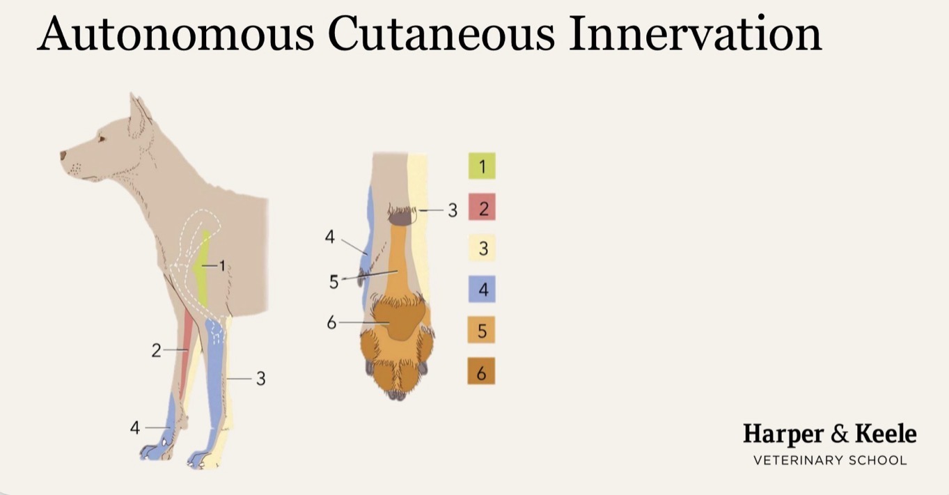
What nerve innervates the area of skin labeled 2?
Musculocutanous

What nerve innervates the area of skin labeled 3?
Ulnar

What nerve innervates the area of skin labeled 4?
Radial nerve

What area of skin innervates the area of skin labeled 5?
Median nerve

What nerve innervates the area of skin labeled 6?
Median and ulnar
Briefly describe a cranial brachial plexus avulsion:
Weak shoulder extension, little to no shoulder flexion, no elbow flexion. Sensation and deep pain.
Briefly describe a caudal brachial plexus avulsion:
Proprioceptive deficit, dropped limb, knuckling, limited sensation and panniculus reflex, may have partial horners syndrome.
Briefly describe a complete brachial plexus avulsion:
Non-weight baring, no pain sensation in the distal limb
Briefly describe why horners syndrome is commonly seen alongside a caudal avulsion:
The sympathetic supply to the head often get damaged at the same time leading to clinical signs being seen in the face.
Where does the lumbosacral plexus extend from?
L4-S3
What are the main nerves that form the lumbosacral plexus?
Sciatic, femoral, saphenous and obturator.
Briefly describe the location of the sciatic nerve:
L6-S1. Crosses the hip deep to the gr. trochanter. Lies beneath the biceps femoris. Gives rise to the peroneal and tibial nerves above the stifle.
What is the function of the sciatic nerve?
Innervates the hamstrings
What are two ways in which the sciatic nerve can be injured?
during hip surgery
IM injections
Briefly describe the location of the femoral nerve:
L4-6. Passes through the psoas muscles between the sartorius and pectineus. Gives rise the the saphenous nerve.
What is the function of the femoral nerve?
Supplies the quadriceps
What is the function of the saphenous nerve?
Supplies the sartorius and continues down the metatarsus to supply the skin.
Briefly describe the location of the obturator nerve:
L4-6. Passes through the obturator foramen.
What is the function of the obturator nerve?
Innervates the medial thigh muscles (adductors)
When is the obturator muscle commonly injured?
In cows/horses during parturition.

What nerve innervates the area of skin labeled 1?
Caudal cutaneous femoral
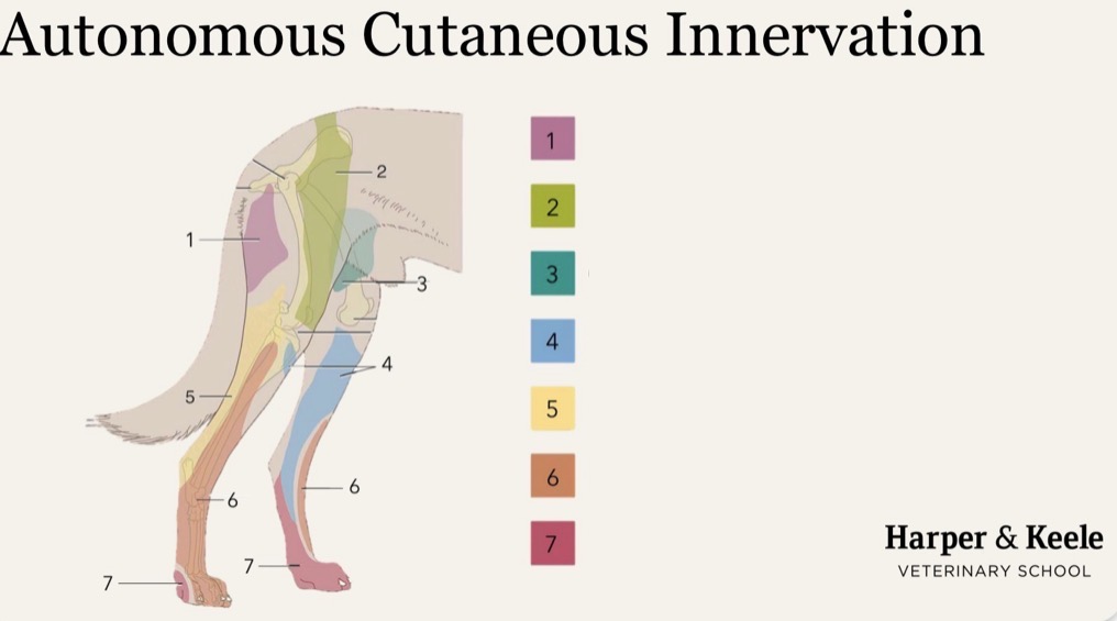
What nerve innervates the area of skin labeled 2?
Lateral cutaneous femoral

What nerve innervates the area of skin labeled 3?
Genitofemoral

What nerve innervates the area of skin labeled 4?
Saphenous

What nerve innervates the area of skin labeled 5?
Sciatic

What nerve innervates the area of skin labeled 6?
Peroneal

What nerve innervates the area of skin labeled 7?
Tibial