Functional and microanatomy of endocrine glands
1/24
There's no tags or description
Looks like no tags are added yet.
Name | Mastery | Learn | Test | Matching | Spaced | Call with Kai |
|---|
No analytics yet
Send a link to your students to track their progress
25 Terms
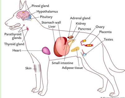
what are endocrine organs
Preserve Homeostasis
Ductless organs, derived from the 3 germ layers
Secrete hormones (chemical messengers) directly to blood, lymph or tissue fluid
Great communicator, feedback control mechanisms
Endocrine and nervous systems are integrated (neurohormonal system)
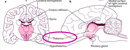
hypothalamus
Coordinates the activity of the pituitary gland through the secretion of peptides and amines
Located at the ventral part of the brain
Regulator of endocrine and nervous system
Regulates temperature, thirst, hunger, sexual behaviour, blood volume etc. maintain homeostasis
Hormones produced are of two types=releasing and inhibitory
nuclei in hypothalamus
Clusters of neurons in the Hypothalamus
Sends axons to the posterior pituitary
Secretes releasing hormones to the anterior pituitary
Intergrade and regulate vital body functions
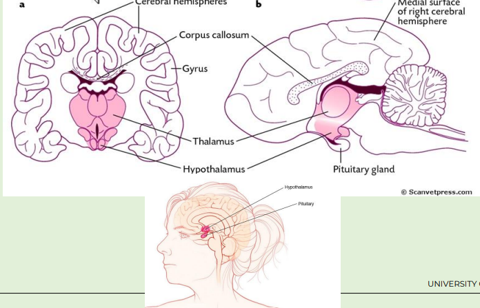
pituitary gland (hypophysis)
Structurally and functionally integrated with hypothalamus
Suspended below the hypothalamus by a narrow stalk (infundibular or hypophysial stalk)
Lies within bony cavity called the Hypophysial fossa (Sella Turcica) of the sphenoid bone
Small , oval gland
Between rostrally optic chiasm and the caudally mammillary bodies
Controlled by the CNS and feedback from target organs

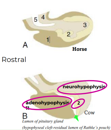
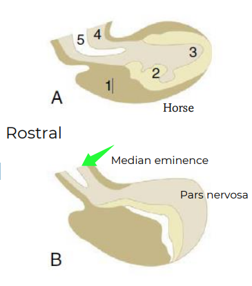
transverse and cross section of pituitary gland
the two anatomically and fuctionaly distinct lobes of pituitary gland
Adenohypophysis (Anterior lobe)- (oral ectoderm)
Outgrowth of pharynx (root of mouth)- adeno (glandular)
three parts (or two in some)
Neurohypophysis (Posterior lobe) –(neural ectoderm)
Diencephalon- outgrowth of the brain hence consist of
nervous tissue from hypothalamus
three parts
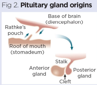
development of pituitary gland
During embryonic development, the roof of the mouth bulges upwards
(invaginates) to form, structure known as Rathke’s pouch,
Cells from posterior gland migrate down from brain
Rathke’s pouch, also known as the hypophyseal diverticulum, is an ectodermal outpouching of the stomodeum
Pouch normally closes early in fetal development
main parts of pituitary gland
1, Adenohypophysis
2, Pars Intermedia
3, Neurohypophysis
4, Hypophysial stalk
5, Recess of third ventricle
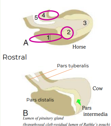
adenohypophysis
Adenohypophysis (anterior lobe)
• Pars distalis (1)
• Pars intermedia* (2)
• Pars tuberalis (forms the stalk)
*Pars intermedia is rudimentary in humans, lacking in birds
adenohypophysis pars distalis
Largest part of pituitary gland- many cell types
–Chromophils and Chromophobes
Chromophils (Acidophils and Basophils)
• Acidophils (1) Cells that contain the polypeptide hormones-will stain red or orange
• Somatotrophs
Growth hormone (GH)
somatotropin
• Lactotrophs
Prolactin (PRL)
Basophils in pars distalis of adenohypophysis of pituitary gland
• Basophils (2) Cells that contain the glycoprotein hormones-will stain bluish color
Thyrotrophs
Thyroid stimulating hormone (TSH)
Gonadotrophs
Follicle stimulating hormone (FSH)
Luteinizing hormone (LH)
Corticotrophs
Adrenocorticotropic hormone (ACTH)
Chromophobes in pars distalis of adenohypophysis of pituitary gland
3
• Chromophobes have cytoplasm that stains
very poorly
• • ? Postsecretroy acidophils and basophils
• • ? Stem cell
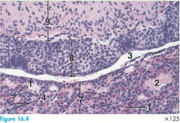
adenohypophysis - pars intermedia
Closely associated with pars nervosa
This part of the pituitary shows variation in size among species
Pars intermedia is a thin zone of basophilic cells (8)
Primary function is color change regulation
• Melanotrophs
• α-melanocyte stimulating hormone (α-MSH)
adenohypophysis - pars tuberalis
Provides scaffold for the capillary network of the hypophyseal portal system
Capillaries in this gland are fenestrated, to enable passage of hormones from the secretory cells into the bloodstream
Cells form folding sheets (folds) and occasional cysts
neurohypophysis
Median eminence (base of the hypothalamus)
Infundibular stalk (4)- nerve tract
Pars nervosa (3) bulk of the neurohypophysis
Neurohypophysis has a lumen (5) that's continuous with the lumen of the brain's third ventricle
neurohypophysis cont
Neuron cell bodies clustered in the hypothalamus (nuclei)
Send axons to the neurohypophysis
• Axons of hypothalamic neurons (unmyelinated)
• Central gliocytes (pituicytes-neuroglial cells)
• No neuron cell bodies
• Secretory vesicles along the axons = Herring bodies
• Oxytocin and Antidiuretic hormone (ADH) = vasopressin - promotes fluid reabsorption by kidneys (ADH)
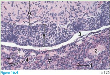
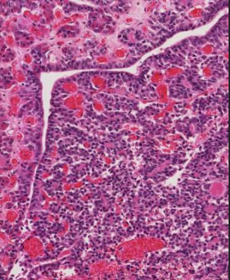
histology of neurohypophysis
9 pars nervosa
8 pars intermedia
3 cavity of Rathke's pouch
2 blood vessel
7 pars distalis
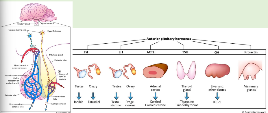
anterior pituitary hormones
Hypothalamus produces peptides and amines that influence the pituitary gland to produce tropic hormones* e.g. corticotropin which in turn influences production of cortisol by peripheral target tissues
They are produced by neurosecretory cells in the several hypothalamic nuclei
Most tropic hormones are produced and secreted by the anterior pituitary gland
Adenohypophysis produces several hormones designated by acronyms as growth hormone (GH), follicle-stimulating hormone (FSH)
*trope, turning-because they turn on endocrine glands or support
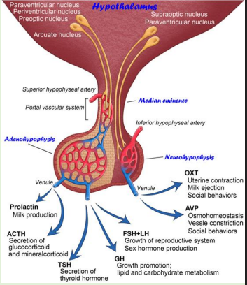
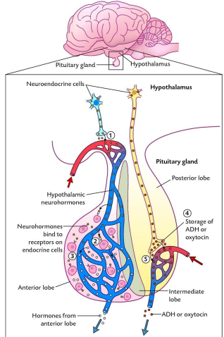
hypophysial portal system
Blood vessels in the brain that connects the hypothalamus with the adenohypophysis
It begins at the base of the hypothalamus
Arterial blood reaching hypothalamus branches to capillaries
Here joins venous blood and makes small portal vein that pass through the stalk into the adenohypophysis
Here they branch to anterior capillaries which in turn drain into the venous system
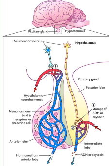
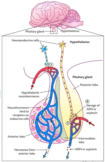
Adenohypophysis
1- Neurohormones from the hypothalamus are released to the Hypothalamic-pituitary portal system
2-Neurohormones bind to receptors on endocrine cells
3- Regulate their hormonal secretion (releasing or inhibiting)
HYPOTHALAMIC -HYPOPHYSIAL TRACT
Neurohypophysis
Neurohypophysis has cell bodies that originate in the hypothalamus
Secretes two neurohormones -ADH and oxytocin
These neurohormones are placed in vesicles and transported and stored each terminal stores either vasopressin or oxytocin
Hormones conveyed along axons and released into neurohypophyseal capillary bed
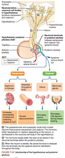
neurohypophysis
Oxytocin: Stimulates uterine contractions, milk let-down (hormone of love!)
Anti-diuretic hormone(ADH) or Vasopressin: Stimulates vasoconstriction and
promotes fluid reabsorption by the kidneys; constricts vessels to raise blood
pressure (arterioles)
Synthesized in the hypothalamus but released into the bloodstream in the pars
nervosa
Neurohypophysis is connected to hypothalamus by neural pathway, whereas adenohypophysis is connected by a vascular link
Unlike the neurohypophysis, which releases hormones that are synthesized by the hypothalamus, adenohypophysis itself synthesizes the hormones that it releases into the blood
Neurohypophysis of the pituitary does not have a portal system. Neurohormones are deposited directly into the capillaries
epiphysis cerebri (pineal gland)
Part of the epithalamus, located mid brain
Small organ, shaped like a pinecone
Attached to the caudal end of the roof of the third ventricle and directly before the rostral colliculi
Only endocrine gland directly influenced by the external environment via the retina
Secretes the hormone melatonin (derived from aa tryptophan)
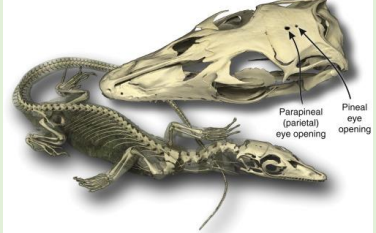
A vestigial eye (Many fish, and amphibians have a median pineal eye and/or parietal eye/third eye)
In lower vertebrates, the pineal gland is directly photosensitive
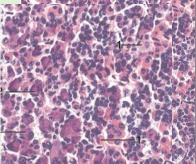
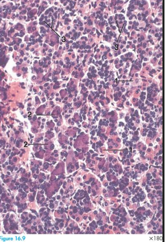
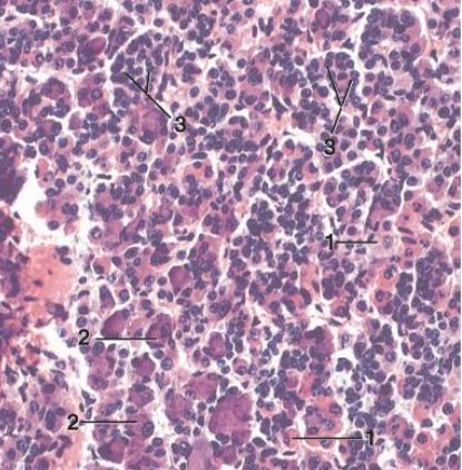
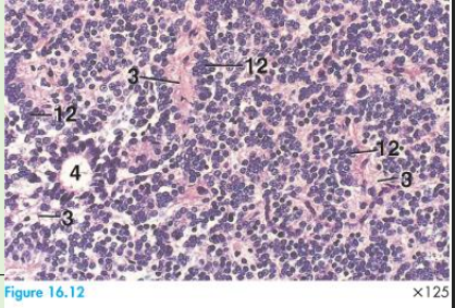
histology of epiphysis (pineal gland)
Pineal gland contains pinealocytes (12), astrocytes (3)*
Pinealocytes are responsible for secreting melatonin
Pinealocytes are arranged like cords, follicles
Human pineal gland contains characteristic, extracellular concretions called corpora arenacea (brain sand)
role of pineal gland
Secretes a hormone called Melatonin- is the hormone of darkness
Melatonin is important for regulating biological rhythms
Circadian rhythms & season effects on breeding, sleep etc. also
innate immune response
Master biological clock that serves as the pacemaker for circadian
rhythms is located in the hypothalamic suprachiasmatic nucleus
Its rhythm controls the rhythm of melatonin secretion by the pineal
gland by a polysynaptic pathway
retiono-pineal pathway
Light enters the retina
Suprachiasmatic nuclei
Paraventricular nuclei
Intermediolateral cell column of the spinal cord
Cervical ganglia
Pineal gland
Your eyes detect light, which sends signals through a specific pathway in your brain that ultimately controls melatonin production in the pineal gland. This is how light regulates your sleep-wake cycle.
The Pathway Step-by-Step:
Light enters your eye and hits the retina, where special cells detect it
Suprachiasmatic nucleus (SCN) - This is your brain's "master clock" located just above where the optic nerves cross. It receives the light information and acts as the central timekeeper for your body's circadian rhythms
Paraventricular nucleus (PVN) - The SCN sends signals here, which acts as a relay station
Spinal cord - Specifically the intermediolateral cell column, which carries the signal down from your brain
Superior cervical ganglion (SCG) - A cluster of nerve cells in your neck that receives the signal from the spinal cord
Pineal gland - Finally, the signal reaches this small gland deep in your brain, which produces and releases melatonin
![<p>Light enters the retina</p><p></p><p>Suprachiasmatic nuclei</p><p></p><p>Paraventricular nuclei</p><p></p><p>Intermediolateral cell column of the spinal cord</p><p></p><p>Cervical ganglia</p><p></p><p>Pineal gland</p><p></p><p>Your eyes detect light, which sends signals through a specific pathway in your brain that ultimately controls melatonin production in the pineal gland. This is how light regulates your sleep-wake cycle.</p><p class="font-claude-response-body break-words whitespace-normal leading-[1.7]"><strong>The Pathway Step-by-Step:</strong></p><ol><li><p><strong>Light enters your eye</strong> and hits the retina, where special cells detect it</p></li><li><p><strong>Suprachiasmatic nucleus (SCN)</strong> - This is your brain's "master clock" located just above where the optic nerves cross. It receives the light information and acts as the central timekeeper for your body's circadian rhythms</p></li><li><p><strong>Paraventricular nucleus (PVN)</strong> - The SCN sends signals here, which acts as a relay station</p></li><li><p><strong>Spinal cord</strong> - Specifically the intermediolateral cell column, which carries the signal down from your brain</p></li><li><p><strong>Superior cervical ganglion (SCG)</strong> - A cluster of nerve cells in your neck that receives the signal from the spinal cord</p></li><li><p><strong>Pineal gland</strong> - Finally, the signal reaches this small gland deep in your brain, which produces and releases melatonin</p></li></ol><p></p>](https://knowt-user-attachments.s3.amazonaws.com/419992c6-4de0-4d39-b8a1-a8372166be25.png)