Study until 2/3/26
1/120
There's no tags or description
Looks like no tags are added yet.
Name | Mastery | Learn | Test | Matching | Spaced | Call with Kai |
|---|
No study sessions yet.
121 Terms
What are the two basic types of cells in nature?
The two basic types of cells in nature are prokaryotic cells (simple cells without a nucleus) and eukaryotic cells (complex cells with a nucleus and membrane-bound organelles).
What is a prokaryotic cell?
A prokaryotic cell is a simple cell that lacks a nucleus (a membrane-bound structure that holds DNA) and membrane-bound organelles (specialized compartments), and includes Bacteria and Archaea.
What is a eukaryotic cell?
A eukaryotic cell is a complex, compartmentalized cell that contains a nucleus and membrane-bound organelles, and includes fungi, protozoa, algae, plants, and animals.
Why are viruses not classified as prokaryotic or eukaryotic cells?
Viruses are not considered cells because they lack cellular structures and can replicate only inside a living host cell.
Which domains of life contain microbes?
Microbes are found in all three domains of life: Bacteria, Archaea, and Eukarya.
How does DNA organization differ between prokaryotic and eukaryotic cells?
Prokaryotes contain a single circular chromosome located in the nucleoid region (DNA area not surrounded by a membrane), whereas eukaryotes contain paired linear chromosomes enclosed within a nuclear membrane.
Do prokaryotic cells contain histones?
Prokaryotic cells generally lack histones (proteins that help organize DNA), while eukaryotic cells contain histones.
How do prokaryotic and eukaryotic cells divide?
Prokaryotic cells divide by binary fission (one cell splits into two identical cells), while eukaryotic cells divide by mitosis.
Where is DNA located in a prokaryotic cell?
DNA in a prokaryotic cell is located in the nucleoid region, which is not surrounded by a membrane.
What structures are found in all prokaryotic cells?
All prokaryotic cells contain DNA, a plasma membrane, cytoplasm, and 70S ribosomes (protein-making structures).
Do prokaryotic cells have membrane-bound organelles?
Prokaryotic cells do not have membrane-bound organelles such as mitochondria or a nucleus.
What is the average size of a bacterial cell?
Most bacterial cells are very small, measuring about 0.2-2.0 \mu m in diameter and 2-8 \mu m in length.
What does monomorphic mean in bacteria?
Monomorphic bacteria maintain the same shape regardless of environmental or physiological conditions. (Which are most bacteria)
What does pleomorphic mean in bacteria?
Pleomorphic bacteria vary in shape and do not have a single characteristic form. (Few bacteria)
What is a coccus, and what is its typical size
Coccus: a spherical-shaped bacterium
Size: approximately 0.5–1.0 µm in diameter
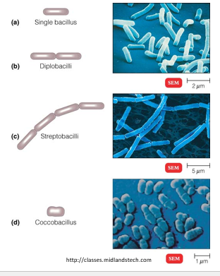
What is a bacillus? (Plural for bacilli)
A bacillus is a rod-shaped bacterium (bacterial cell)
What is a coccobacillus?
A coccobacillus is a very short rod-shaped bacterium that may appear spherical.
What are spiral bacteria and how do spirilla differ from spirochetes?
Spiral bacteria include spirilla (rigid, thick spirals) and spirochetes (thin, flexible spirals that move by twisting).
What are curved or comma-shaped bacteria called?
Curved or comma-shaped bacteria are called vibrios.
What determines the arrangement of bacterial cells?
Bacterial arrangements are determined by the plane of division during binary fission and whether cells remain attached after division.
What are diplococci and diplobacilli, and how are they arranged?
Diplococci: spherical bacteria that divide in one plane and remain in pairs.
Diplobacilli: rod-shaped bacteria that divide in one plane and remain in pairs.
Memory tip: think “diplo = pair” for both shapes.
How do streptococci and streptobacilli grow, and what are their arrangements?
Streptococci: spherical bacteria that divide in one plane and remain in chains.
Streptobacilli: rod-shaped bacteria that divide in one plane and remain in chains.
Optional tip for memorization: think “strepto = chain” for both shapes.
What are tetrads?
Tetrads are spherical bacteria that divide in two planes and form groups of four.
What are sarcinae?
Sarcinae are spherical bacteria that divide in three planes and form cube-like groups of eight.
First division: 2 cells
Second division: 4 cells
Third division: 8 cells.
Always spherical.
What are staphylococci? (Staphylococcus = singular)
Staphylococci are spherical bacteria that divide in multiple planes and form grape-like clusters.
How do bacilli usually divide and arrange?
Bacilli divide across their short axis (perpendicular to the long rod, divide across their width) in one plane, leading to fewer arrangement patterns.
What is the primary function of the bacterial cell wall?
The bacterial cell wall provides structural support, maintains cell shape, and protects the cell from osmotic lysis (bursting due to water pressure).
What is peptidoglycan?
Peptidoglycan is a strong, mesh-like polymer that forms the bacterial cell wall and gives it rigidity.
What molecules make up peptidoglycan?
Peptidoglycan is made of NAM and NAG molecules linked together by β-1,4 glycosidic bonds, forming long polymer chains, which are then cross-linked to each other by peptide bonds to create the rigid peptidoglycan layer of bacterial cell walls.
What defines a Gram-positive bacterial cell wall?
Gram-positive bacteria have a thick peptidoglycan layer and no outer membrane.
What are teichoic acids and what is their function?
Teichoic acids are negatively charged polymers found only in Gram-positive bacterial cell walls. They help regulate ion movement (especially cations like Mg²⁺) across the cell wall and contribute to antigenic specificity, allowing immune recognition.
The thick peptidoglycan layer of Gram-positive bacteria provides the structural framework that anchors and accommodates teichoic acids.
What is lipoteichoic acid?
Lipoteichoic acid is a teichoic acid that anchors the cell wall to the plasma membrane.
What defines a Gram-negative bacterial cell wall?
Gram-negative bacteria have a thin peptidoglycan layer, an outer membrane, and a periplasmic space.
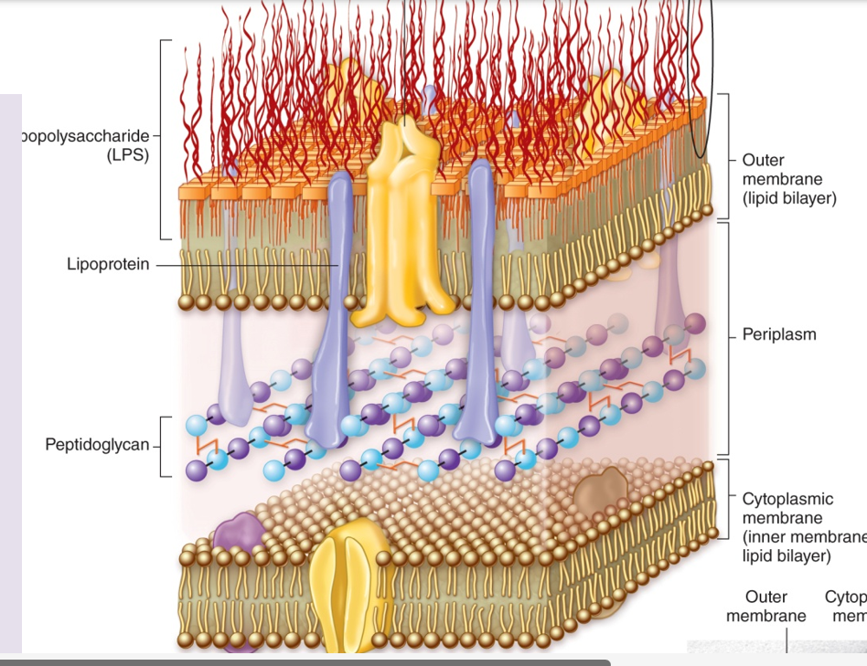
What is the outer membrane made of in Gram-negative bacteria?
The outer membrane is made of phospholipids, lipopolysaccharides (LPS), and proteins.
What is the periplasmic space?
In Gram-negative bacteria, the periplasmic space is between the inner and outer membranes, contains a thin layer of peptidoglycan, and also houses enzymes and transport proteins.
What are porins and why are they important?
Porins are protein channels in the outer membrane of Gram-negative bacteria that allow the passage of small molecules into the cell.
Only in gram-negative bacteria
What is lipopolysaccharide (LPS)?
Lipopolysaccharide is an endotoxin (toxin released from the bacterial cell wall) found in Gram-negative bacteria that contributes to disease. It is one of the components of the outer membrane of Gram-negative bacteria.
What are the three components of LPS?
LPS\left(Lipopolysac\operatorname{ch}aride\right) LPS (Lipopolysaccharide) is made of Lipid A (the component that releases endotoxin when the polysaccharide dies) and a core oligosaccharide, and the other component of LPS is O-antigen.
Which part of LPS causes toxic effects in humans?
Lipid A is responsible for the toxic effects of LPS.
What is the function of the O-antigen?
The O-antigen provides antigenic variability, helping bacteria avoid immune detection.
What are acid-fast bacteria?
Acid-fast bacteria have waxy cell walls containing mycolic acids, making them resistant to staining and antibiotics.
The acids make the cell wall hydrophobic and impermeable; the staining process cannot easily penetrate. Mycolic acids also provide a barrier to make it hard for antibiotics to penetrate.
What is mycolic acid?
Mycolic acid is a long-chain fatty acid that strengthens the cell wall and increases resistance.
In gram-positive bacteria.
What is cord factor and why is it important?
Cord factor is a glycolipid virulence (how capable it is of causing disease, higher virulence more severe infection), a factor that causes bacteria to grow in rope-like bundles (harder for immune sytem to detect and attach to host) and helps them evade the immune system.
Which bacteria lack a cell wall entirely?
Mycoplasma species lack cell walls and instead contain sterols in their plasma membranes.
How are archaeal cell walls different from bacterial cell walls?
Archaeal cell walls may lack walls or contain pseudomurein, which lacks NAM (component of the peptidoglycan cell wall layer) and D-amino acids.
What does lysozyme do to bacterial cells?
Lysozyme breaks down peptidoglycan, weakening the bacterial cell wall leading to lysis,
How does penicillin affect bacterial cell walls?
Penicillin inhibits the enzyme that forms peptide cross-links between NAM subunits in peptidoglycan, weakening the cell wall and causing bacterial lysis.
What is a protoplast?
A protoplast is a Gram-positive bacterium that has completely lost its cell wall, usually due to enzymes (like lysozyme) or laboratory treatment.
-Protoplasts are formed artificially (in the lab) using enzymes like lysozyme or chemical treatments to remove the cell wall.
What is a spheroplast?
A Gram-negative bacterial cell that lost its cell wall due to certain conditions. Partially stripped down version of the og Gram-negative cell.
(partially wall-less)
What are L-forms?
Bacteria (Gram-positive or Gram-negative) that lose their cell wall under stress (like antibiotics) ,can survive without it, and have irregular shapes. So protoplasts sphereoplast can become L-forms, but not all L-forms are protoplasts and sphereoplast.
What bacterial shapes fall under the spiral category?
Vibrio – comma-shaped
Spirillum (spirilla plural)– rigid spiral
Spirochete – flexible, corkscrew-shaped.
How do spirillum and spirochetes differ?
Spirillum: rigid spiral shape (thicker)
Spirochete: flexible, corkscrew motion.
What other shape can bacterial cells be?
Star-shaped (0.5 microns) and rectangular (1 micron). (They’re less frequent shapes in low-nutrient aquatic environments.)
What are the different cell shapes of Archaea, and give examples?
Archaea exhibit diverse and unusual shapes, including:
Pleomorphic (variable shapes)
Branched
Curved rods
Spiral
Flat, square shapes
Examples:
Haloquadratum walsbyi → flat, square-shaped
Thermoproteus tenax → thin, branched/filamentous rod
What are the different arrangements of cocci bacteria and how are they formed?
Diplococci: spherical bacteria that divide in one plane → remain in pairs
Streptococci: spherical bacteria that divide in one plane → remain in chains
Tetrads: spherical bacteria that divide in two perpendicular planes → form groups of 4
Sarcina: spherical bacteria that divide in three perpendicular planes → form cubical packets of 8, 16, or more cells
Memory tip:
“Diplo = pair”
“Strepto = chain”
“Tetra = 4”
“Sarcina = cube/stack”
What determines the characteristic arrangement of bacterial cells?
The arrangement of bacteria (pairs, chains, clusters, packets, etc.) is determined by the plane(s) in which they divide and whether the cells remain attached after division.
Examples:
Diplococci → divide in one plane → pairs
Streptococci → divide in one plane → chains
Tetrads → divide in two planes → groups of 4
Sarcina → divide in three planes → cubical packets
Staphylococci → divide in multiple planes → clusters
What is the shape and size range of Bacillus bacteria?
Shape: Rod-shaped
Size: 0.5–1.0 µm wide × 1.0–4.0 µm long
What bacterial features are important for antibacterial treatment, identification, and pathogenesis?
Potential targets for antibacterial treatments – features that allow selective killing of bacteria.
Unique features for identification – structures or molecules specific to certain bacteria that help identify the infectious agent.
Structures aiding pathogenesis – many bacterial components contribute to disease-causing ability.
Name the structures external to the bacterial cell wall.
Glycocalyx – slime layer or capsule; helps in protection and adhesion
Flagella – for motility
Axial filaments (endoflagella) – found in and allow spirochetes to move in a corkscrew motion. anchored at one end of a cell.
Pili—for conjugation (DNA transfer). involved in motility (type 4 pili-twitching motility).
Fimbriae—short, hair-like projections for attachment to host cells or each other, most common in gram-negative bacteria
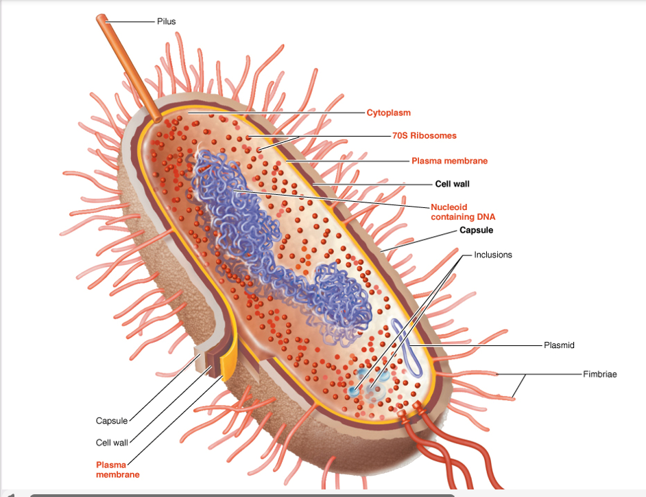
What is the glycocalyx, its types, and their roles in virulence?
Definition: The glycocalyx is a sticky, jelly-like layer outside the cell made of sugars (polysaccharides) and/or proteins (polypeptides).
Types:
Capsule – neatly organized, firmly attached
Prevents phagocytosis (engulfment by immune cells)
Aids in strong surface attachment and immune evasion (avoiding host defenses)
Slime layer – unorganized, loose
Contributes to biofilm formation (bacterial communities on surfaces) and surface adhesion
Both types enhance bacterial virulence (disease-causing ability)
Both Gram-positive and Gram-negative bacteria can have a glycocalyx.
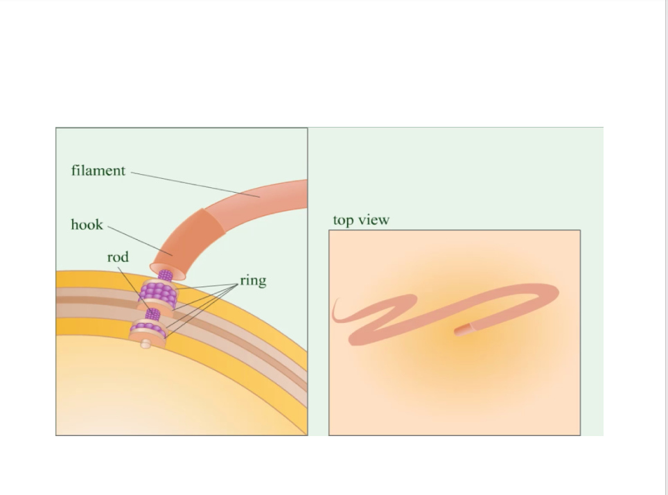
What are the structure and function of bacterial flagella?
Structure (3 parts):
Filament – outermost region
Hook – attaches filament to basal body
Basal body – rod and pairs of rings; anchors flagellum to cell wall and membrane
Composition: Made of flagellin and other proteins
Function: Filamentous appendages external to the cell; propel bacteria (motility)
What are twitching and gliding motility in bacteria?
Twitching: Jerky movement via Type IV pili, powered by ATP; pili extend, attach, retract → pull cell forward
Examples: Pseudomonas aeruginosa, Neisseria gonorrhoeae, Myxococcus xanthus
Gliding: Smooth movement via motor proteins & adhesins/slime, powered by proton motive force (PMF)
Examples: Myxococcus xanthus, Cytophaga
What is the O antigen component of LPS?
The O antigen is the outermost component of LPS and is made of repeating sugar molecules.
What is the function of the core oligosaccharide in LPS?
The core oligosaccharide connects the O antigen to Lipid A in the LPS molecule.
What is the function of the Lipid A molecule in the LPS?
-Anchors the LPS molecule in the outer membrane
-Portion of LPS recognized by immune system
What are haponoids?
Haponoids are lipid molecules found in prokaryotic cell membranes that help stabilize membrane structure, similar to sterols in eukaryotes.
What is microbiology?
Microbiology is the study of microorganisms, which are organisms too small to be seen with the unaided eye.
What is a microorganism (microbe)?
A microorganism is a microscopic living organism that cannot be seen without a microscope.
What is a micron?
A micron (\mu m) is one millionth of a meter (10^{-6} m).
A micron is the same thing as a micrometer.
What are the major groups of microorganisms?
The major groups include bacteria, archaea, fungi, protozoa, algae, viruses, and multicellular animal parasites (helminths).
Which domains of life contain microorganisms?
Microorganisms are found in all three domains of life: Bacteria, Archaea, and Eukarya.
The domains of life are the broadest level of biological classification, grouping organisms based on major differences in cell structure and genetics.
Bacteria – prokaryotic microorganisms with peptidoglycan cell walls
Archaea – prokaryotic microorganisms that are genetically different from bacteria and often live in extreme environments
Eukarya – organisms with eukaryotic cells; includes microorganisms like fungi, protozoa, and algae, as well as plants and animals
Which microorganisms are prokaryotic?
Bacteria and Archaea are prokaryotes because they lack a nucleus.
Which microorganisms are eukaryotic?
Fungi, protozoa, algae, and multicellular parasites are eukaryotes because they have a nucleus and membrane-bound organelles.
What are the key characteristics of bacteria?
Bacteria have the following characteristics:
They are single-celled prokaryotic organisms.
They lack a true nucleus.
They have cell walls made of peptidoglycan.
They reproduce by binary fission (an asexual process where the cell replicates its DNA and divides into two genetically identical cells).
They obtain nutrients from organic or inorganic chemicals, and some perform photosynthesis.
Some bacteria can move using flagella.
How are archaea different from bacteria?
Archaea are prokaryotes that lack peptidoglycan in their cell walls and often live in extreme environments.
What are the characteristics of fungi?
Fungi are eukaryotes with chitin cell walls that absorb organic chemicals for energy and include yeasts (unicellular) and molds/mushrooms (multicellular).
What are protozoa?
Protozoa are single-celled eukaryotes without cell walls that ingest or absorb organic chemicals and may move by pseudopods (temporary extensions of the cytoplasm), cilia, or flagella. Reproduce sexually or asexually.
What are the characteristics of algae?
Algae are photosynthetic eukaryotes with cellulose cell walls that produce oxygen and carbohydrates. They’re found in freshwater, saltwater, and soil.
Why are viruses not considered living cells?
Viruses are acellular, lack metabolism, and can only replicate inside a living host cell.
What are viroids?
Viroids are small infectious single-stranded RNA molecules that infect plants through wound sites.
What are prions?
Prions are infectious protein particles that lack nucleic acids and cause fatal neurological diseases. Transmissible only within species.
Where are microorganisms found?
Microorganisms are found in nearly every environment on Earth, including soil, water, ice, hot springs, salt lakes, and the human body.
What is normal microbiota?
Normal microbiota are microorganisms that normally live on or in the human body without causing harm.
What is transient microbiota?
Transient microbiota are microorganisms that are present on the body for a short time.
What is the microbiome?
The microbiome is the total collection of all genetic material (all genes, both unique and shared) from all the microorganisms living in a specific environment.
What is spontaneous generation (abiogenesis)?
Spontaneous generation is the incorrect theory that life arises from nonliving matter.
What is biogenesis?
Biogenesis is the theory that living cells arise only from preexisting living cells.
What did Robert Hooke contribute to microbiology?
Robert Hooke was the first to use the term “cell” in 1665 after observing thin slices of cork under a microscope. He saw tiny, box-like compartments that looked like small rooms (cellula in Latin), which led him to call them cells.
Who first observed bacteria?
Anton van Leeuwenhoek was the first to observe and describe bacteria.
Who disproved spontaneous generation for large organisms?
Francesco Redi showed that maggots come from fly eggs, not decaying meat.
Who supported spontaneous generation using broth experiments?
John Needham supported spontaneous generation after microbes grew in boiled broth.
Who challenged Needham’s support of spontaneous generation, and how?
Lazzaro Spallanzani argued against Needham by showing that microorganisms do not arise spontaneously.
He boiled nutrient broth to kill microbes and then sealed the flasks tightly so nothing from the air could enter. The broth remained sterile (no microbial growth).
This showed that microbes came from contamination in the air, not from nonliving broth.
Who conclusively disproved spontaneous generation?
Louis Pasteur disproved spontaneous generation (the idea that life, like microbes, could arise from nonliving matter, like broth on its own) by using a swan-neck flask experiment that showed microorganisms come from existing microbes in the air, not from nonliving matter.
What does the germ theory of disease state?
The germ theory states that microorganisms are the cause of many diseases.
Who provided strong evidence for germ theory?
Robert Koch proved that specific microbes cause specific diseases and developed Koch’s postulates (he created a systematic set of criteria that help scientists prove that a particular microorganism is the cause of a specific disease)
What is pasteurization?
Pasteurization is heating beverages briefly to kill harmful microbes without damaging the product.
Who developed the first vaccine?
Edward Jenner developed the smallpox vaccine in 1796 using cowpox. Jenner used cowpox material, a much milder disease, and introduced it to someone; they were exposed to smallpox later and were and immune to it.
Who introduced antiseptic surgery?
Joseph Lister introduced the use of the chemical phenol (carbolic acid) as an antiseptic in surgery. He used it to sterilize surgical instruments and clean wounds, which greatly reduced infections after operations.
His work helped establish antiseptic techniques and supported the germ theory of disease.
Who discovered penicillin?
Alexander Fleming discovered penicillin, the first antibiotic.
Who showed handwashing prevents disease?
Ignaz Semmelweis demonstrated that handwashing prevents puerperal fever (childbed fever).