Structure and development of the cardiovascular system
1/44
There's no tags or description
Looks like no tags are added yet.
Name | Mastery | Learn | Test | Matching | Spaced | Call with Kai |
|---|
No analytics yet
Send a link to your students to track their progress
45 Terms
development of heart
-day 15: cardiogenic precursors have formed a crescent
-day 21: 2 arms of crescent fuse along midline to give a linear heart tube
-gets elongated and becomes patterned with the regions and chambers of the mature heart
-day 28: after looping these regions are disposed approx in their eventual positions
-day 50: formation of valves
what are the 2 circulatory systems the heart pumps into
-pulmonary
-systemic
pulmonary circuit
-starts and ends at heart
-closed system
-carries blood from heart to lungs
-return oxygenated blood to heart
systemic system
-transports oxygenated blood around the body and returns the blood to heart
how much blood in total do both systems contain
5 litres
how is the pressure and distribution controlled in these systems
-by blood vessels
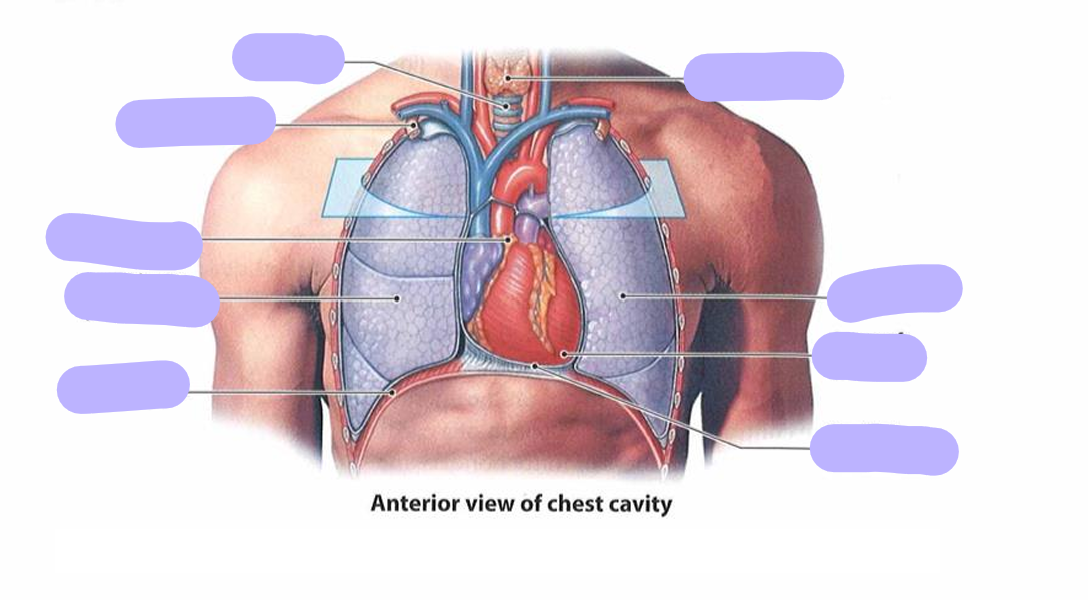
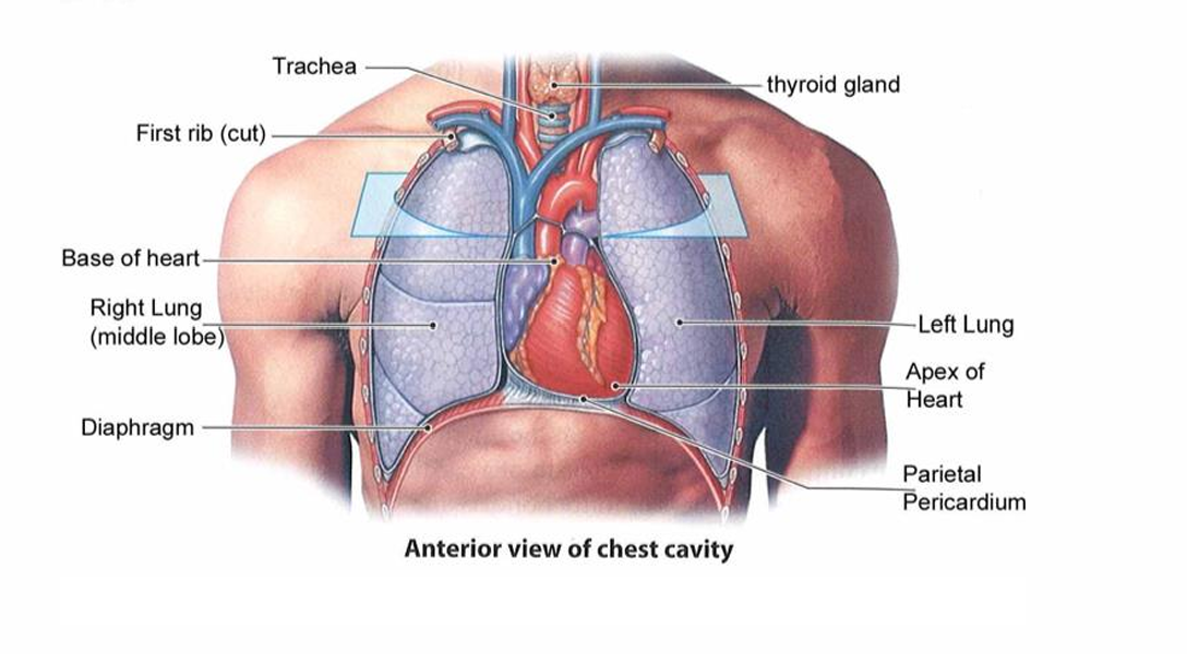
what is the outer structure of the heart
-heart is enclosed in pericardium
-which is connective tissue sheath
-ensures it is maintained in position in thorax, and actions are friction free
what ais the structure of the pericardium
-outer fibrous layer
-inner layers: visceral and parietal pericardium
-visceral layer (epicardium) adheres to heart
-small fluid filled space separates visceral and parietal percardia
what is the function of the outer fibrous layer of the pericardium
-anchors heart to diaphragm and great vessels and anterior chest wall
-maintaining its position
what is the function of parietal pericardium lines
-lines inner surface of fibrous layer
what is the function of small fluid filled space that separates visceral and parietal percardia
-reduces friction between surfaces as the heart beats
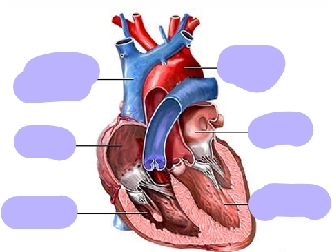
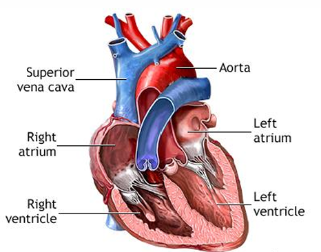
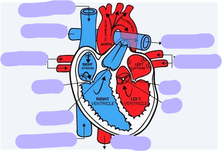
what are the chambers of the heart
4 chambers
-2 atria
-2 ventricles
what is the atria
-receiving chambers
-right: receives venous blood from body
-left: receives arterial blood from lungs
what are ventricles
-expels blood from heart
-right: expels blood directed to lungs
-left: expels blood to aorta to be distributed throughout body
flow of blood through heart (start at right atrium)
-Blood enters into the right atrium
-The right ventricle pumps the blood to the lungs where it becomes oxygenated
-The oxygenated blood is brought back to the heart by the pulmonary veins and enters the left atrium
-The left ventricle pumps the blood to the aorta and oxygenated blood is distributed around the body
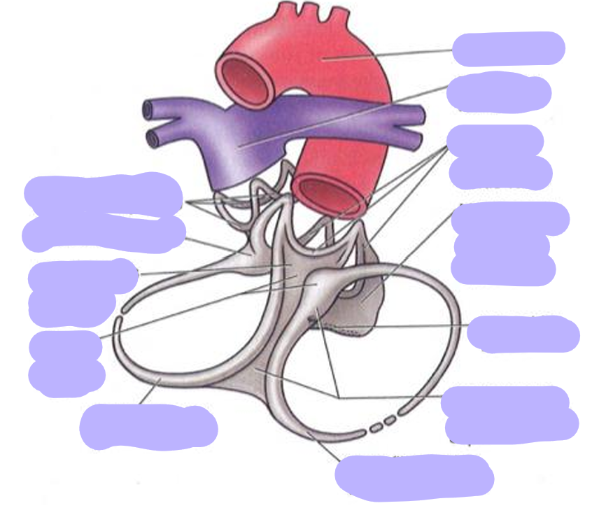
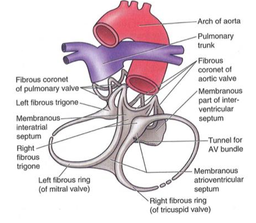
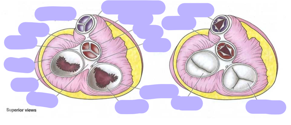
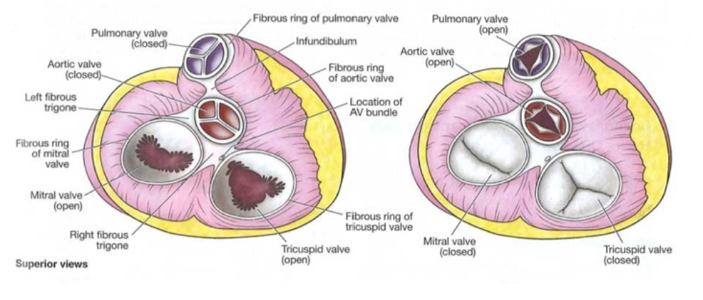
what is the fibrous skeleton of the heart
-dense connective tissue
-separates atria from ventricles
-provides critical support
-separates flow of electrical impulses
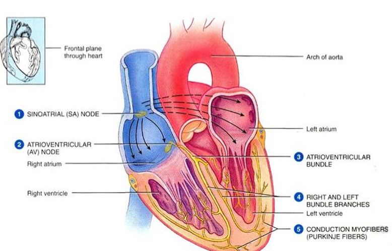
what is the cardiac conduction system
1. Excitation signal is created by sinoatrial node, wave of excitation spreads across the atria causing them to contract
2. Once reaches atrioventricular node, signal is delayed
3. Conducted into the atrioventricular bundle (bundle of His)
4. Down the left and right bundle branches
5. Wave pulses are spread along the ventricles causing them to contract

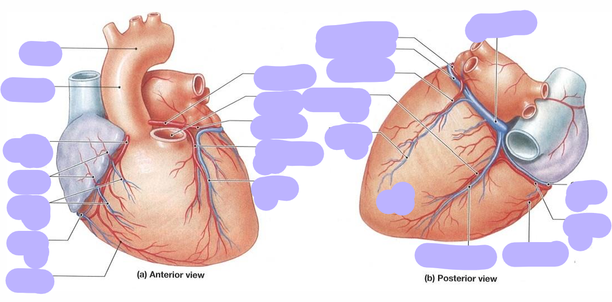
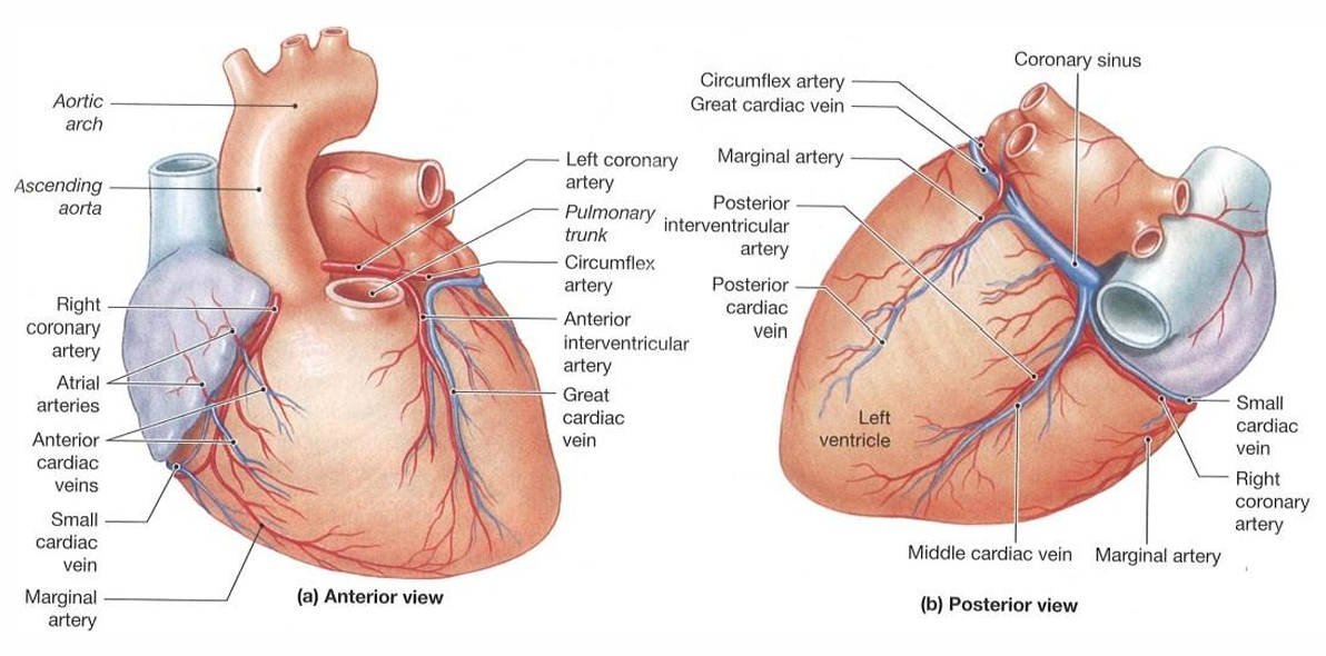
how does the heart receive blood supply
-through coronary arteries and cardiac veins
what are the cardiac arteries
-2 main arteries supply the heart: left and right coronary artery
-arise from aortic sinuses within the aorta
-when heart is relaxed back flow of blood fills these pockets and blood enters the coronary arteries
what are the cardiac veins
-venous drainage is by the cardiac veins
-several drain into the great cardiac vein
-this empties into the coronary sinus which enters the right atrium
why does heart have valves
-to prevent backflow as ventricles relax
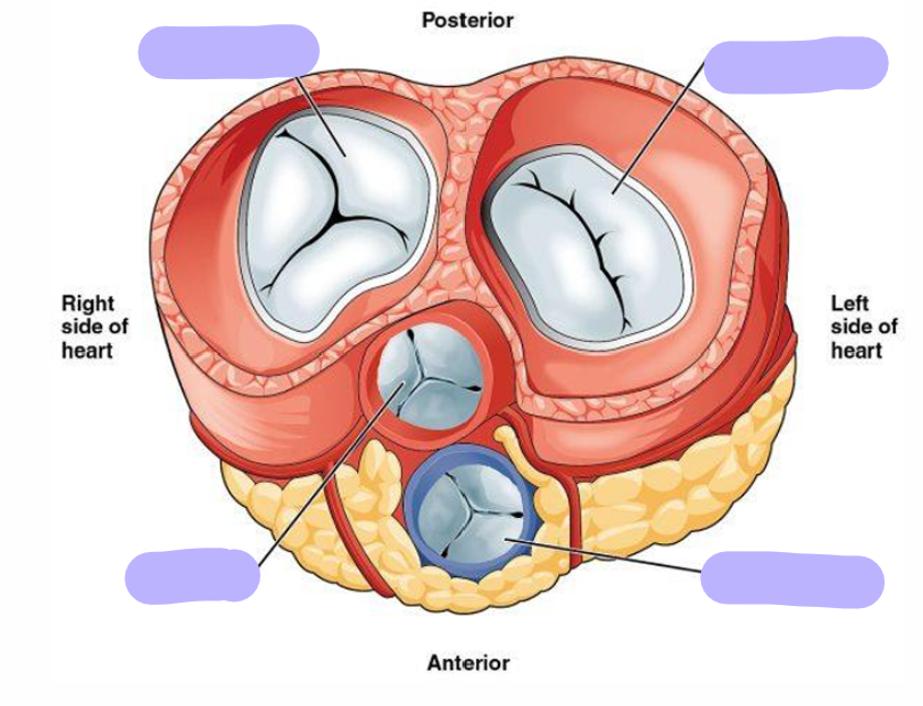
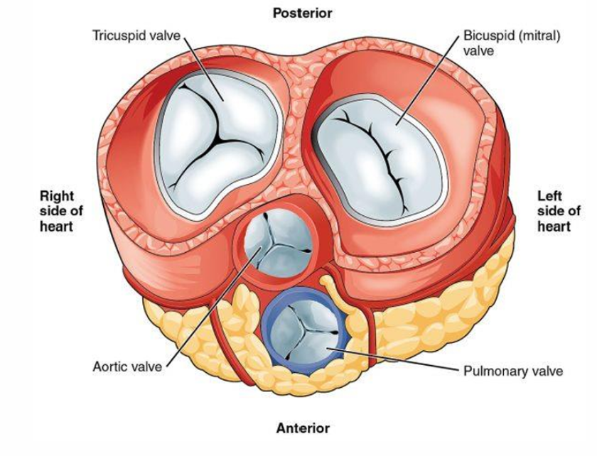
what are the atrioventricular valves
-prevent backflow between ventricle to atria, when ventricle contracts
-tricuspid & bi cuspid
what are the 3 elements to valves
-papillary muscles
-chordae tendineae
-cusps of valves
how do av valves work
-when ventricle is relaxed: cords are loose and valve offers no resistance to the flow of blood from atrium to ventricle
-when ventricle contracts, papillary muscles tenses the cords which are attached to cusps
-cusps swing together preventing backflow of blood
-tense cords prevent them for everting
what are semilunar valves
-located between ventricles and outflow vessels
-in the walls of blood vessels
-pulmonary valve: between right ventricle and pulmonary trunk
-aortic valve: between left ventricle and ascending aorta
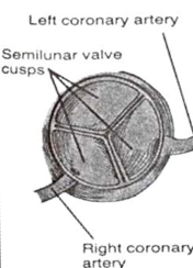
what is the structure of the semilunar valves
-as they are in bv; position is stable
-consist of thin layer of connective tissue covered by endothelium
-connective tissue is thickened along its free margin where it forms a nodule
-semilunar valves are forced apart by ejection of blood from contracting ventricles
-elastic recoil of vessels forces blood back to ventricles
-results in ballooning out of each valve so free margins are forced together to form a Y
semilunar valve diagram
what are heart sounds
-heart beat caused by closing of valves
what are the 4 heart sounds
-S1 is the closing of AV valve as ventricle contracts (lubb)
-S2 is the closing of sl valve (ventricle relaxes and beings to fill) (dubb)
-S3 & S4 are fainter and difficult to hear
-S3 is filling of ventricle
-S4 is atrial contraction
what does a heart murmur sound like
-when AV valve doesnt close properly
-regurgitation can occur creating gurgling sound
what is diastole
-av valve opens
-sm valve closes
-dubb sound
what is systole
-av valve closed
-sl valve opens
-lubb sound
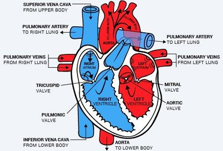
what are the 5 vessels that enter and leave the heart
-superior vena cava
-inferior vena cava
-pulmonary artery
-pulmonary vein
-aorta
what are the superior/inferior vena cava
-return deoxy blood from circulation in the body and empty it into the right atrium
what is the pulmonary artery
-carries deoxy blood from right ventricle to lungs
-for oxygenation
what is the pulmonary vein
-carries oxy blood form lungs to left atrium
-where it is returned to systemic circulation
what is the aorta
-largest artery in the body
-carries oxygenated blood from left ventricle of the heart into systemic circulation