Exam 2 - HD
1/128
Earn XP
Description and Tags
Cardiovascular System - Urogenital System - CNS/PNS - Pregnancy/Birth - etc.
Name | Mastery | Learn | Test | Matching | Spaced | Call with Kai |
|---|
No analytics yet
Send a link to your students to track their progress
129 Terms
Explain the blood flow of the heart postnatally.
Deoxygenated blood goes from right atrium → right ventricle → via pulmonary arteries → lungs
Oxygenated blood → pulmonary veins → left atrium → left ventricle → Aorta → Body Tissue
Distinguish between Arteries, Veins and Cppilaries,
Arteries - oxygen rich blood travels from heart → body
Veins - oxygen poor blood travels back to heart
Cappilaries - oxygen and nutrients given to cells in body
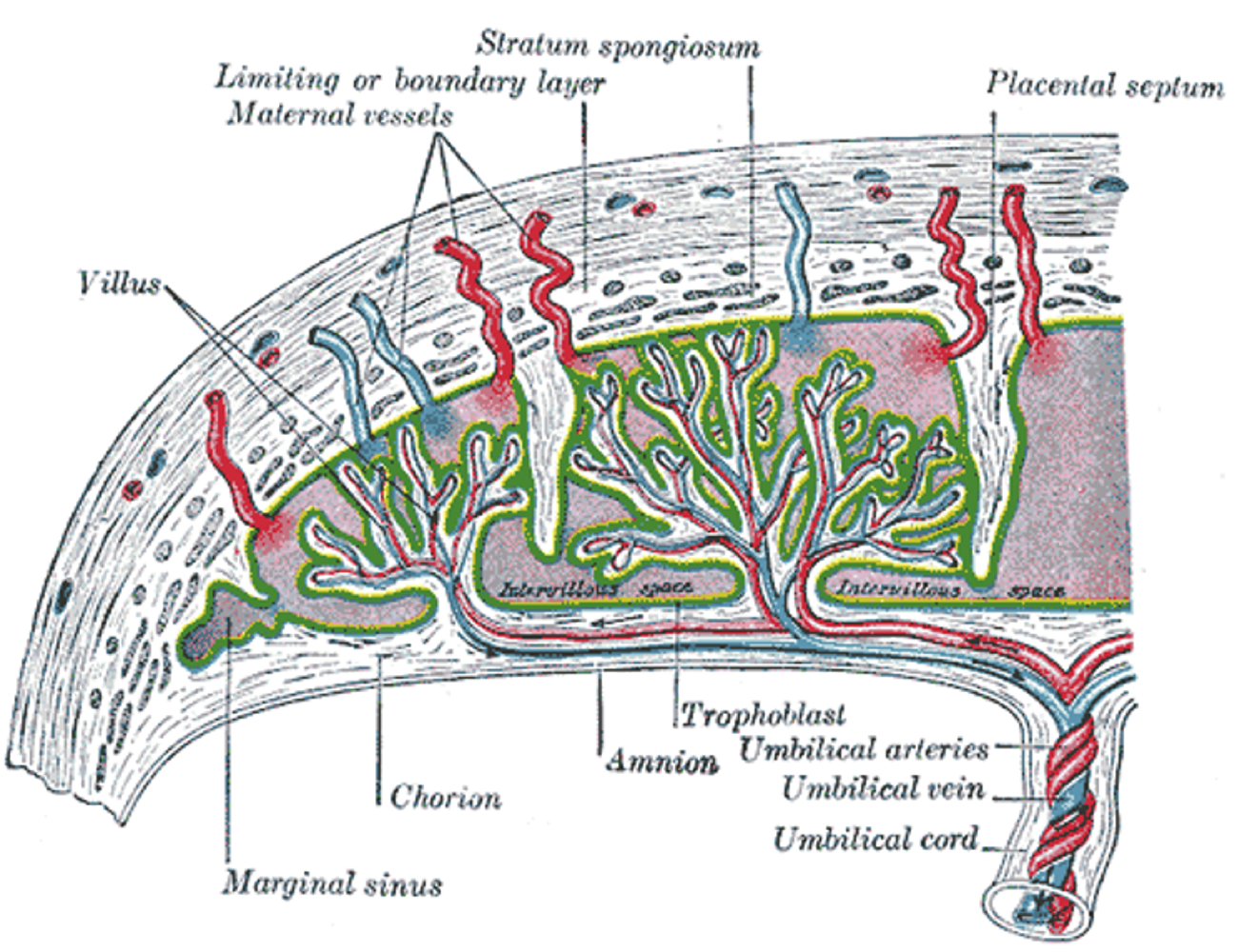
Prenatal Blood-Flow
umbilical arteries carry deoxygenated blood from fetus to placenta
umbilical veins carry oxygenated blood from placenta to fetus
What veins and arteries does the Fetus have??
Fetus has vein (to heart) and 2 arteries (from fetal heart) to the placenta.
Single umbilical disorder
This is a disorder where a fetus has only one artery
Can occur together with Down Syndrome & Failure of Kidneys

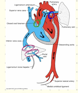
What are the two openings in the fetal heart which change from prenatal to postnatal?
In fetal heart blood is detoured from lungs via two openings.
Lungs are not needed because placenta provides oxygen.
The two openings are ductus arteriosis and foramen also the umbilical vein and ductus venosous also closes.
Explain the mechanism of cephalocaudal folding.
Folding around the formed axis.
Embryonic disk grows not yolk sac.
Movement of the heart crainally
Heart is folded into embryo at the ventral part/
Which mesoderm forms cardiogenic mesoderm?
intra-embryonic mesoderm
How does the cardiac region of the embryo form??
Embryo is a disk shape (horseshoe gap in cranial region)
The pericardial plate is located above the cardiogenic plate infront of the embryo.
Right now no connection between cavity and extraembryonic coelum.
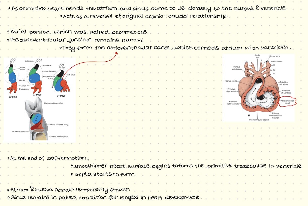
Cranial pericardium undergoes 180 rotation and now the cardiac primordium lies dorsally to the perochardial cavity.
The pericardial cacity is bent over forward and has enclosed cardiac primordium in the front (ventrally)
Dorsally the cardiac primordium forms two layers of the bilaminar structure.
How does the vitelline duct form??
Takes place when yolk sac becomes part of the gut tube and then forms the duct.
How do allantois form??
Form from within the connecting stalk.
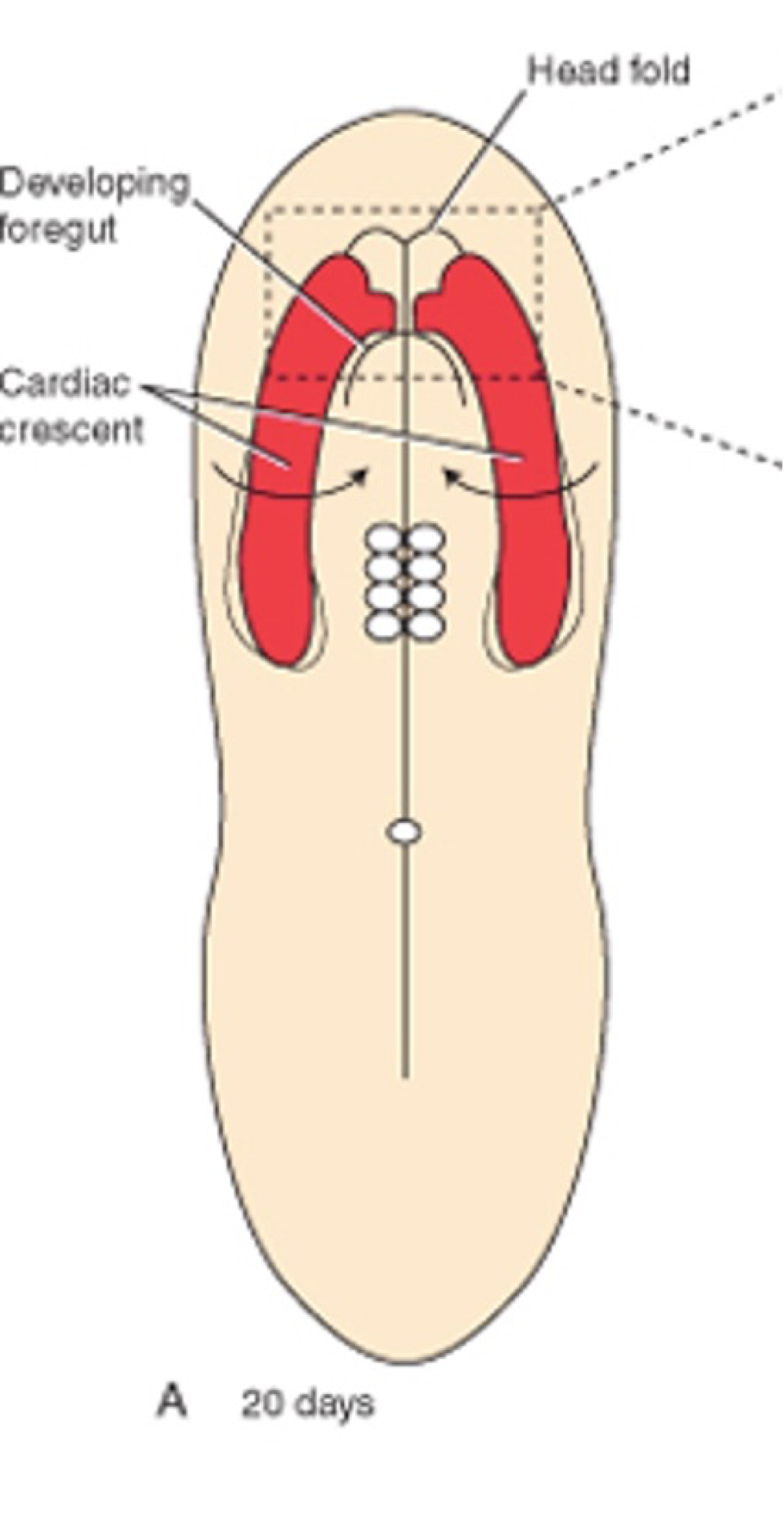
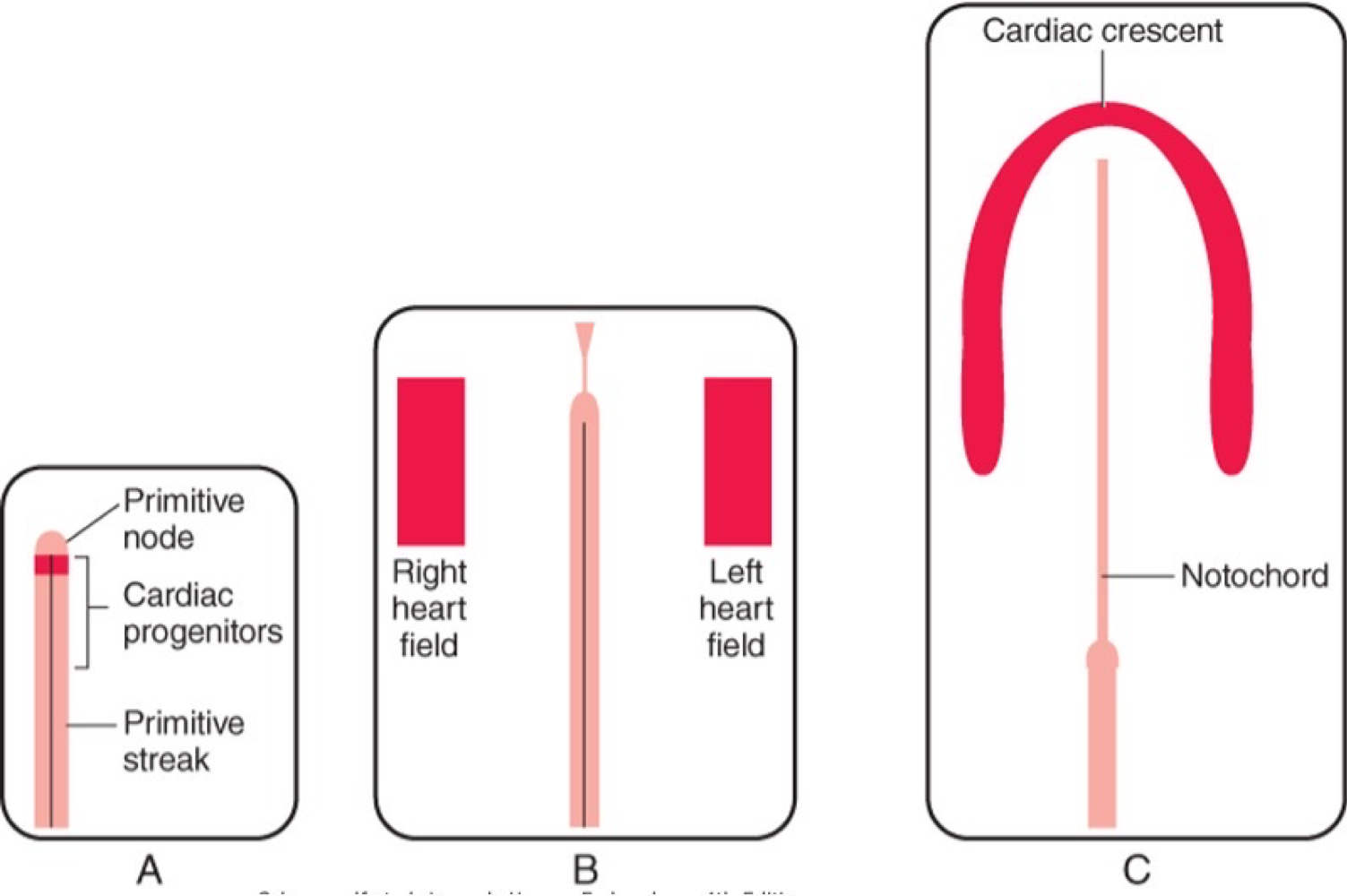
How do the mesoderm cells begin to form the heart??
Cells from mesoderm migrate through the primitive streak
Some of which become cardiac progenitor cells
Move towards head of embryo.
Cardiac cells organise into left/right heart fields and curve around embryo (front) to form a cardiac crescent.
BMP, Activin, TGF-B indicate mesoderm cells to differentiate into heart cells.
From these cells two thin walled endocardial tubes emerge which then fuse at the anterior sides.
What forms the myocardium (muscle)?
Splanchnic/Visceral mesoderm
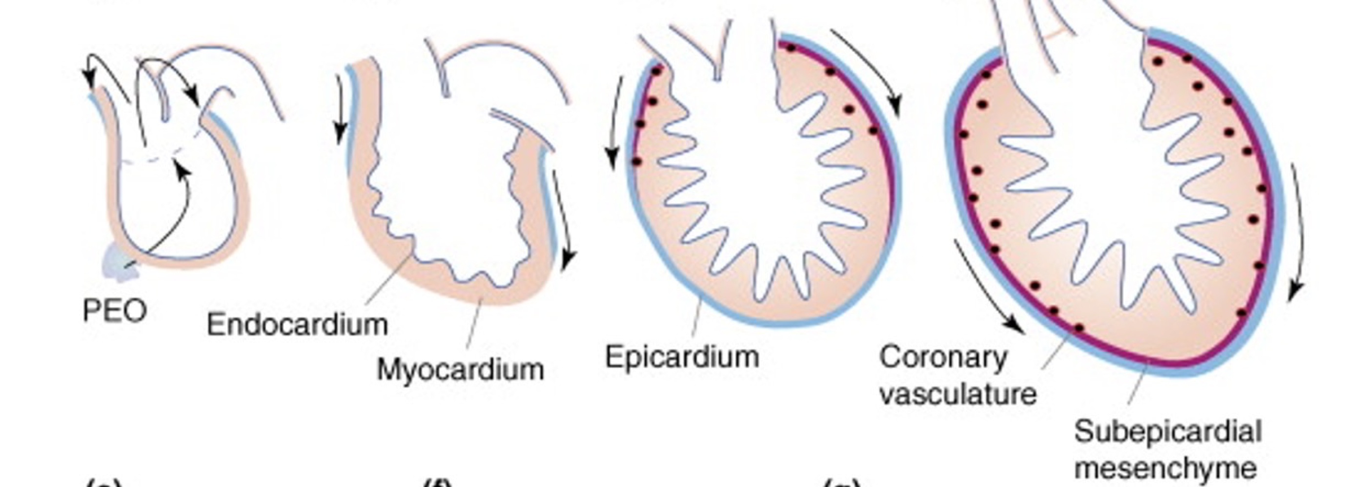
What forms the epicardium?
Surrounding mesenchyme cells thicken to form epicardium (serves as a progenitor source)
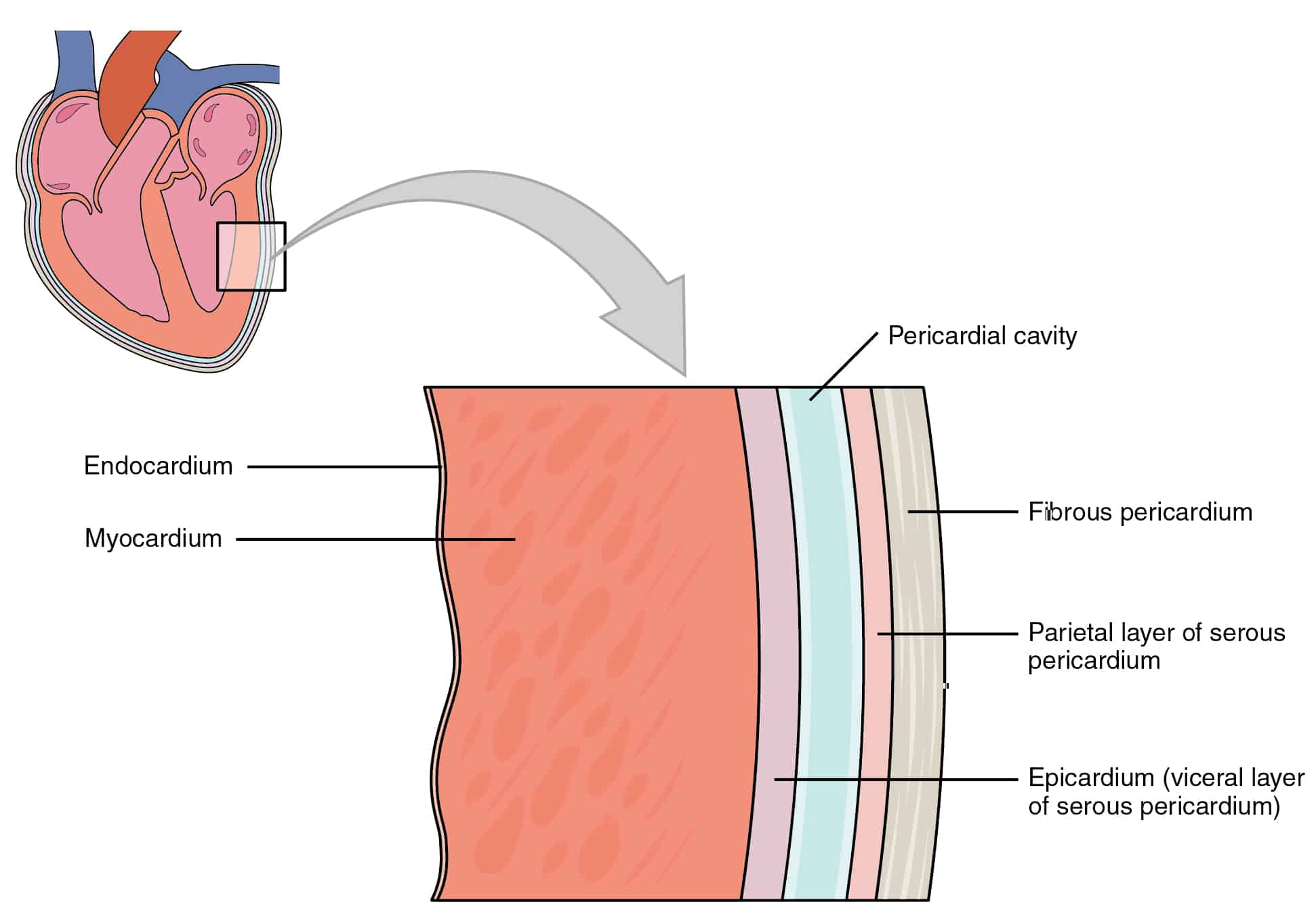
What is the purpose of the epicardium and pericardium?
Epicardium - innermost layer of perocardium (outermost layer of heart itself)
Pericardium - fluid filled sac which surrounds and protects the heart.
Between which layers does the coronary vasculature form?
Myoepicardium Layer: Myocardium & Epicardium
What is cardiac jelly??
Extracellular matrix between epimyocardium and endocardial tubes
Made from glycoproteins and collagen
Involved in cardiac looping
What are endocardial cells?
Cells that coat the cardiac lumen surrounding the cardiac jelly.
Briefly explain how sacculations form?
When tubular heart elongates and then forms the dilations.
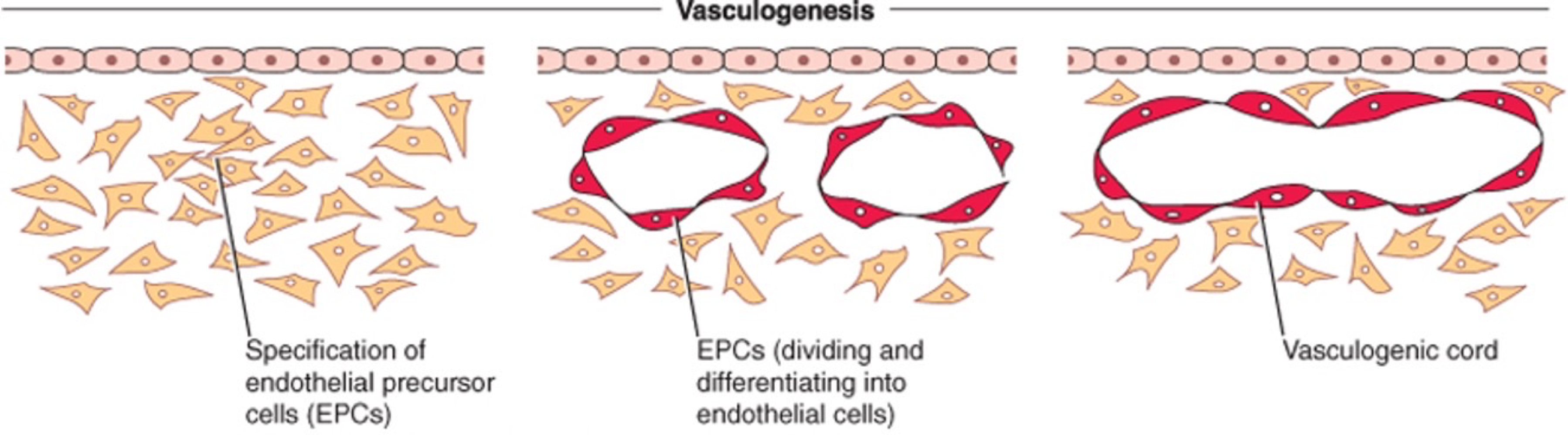
What is vasculogenesis??
Takes place in the yolk sac (early development)
Clusters of mesodermal cells form Blood islands, which develop into:
Primitive blood cells
Endothelial cells - blood vessel walls
What are Angioblasts and what are they formed from??
Arise from mesoderm (splanchnic and chorionic)
They are mesenchymal cells from the yolk sac and umbilical cord
Differentiate into blood islands
Eventually, which form blood/blood vessels
How is the first circulatory loop formed??
Vessels from yolk sac begin to connect with vessels in embryo.
Include: dorsal aorta, umbilical arteries/veins
What is the situation of heart at 25-27 days?
The primitive heart formed
Looping & rotating of heart has started
Early circulation starts
→ Heart can pump blood through the dorsal aorta and white vessels bring blood back from yolk sac to embryo.
At what stages does hematopoiesis happen in what areas of fetus??
Formation of bloos cells
Yolk sac 3-8 weeks
Liver 8-22 weeks
Then bone marrow
Thiabendazole
Compound that can lead to blood vessels falling apart.
When does the development of the aorta begin and what does it look like??
In week 3, when main organ systems are established.
First blood vessels are solid angioblasts then they aquire lumen to form longitudinal vessels.
Pharingeal Arches
Aorta forms from these
5 pairs (1-4 & 6 - no 5)
Have a craniocaudal development
Form as masses of mesenchymal tissue which gets invaded by cranial neural creast cells.
Each arch externally covered by ectoderm and internally endoderm
Pharygneal Clefts
Separate pharygneal arches
What happens to each of the Arches??
Arch 1&2 - disappears by day 29
Arch 3 - carotid arteries (each side of head providing blood to face neck brain & meninges)
Arch 4 - aortic arch (left), subclavian artery (right) - arms, head, thorax
Arch 5 - doesnt form
Arch 6 - pulmonary arch
How do we go from pharyngeal arches to aorta?
Due to vascularization of arches
Run ventrally and form arteries of head neck and upper thorax on the way.
→ formation of angiogenic cluster (primordial heart)
What are the progenitor cells of coronary artery??
Epicardial cells / sinus venousus which differentiate
What does the arterial system consist of at 5 weeks??
Left Aorta and aortic aches (3 carotid LR, 4R subclavian, 6LR pulmonary)
Coronary
Vitelline
2x Umbilical
What forms the venous system??
Sinus horn
What are the 3 systems of paired veins in the heart??
Vitelline Veins (blood from yolk sac back to fetus, drainage of gastrontestinal tract
Umbilical Veins (oxygenated blood from chorion and placenta to fetus - right one regresses)
Cardinal Veins (returning blood from body)
How does the sinus venousus become part of right atrium?
Heart starts with two equal inflow sides
Blood shifts to the right
Left side shrinks
Right side becomes part of the right atrium
Major veins (SVC, IVC) form from these structures
How do vitelline veins and the umbilical veins interact with the liver??
Vitelline veins surround gut → form a network
Liver grows into this network
Network becomes hepatic sinusoids
Umbilical veins connect to this system
Parts of veins disappear → cleaner circulation forms
What happens to the heart after the fusion of the tubes??
Heart tube sprouts aortic arch from outflow region
Elongation and formation of sacculations
New tissue growth into 4 chambers
How do the 4 chambers of the heart form?
Within the atria ventricles form
Openings on left & right to ventricles form
Division between left & right atrium
Ventricular septa forms
Cardiac Looping
How does the cardiac septa form?
4th week using two methods:
Tissue growth: fusiion of two or more actively growing masses of tissue which approach eachother
Fuse a single chamber into two
Overgrowth: growth of chamber except one narrow strip which leaves a small canal connecting two chambers.
How does the atrioventrical septum form (4th week)??
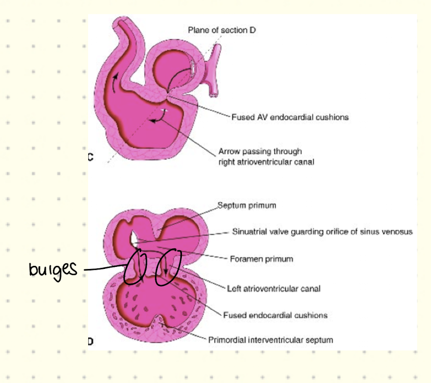
Bulges form on dorsal & ventral walls of the AVv canal which forms the endocardial cushion septum
Eventually, the ventricles begin to divide.
How does the atrial septa form??
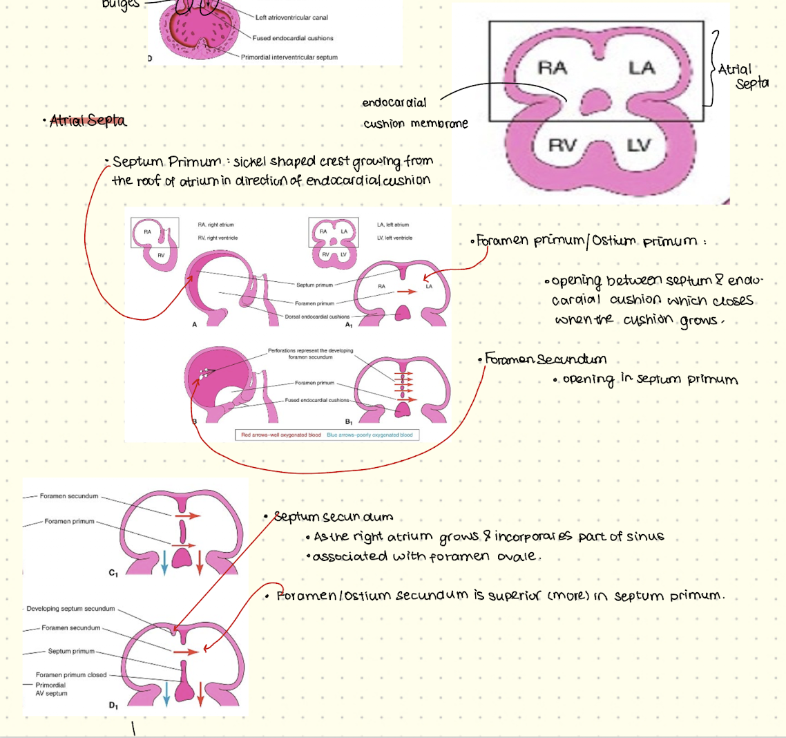
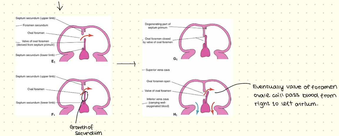
Foramen Ovale
Needs to close in newborn when air inhaled
If open low-oxygen blood enters the left atrium
Shunts blood from the right to the left atria via the ostium secundum
Mostly blood via inferior vena cava and bypasses the lungs in fetus
Associated with septum secundum
At birth, it’s pressed against the septum premium which seals the opening.
Can close within 18 months afterbirth otherwise surgery
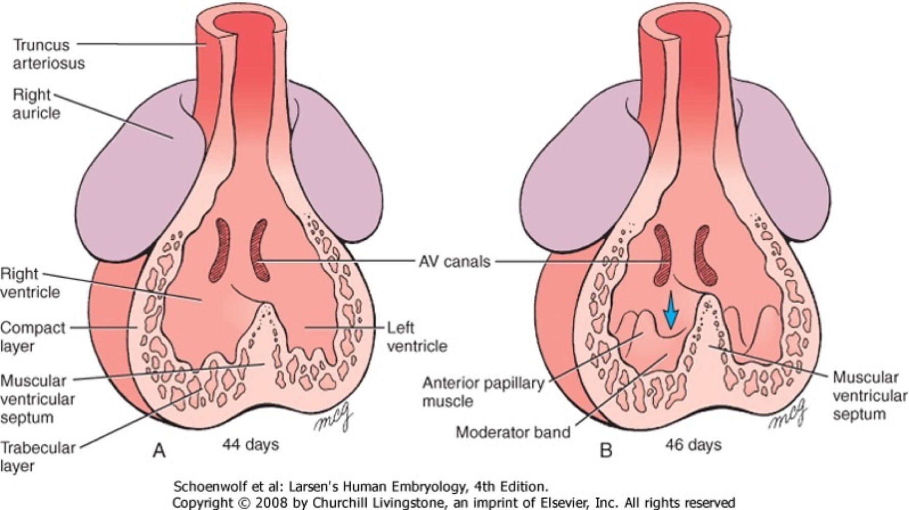
Overview Steps of Ventricular Septa
Division of ventricles
Division of blood stream into pulmonary vein and aorta
Starting point of ventricular septa formation
One common ventricluar chamber
By end of week 4 splits into left & right ventricles
Step 1 of Ventricular septum formation
Growth of ridge from apex towards endocardial cushion → intraventricular septum
By week 7, the ventricles close
Step 2 of ventricular septum formation
Begin with truncus arteriousus → splits into aorta & pulmonary trunk
How:
Inside, the truncus conotruncal ridges form which grow towards eachother
Ridges fuse to form spiral wall called Aorticopulmonary septum.
Connects aorta to left ventricle and pulmonary trunk to right ventricle
Spiral Septum then forms aorta and pulmonary trunk.
Primitive Heart Contractions
Begin at day 22 - unidirectional flow
End of week 4 - rhythmic contraction
Switch from prenatal to postnatal circulation
Ductus arteriosus closes due to muscle contractions
Raises pressure in the left atrium
Stops placental blood flow and decreases pressure in right atrium
Foramen Ovale closes
umbilical cord cut so umbilical vein and ductous venousus closes
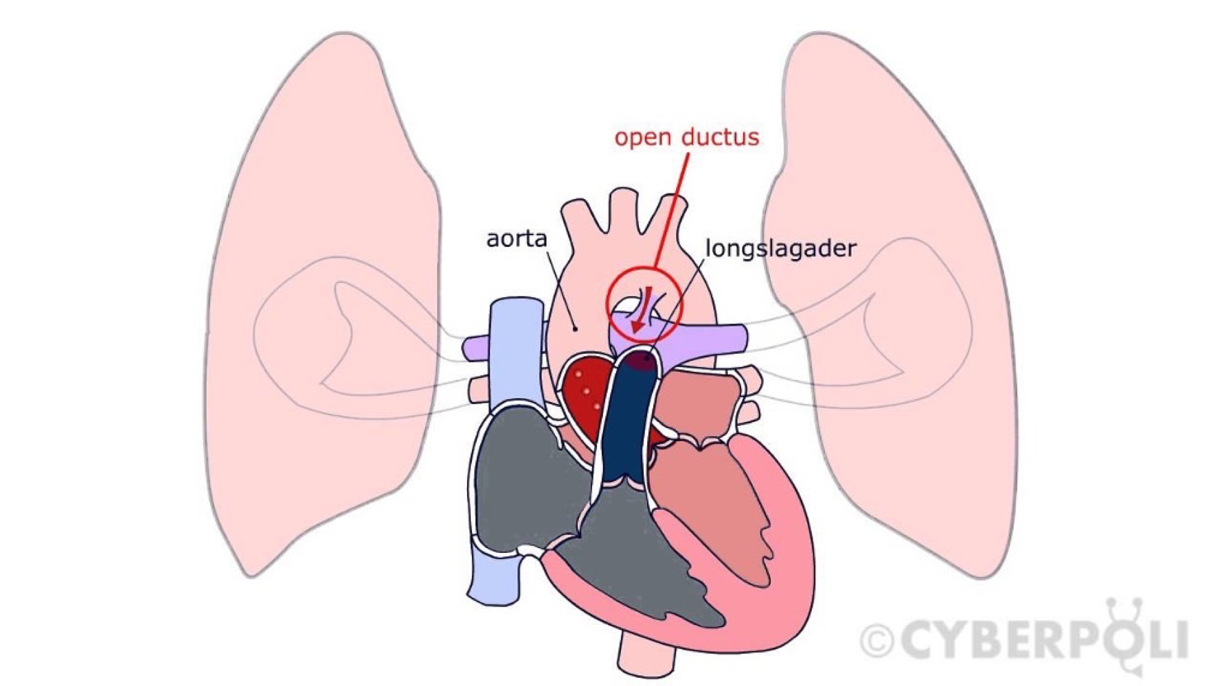
Ductus Arteriosus
Closure in newborn
If it’s open mixed level of oxygenated blood enters the lungs
Can close within 6 months after birth naturally if not then surgery is needed otherwise oxygen problems.
Atrial septum defects
opening below foramen ovale
opening at foramen ovale
Urogenital - What are reproductive organs divided into??
Genitalia
Subdivided into internal and external
Function of penis
Transport of urine via urethra & ejaculating sperm
What are the parts of the urethra??
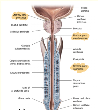
Pars prostatica (prostatic part)
Pars membranous (membranous part)
Pars spongiosa (spongy part)
What are some characteristics of the Urethra??
Disadvantage at length
Smaller risk of bladder infection
Higher risk of uretral blockage
What is prostate enlargement??
Frequent in men 40-50
Growth of unknown origin
Symptoms:
complaints in urination - inward swelling
obstruction of urethra
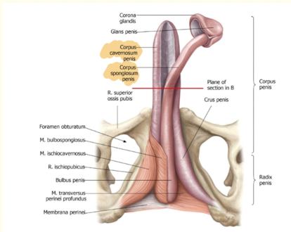
What are the two swelling bodies of the penis and what are their functions??
2x corpera cavernosa (dilation of vessels for more blood flow)
1x corpus spongiosum (formation of the glans penis, less swelling as that would block the urethra)
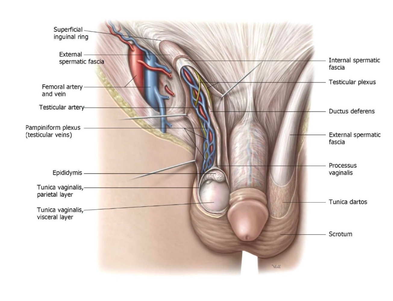
Structure of Scrotum
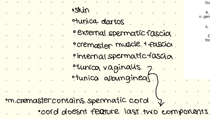
Spermatic Cord (function of vas deferens)??
Tube like structure and contains vas deferens, blood vessels, and nerves
Functions are: transport of the sperm cells from the inside to the outside.
Start in scrotum.
Internal Reproductive Organs
Follows the trajectory of sperm which is testicles → epididymus → vas defers → glands.
Testicles
Located under the epididymus
Production of sperm cells happens here
Connective tissue → tunica albuniginea (capsule)
Spermatogenesis
Sperm cells in seminiferous tubules
Leydig Cells → testosterone
convulated tubules → straight tubules
Form the rete testis
What is the structure and function of the epididuymus??
Contains the efferent ductules
Subdivided in caput & corpus, cauda
Function: storage of sperm cells, recycling damaged sperm cells and production of fluid.
What are the three glands in the male reproductive system??
Seminal vesicle
Prostate Glands
Cowpers Glands
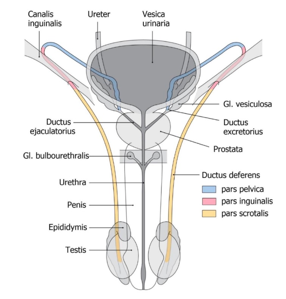
Ductus Deferens
Crosses the ureter dorsally
Drains in the ejaculatory duct and joins to form the urethrta
Seminal Vesicle
Paired structure of 3-5cm
Adds unto 70% fluid to semen, which contains fructose & prostaglandines.
Prostate Gland
30% semen
Neutralises the acidity of the semen and improves motility.
Cowper’s Glands
Situated near bulb of penis
Size of a pea
Produces alkaline fluid (lubrication) and neutralises urine
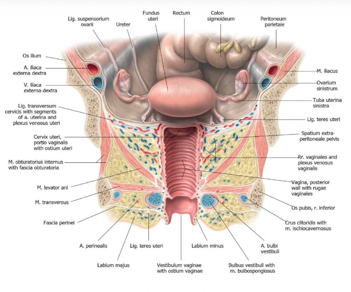
Internal vs External Genitals of Female
External - vagina, labia, pubic mound, glands
Internal - womb, ovaries, fallopian Tubes, vagina
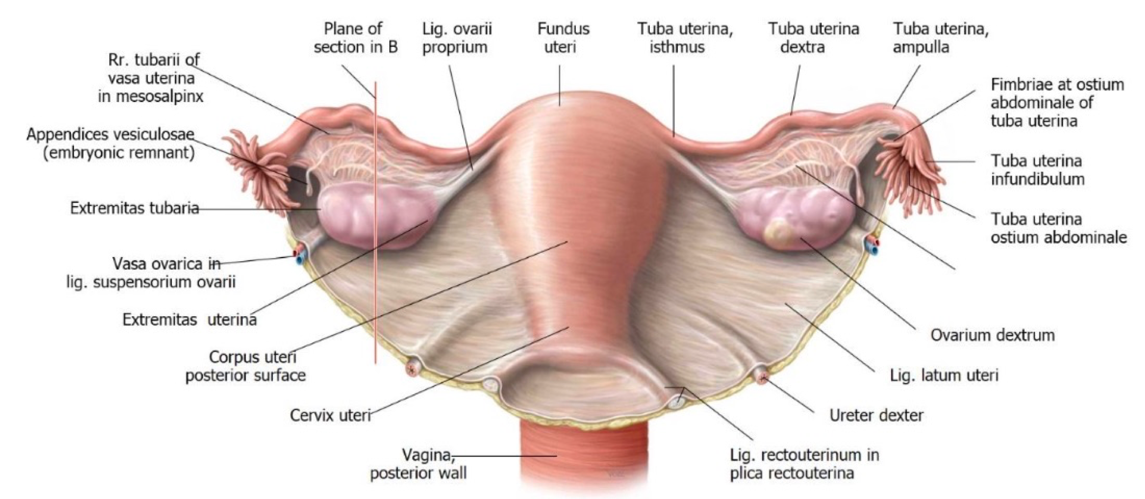
Ovary
Storage of egg cells
Connected to uterus via the Fallopian Tubes
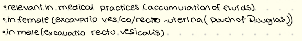
Peritoneal Cavity
Eggs travel accross the peritonieal cavity
Can increase risk of extrauterine pregnancy
Possible infection
Ligaments of Ovaries
Suspensory ligaments - contains ovarian artery
Proper ovarian ligament- embryonic rest
Mesovary - (part of broad ligament) fold of the peritoneal membrane.
What is the further trajectory of the egg??
Travels through the tube
Conception occurs in ampulla
Implantation into the uterine wall
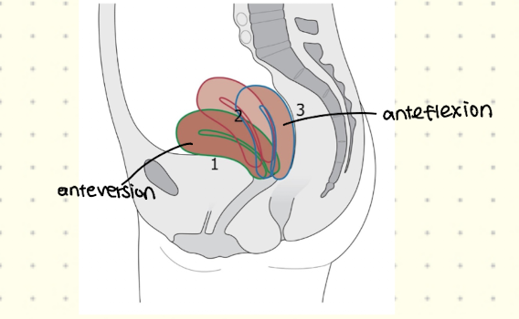
Uterus - Womb
Hollow organs with tubes at both ends
can be in specific positions (antiversion & anteflexion)
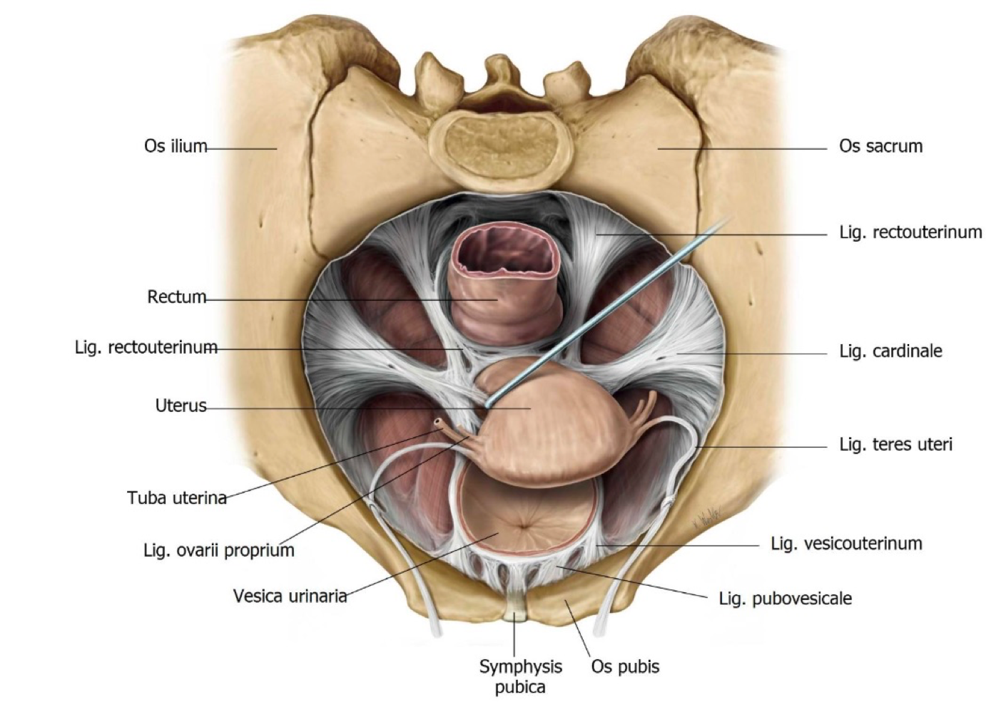
Ligaments of Uterus
Round Ligaments - embryonic rest
Broad Ligament - fold of peritoneum
Cardinal Ligament - contains the uterine artery
Uteroscaral Ligament
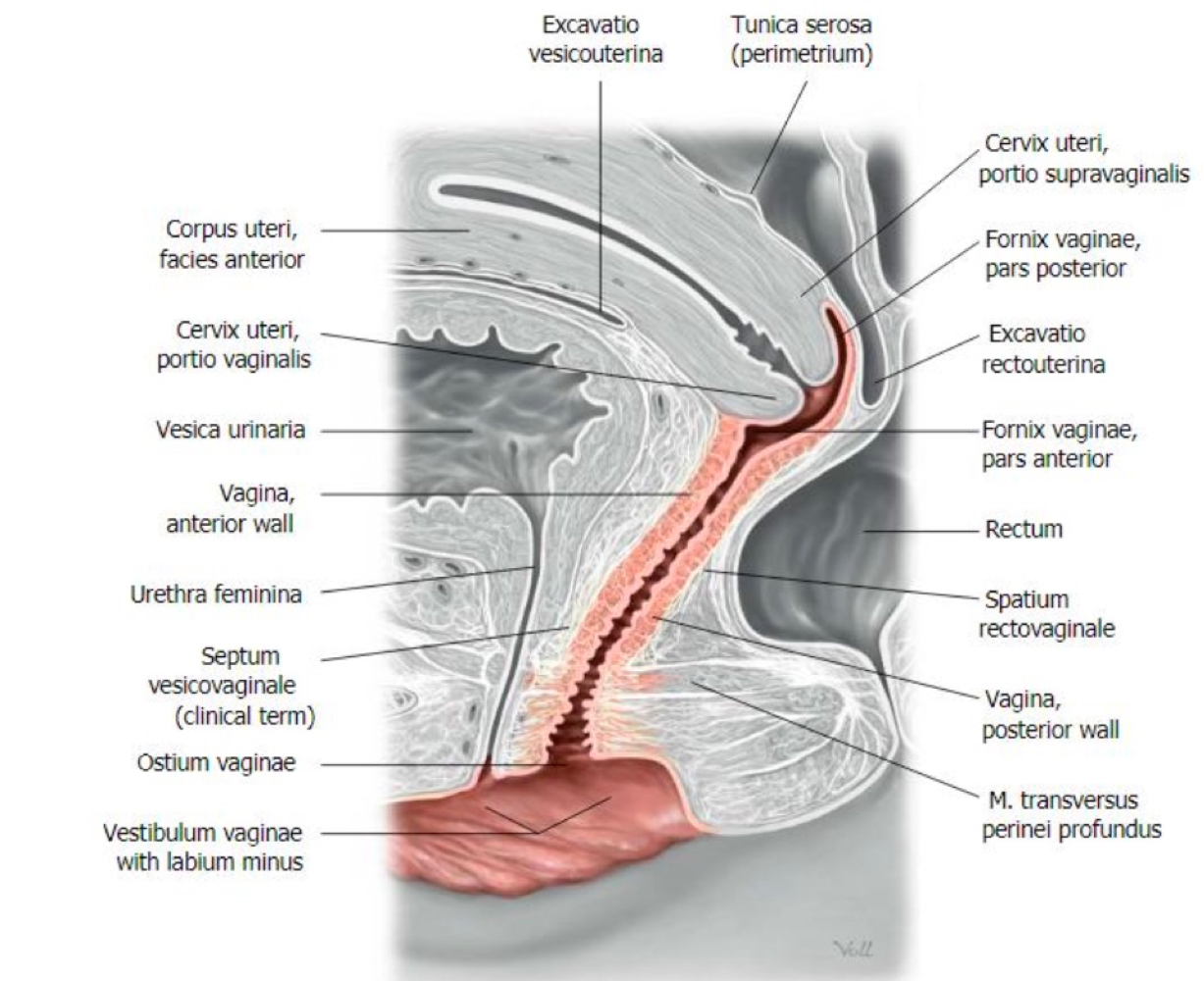
Vagina
Borders the neck of the uterus
Partially internal and external
Bartolin Glands
Secrete mucus for lubricatrion
Skenes Glands
Secrete mucus for lubrication
Clitoris
Female swelling body
What does corpera cavernosa consist of??
corpus clitoridis
2x crus clitoridis
Corpera spongiosa
2x bulbus vestbuli
Glans Clitoridis
Peritonoeal Spaces
How does vascularisation occur in testes and ovaries and uterus??
By testicular & ovaria artery/vein
Both arise from aorta/vena cava (except for renal vein)
Uterus: supplied by internal iliac artery by uterine and vaginal artery
What are the lymphoid structures??
Para aortal, iliac, inguinal
Relevant for metastasis
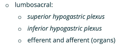
Somatic Innervation
Via sacral plexus
Efferent & Afferent
Autonomous Innervation
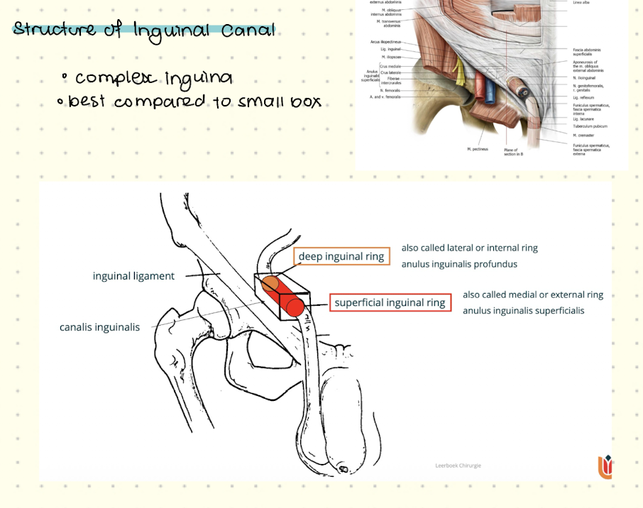
Inguinal Canal
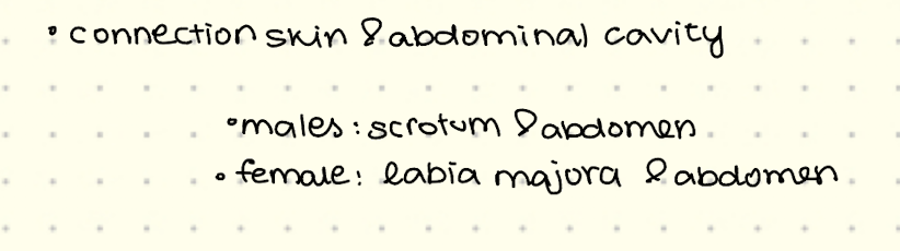
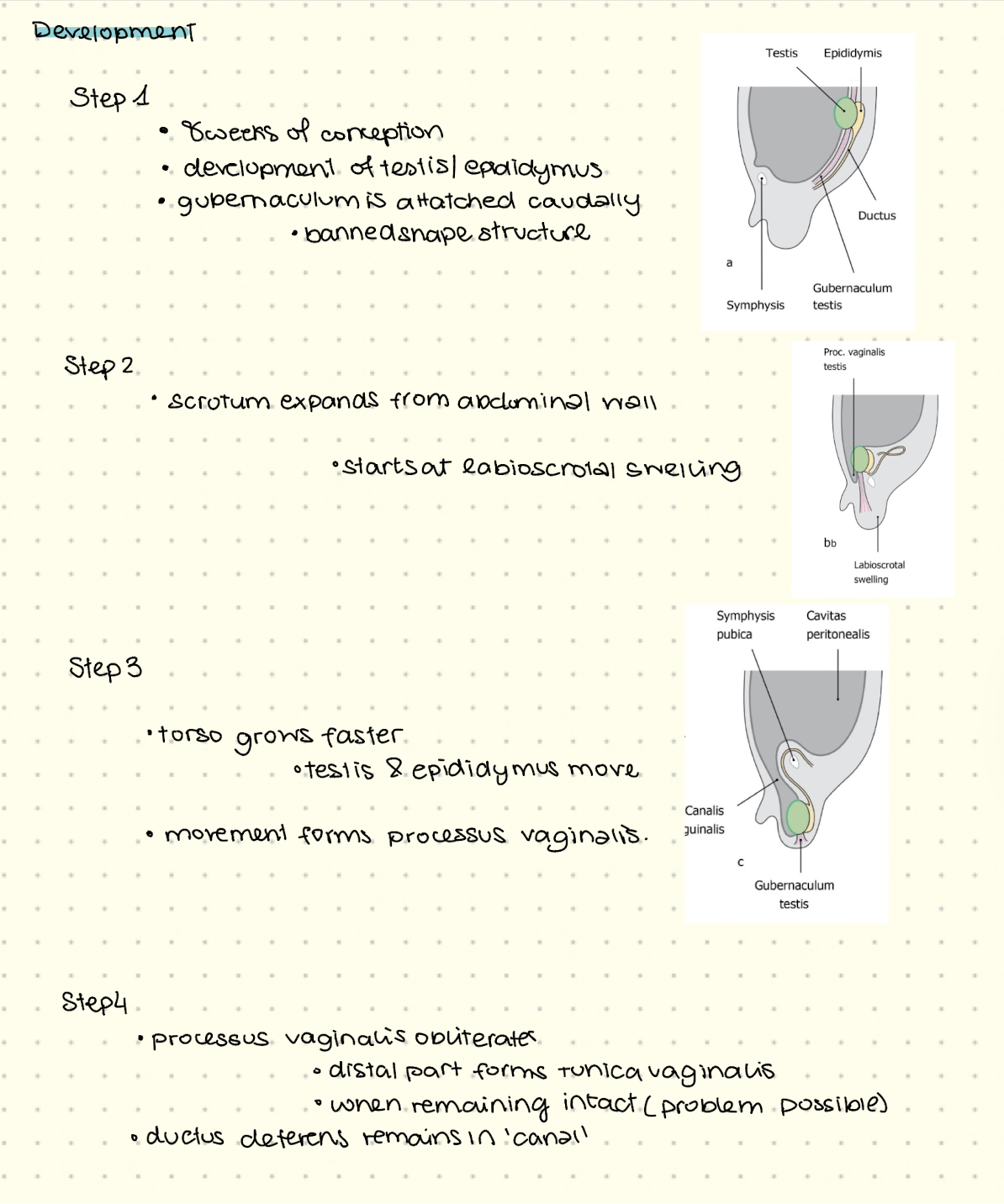
Development of Inguinal Canal
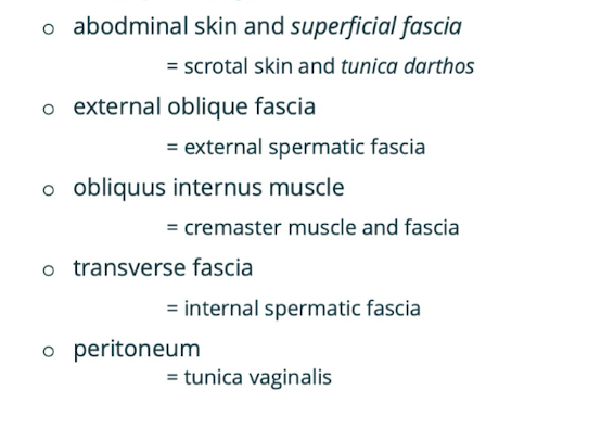
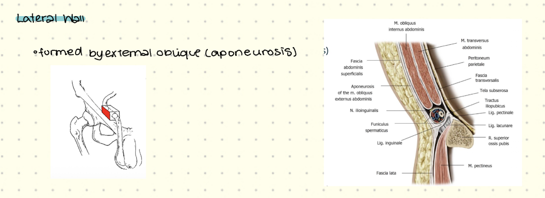
Corresponsing Layers
Structure of Inguinal Canal
Contents of Inguinal Canal (male)
Spermatic cord
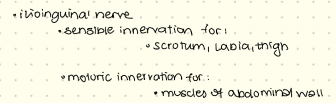
Ilionguinal nerve
Contents of Inguinal Canal (female)
round ligaments of uterus
Ilionguinal nerve
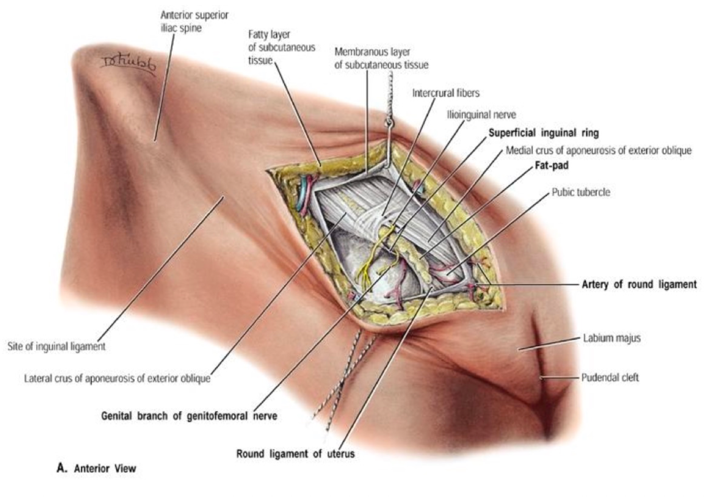
Ilionguinal nerve
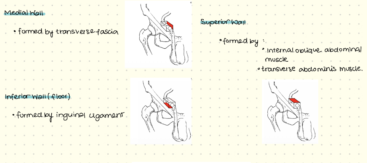
Lateral Wall
Medial, Superior, Inferior Walls
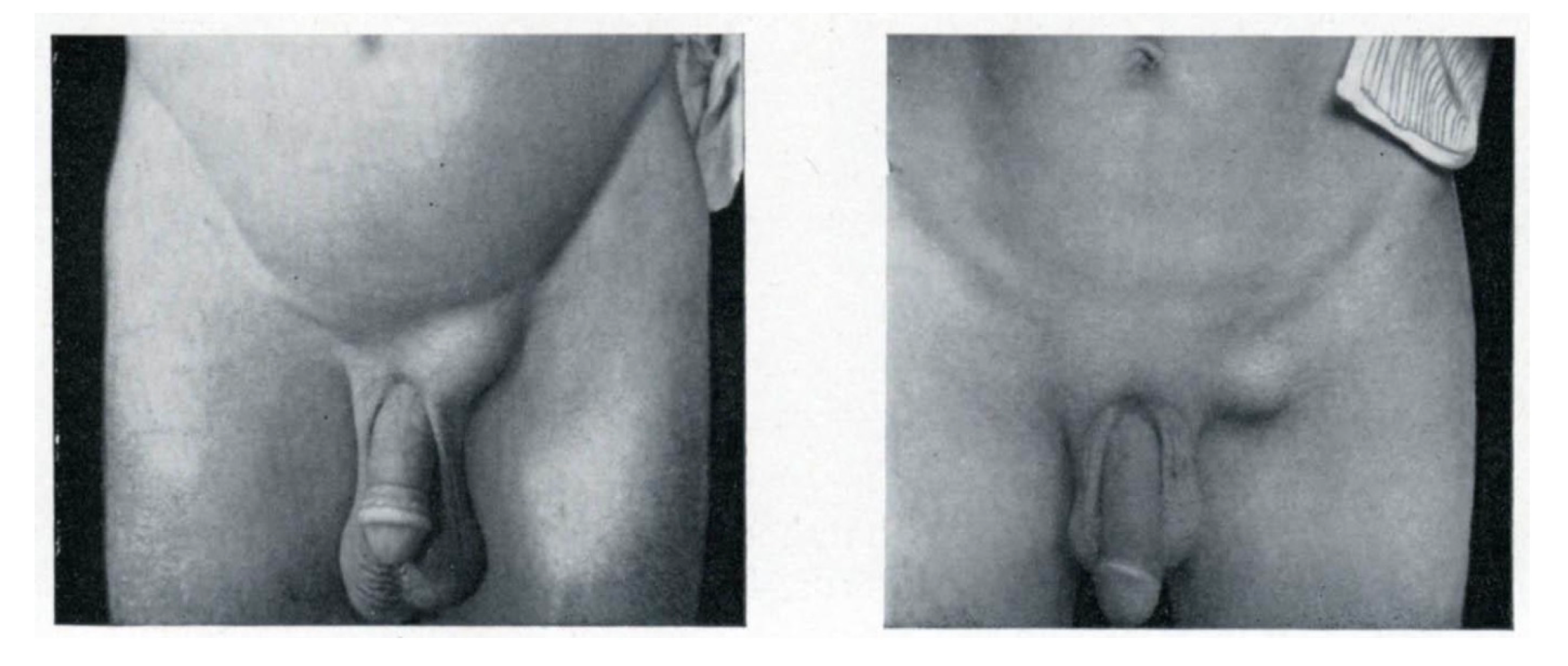
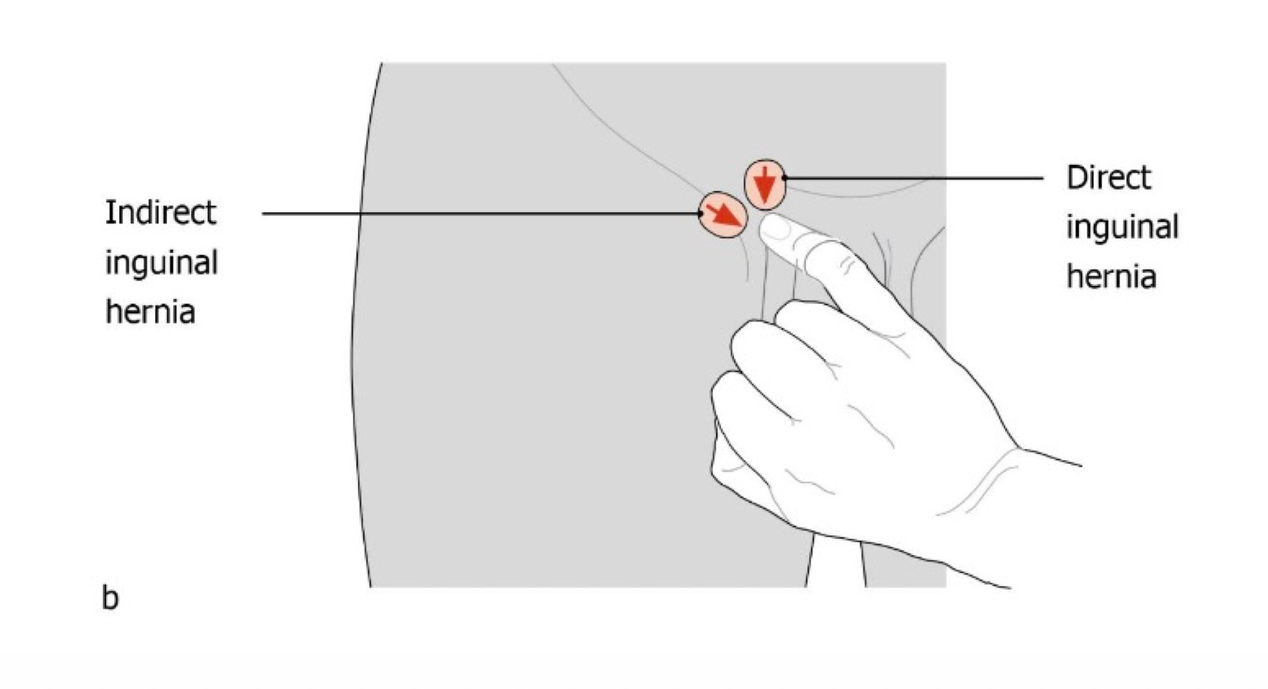
Inguinal Hernia
Protrusions of abdominal contents (through weak spots in wall.
Can be laterl/indirect (through deep inguinal ring)
Or medial direct through transverse fascia.
Symptoms of Inguinal Hernia
Diagnosis of Inguinal Hernia
Physical examination
Use of imaging techniques (ultrasound, MRI etc)
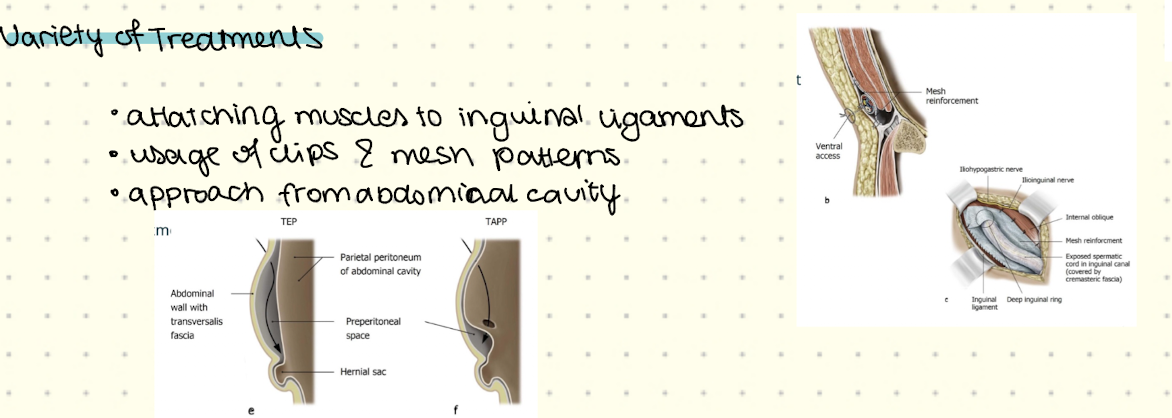
Treatment of Inguinal Hernia
If no strangulations → no treatment
If strangulated then emergency surgery
Variety of Treatments