A&P Chapter 4: Tissue Level of Organization
1/75
Earn XP
Description and Tags
Name | Mastery | Learn | Test | Matching | Spaced | Call with Kai |
|---|
No analytics yet
Send a link to your students to track their progress
76 Terms
[Four Main Groups - Overview]
Epithelial Tissue
Function: Covers body surfaces, lines cavities, forms glands
Structure → Function: Tightly packed cells arranged in sheets → ideal for protection, absorption, and secretion
[Four Main Groups - Overview]
Connective Tissue
Function: Binds, supports, protects, and integrates body parts
Structure → Function: Cells are spread within an extracellular matrix → provides strength, support, and flexibility
[Four Main Groups - Overview]
Muscle Tissue
Function: Movement through contraction
Types: Skeletal (voluntary), smooth, cardiac
Structure → Function: Excitable, contractile cells → generate force and movement
[Four Main Groups - Overview]
Nervous Tissue
Function: Communication via nerve impulses
Structure → Function: Excitable cells that transmit electrochemical signals → enable rapid coordination
3 Major Germ Layers/Embryonic Tissues
Ectoderm (outer): forms nervous tissue and epithelium (skin)
Mesoderm (middle): forms muscle and connective tissue (skeletal, muscular, and circulatory systems)
Endoderm (inner): forms internal epithelial linings (digestive/respiratory tracts)
[Tissue Membranes]
Mucous Membrane
Composed of epithelial tissue.
Line body cavities that are open to outside (digestive tract, respiratory tract, urogenital tract)
Wet/moist membranes because they secrete mucous = reduces friction and facilitates absorption/secretion
[Tissue Membranes]
Serous Membrane
Composed of mesothelium (simple squamous epithelium), supported by areolar tissue
Lines closed body cavities/organs
Never exposed/connected to outside
Secrete serous fluid = reduces friction
Types: pleura (lung), pericardium (heart), peritoneum (abdominal organs)
[Tissue Membranes]
Cutaneous Membrane (skin)
Made of stratified squamos + areolar tissue + supported by dense irregular connective tissue
Covers body surface
Dry, relatively thick, and waterproof
[Tissue Membranes]
Synovial Membrane
Line mobile joint cavities but don’t cover opposing joint surfaces
Secretes synovial fluid
Differs from epithelia because: develops within connective tissue, no basal lamina present, gaps between cells, and synovial fluid/capillaries exchange fluids
[Epithelial Tissues]
Functions of Epithelial Tissue
Provides physical protection from abrasion, dehydration, and destruction
Controls permeability (substances that enter/leave the body)
Provides sensation: sensory nerves detect changes in environment
Absorption/filtration
Can produce secretions (glands)
[Epithelial Tissues]
Structure of Epithelial Tissue
Sheets of tightly packed cells with little extracellular space
Avascular (no blood vessels)
Show polarity: apical surface (free/exposed) and basal surface (attached to basement membrane)
Anchored by basement membrane/basal lamina.
clear layer/lamina lucida: contains glycoproteins + fine protein filaments
dense layer/lamina densa: bundles of coarse protein fibers = strength/filter
[Epithelial Tissues]
Characteristics of Epithelial Tissue
Rapid regeneration (cells divide frequently)
Cellularity: they form an effective barrier
Occluding junctions: form a barrier that isolates surfaces/deeper tissues from the lumen contents. Tight attachments = prevents passage of water/solutes.
Adhesion belt: locks together the webs of cells = strengthens region and prevents distortion/leakage at junctions.
Gap junction: permits chemical communication that coordinate cell activity. Two cells are held together by interlocking proteins (connexons) that serve as channels.
Desmosomes: provide firm attachments between neighboring cells by interlocking cytoskeletons.
Hemidesmosomes: attach basal surface to basement membrane.
CAM: cells adhesion molecules; transmembrane proteins that bind to each other/other materials. Present in adhesion belt/desmosomes.
[Epithelial Tissue]
Naming Epithelial Tissues
Arrangement
simple: one layer thick
stratified: more than one layer
pseudostratified: false layers (look like more than one, but isn’t)
Shape
squamos: thin, flat, irregular. look like fried eggs or pancake with butter (nucleus)
cuboidal: equally wide and tall. hexagonal boxes with spherical nucleus in the center
columnar: more tall than wide, resemble rectangles with elongated nuclei crowded into a narrow band near the basal lamina
[Epithelial Tissue]
Simple Squamos Epithelium
Description: single layer of flattened cells with disc-shaped central nuclei
Function: diffusion/filtration (can secrete lubricant)
Locations: air sacs in lungs, kidney, linings of heart/lymphatic system
[Epithelial Tissue]
Stratified Squamos Epithelium
Description: thick layers of flattened cells (can have keratinized/mitotic layer)
Function: protection
Locations: outer layer of skin, covers organs (mouth/female reproductive organs)
[Epithelial Tissue]
Simple Cuboidal Epithelium
Description: single layer of cube-like cells with large spherical nucleus in center
Function: absorption/secretion
Locations: ovaries, kidneys, glands
[Epithelial Tissue]
Stratified Cuboidal Epithelium
Description: rare in human body
Locations: sweat glands, mammary glands, exocrine glands
[Epithelial Tissue]
Simple Columnar Epithelium
Description: single layer of tall cells with round/oval nuclei (some can have cilia, goblet cells, or microvilli)
Function: absorption, secretion of mucus/enzymes
Locations:
non-ciliated: digestive tract (intestines), gallbladder, glands
ciliated: bronchi, uterine tubes, uterus
[Epithelial Tissue]
Stratified Columnar Epithelium
Description: rare in human body
Locations: lines large ducts (salivary glands/pancreas)
[Epithelial Tissue]
Pseudostratified Columnar Epithelium
Description: single layer of cells of differing heights (nuclei at different heights)
Function: secretion, propulsion by cilia
Locations:
non-ciliated: male reproductive ducts
ciliated: respiratory tract
[Epithelial Tissue]
Transitional Epithelium
Description: resembles both stratified squamos/cuboidal. basal cells are cuboidal/columnar
Function: stretches readily + permits distension
Locations: lines uterus, bladder, and urethra
[Glands]
Endocrine Glands
“ductless” glands that produce hormones
secret directly into interstitial fluids/bloodstream
ex: pituitary gland, adrenal gland, thyroid gland
[Glands]
Exocrine Glands
have ducts
secrete their substance either on body surfaces or within ducts
Different modes
merocrine: most common; secrete products from secretory vesicles by exocytosis (ex: salivary glands)
holocrine: accumulate products until cell ruptures. destroys the cell and must be replaced by cell division (ex: sebaceous glands of skin)
apocrine: products accumulate within the cells then pinches off packets that contain the secretion (ex: mammary glands)
Cellular
unicellular: goblet cells that produce mucin which mixes with water = mucus
multicellular:
simple: single duct, doesn’t branch on its way to secretory cells
compound: duct divides one or more times on way to secretory cells
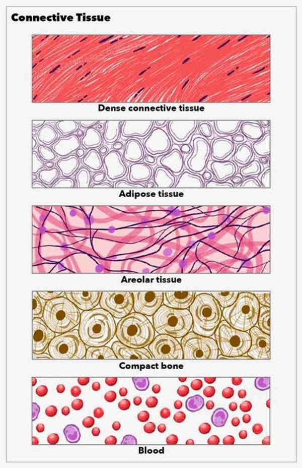
[Connective Tissue]
Characteristics of Connective Tissue
most abundant/never exposed to outside
all originate from mesenchyme
ground substance: fills space between cells + surrounds extracellular fibers
Different kinds
fibroblast cells: produce connective tissue proper
chondrocytes: produce cartilage
osteocytes: produce bone
hemocytoblast cells: produce blood
Fibers
elastic: slender, straight, stretchy
collagen: thick, straight, form bundles = strong and resist stretching
reticular: strong fibers that form branches
[Connective Tissue]
Functions of Connective Tissue
establish structural framework
transport fluids/materials
protect delicate organs
store energy reserves (triglycerides)
defend body from invaders
[Connective Tissue]
Loose Connective Tissues (Connective Tissue Proper)
areolar tissue: most common, packing material. attaches skin to body parts. (AKA superficial fascia)
adipose tissue: fat; found deep in skin, forms layer of padding (made of adipocytes)
reticular tissue: provides support/resistance through tough/flexible network (stroma)
[Connective Tissue]
Dense Connective Tissue (Connective Tissue Proper)
dense regular: fibers oriented parallel to each other = strength along axis of collagen fibers. found in tendons and ligaments
dense irregular: non-parallel = interwoven network. provide strength in many directions
elastic: springy nature that allows extension/recoil
[Connective Tissue]
Ligaments
connect bones to bones
[Connective Tissue]
Tendons
connect bone to muscle
[Connective Tissue]
Fluid Connective Tissue
suspended in watery matrix that contains proteins
blood: flows in cardiovascular system
plasma: watery matrix
hemocytoblasts:
erythrocytes: red blood cells (transport oxygen)
leukocytes: white blood cells (defend body from disease)
thrombocytes: platelets (clotting)
lymph: flows in lymphatic system
forms interstitial fluid
passes through lymph nodes
[Connective Tissue]
Supporting Connective Tissue
protects soft tissues and supports weight of the body
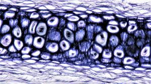
cartilage: solid/rubbery matrix containing chondrocytes. surrounded by perichondrium
hyaline cartilage: support + reduces friction; connects ribs to sternum, covers articular surfaces of bones, forms parts of nose
elastic cartilage: flexible; ear
fibrous cartilage: collagen fibers = strong; found in discs, knee, pubic
bone: solid matrix containing osteocytes. surrounded by periosteum.
hollow, compact bone (osteons) on outside and spongy bone inside
![<p>[Muscle Tissue]</p><p>Skeletal Muscle</p>](https://assets.knowt.com/user-attachments/e37ed168-e0b4-4385-ab18-d45ed4b20f33.png)
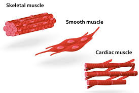
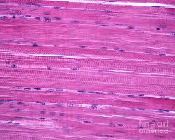
[Muscle Tissue]
Skeletal Muscle
structure: long/cylindrical, striated, multinucleated
location: attached to bones
control: voluntary
function: body movement (locomotion), posture, generates heat (shivering)

![<p>[Muscle Tissue]</p><p>Cardiac Muscle</p>](https://assets.knowt.com/user-attachments/8968d95b-cd42-499a-bd9b-839531744c42.png)
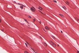
[Muscle Tissue]
Cardiac Muscle
structure: short, branched, striated, single nucleus, connected by intercalated discs
discs: anchoring junctions = hold cells together, gap junctions = signals and coordination
location: heart
control: involuntary
function: pumps blood, contracts in rhythm, acts as one unit
![<p>[Muscle Tissue]</p><p>Smooth Muscle</p>](https://assets.knowt.com/user-attachments/78097d17-2796-48dc-a509-202f22f49495.png)
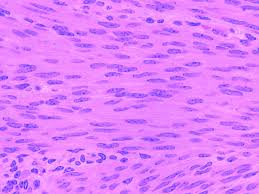
[Muscle Tissue]
Smooth Muscle
structure: spindle-shaped, non-striated, single nucleus
location: walls of organs (digestive tract, blood vessels)
control: involuntary
functions: moves materials through organs (food/urine), blood flow, regulates airways and secretions

[Muscle Tissue]
Composition
vascularized muscle tissue is made of elongated cells (fibers) containing myofilaments (actin/myosin proteins)
[Nervous Tissue]
Perception and Response of Nervous Tissue
input: sensory receptors monitors changes inside/outside the body
integration: processes and interprets the sensory input
motor output: effects a response to the stimulus
maintains homeostasis by acting as regulatory/control center
[Nervous Tissue]
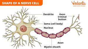
Overview
nervous tissue is made of neurons (branching cells)
neurons are made of cell body (contains nucleus), dendrites (receives signals), and axons (send signals)
function: conduct electrical impulses and maintains homeostasis
neuroglial cells: cells surrounding the neurons that feed/support/protect them
location: brain, spinal cord, nerves
[Tissue Repair]
Capacity for repair
epithelial tissue: replaced by division of stem/undifferentiated cells
connective tissue: bone has continuous capacity; cartilage not as much
muscle tissue: poor capacity for renewal
nervous tissue: poor capacity for renewal
[Tissue Repair]
Characteristics
fibrosis: process of scar formation
if injury is extensive: granulation tissue is formed
clinical connection: adhesions (from scar tissue) causes abnormal joining of adjacent tissues = intestinal obstruction
nutrition is important to tissue repair
proper blood circulation is essential to tissue repair
[Tissue Repair]
Steps/Phases
Cleanup
remove debris, toxins, etc
clot forms (stop bleeding)
scab forms (protects wound)
Tissue Rebuilding
fibroblasts: rebuild collagen/matrix
angiogenesis: new blood vessels form
new vascular tissue develops (granulation)
Remodeling
wound contraction: edges pull together
clot dissolves
scar tissue can form
[Aging and Tissues]
young people’s tissues repair rapidly and efficiently; process slows down with age
younger body is in a better nutritional state, better blood supply, faster metabolic rate
when aging: tissues become thinner, drier, less elastic
collagen decreases
bone loses minerals = reduced height
cartilage deteriorates = joint stiffness
muscle atrophy (loss of mass)
[Disorders]
Homeostatic Imbalances
epithelial tissue disorders are specific to individual organs: skin cancer (epidermis), peptic ulcer disease (epithelial lining of stomach/intestines)
connective tissue disorder: most prevalent is autoimmune disorders (antibodies in immune system fail to distinguish foreign VS self = attacks body tissue)
Systemic lupus erythematosus: chronic inflammatory disease
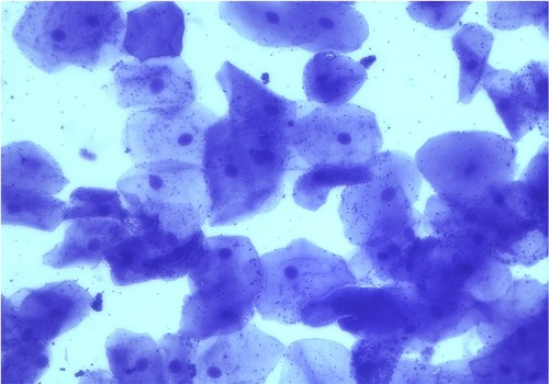
Simple Squamos Epithelium
single layer of flattened cells
diffusion/filtration
air sacs in lungs, kidney, lining of heart/lymphatic system
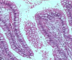
Simple Columnar Epithelial Tissue
single layer of tall cells with round nuclei
absorption, secretion of mucus/enzymes
non-ciliated: digestive tract/intestines, galbladder
ciliated: bronchi, uterus
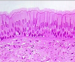
Pseudostratified (ciliated) columnar epithelial tissue
single layer of cells at differing heights so it looks like multiple
secretion, propulsion if it has cilia
non-ciliated: male reproductive ducts
ciliated: respiratory tract
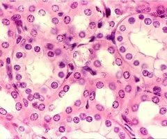
Simple Cuboidal Epithelium
single layer of cube-like cells
absorption/secretion
ovaries, kidneys, glands
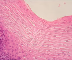
Stratified Squamos Epithelium
thick layers of flattened cells
protection
outer layer of skin, organs
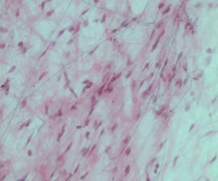
Loose Fibrous Connective Tissue
primary packing material
cushions organs
binds tissues together
provides flexibility
immune defense
just helpful
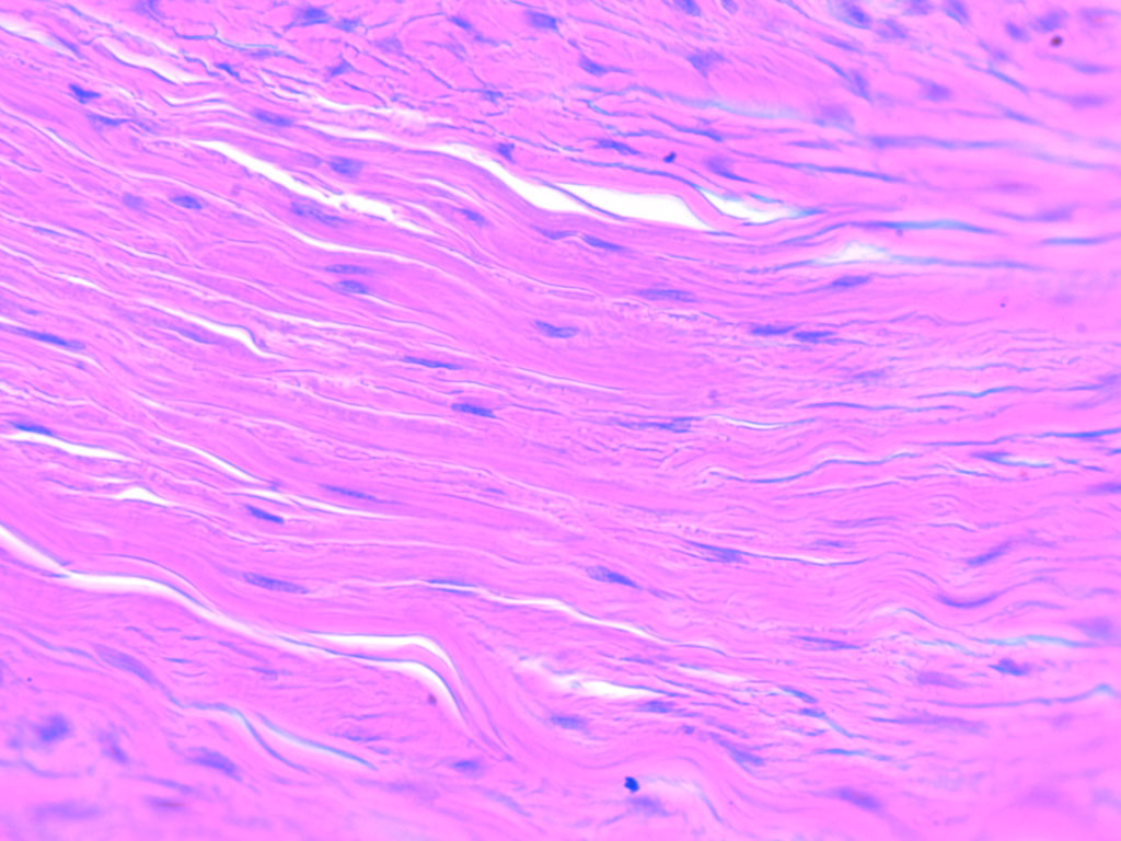
Dense Regular Connective Tissue
fibers are parallel = strength along axis
found in tendons and ligaments
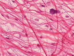
Loose Areolar Connective Tissue
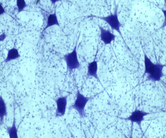
Nervous Tissue
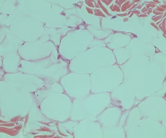
Adipose Connective Tissue
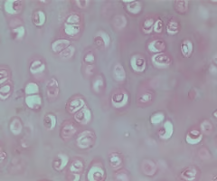
Hyaline Cartilage Connective
support
reduces friction
connections ribs to sternum
covers articular surfaces for bones
forms part of nose
Bone Connective Tissue
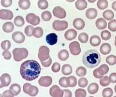
Fluid (blood) Connective Tissue
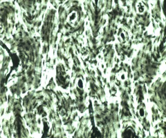
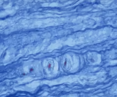
Smooth Muscle Tissue
non-striated, single nucleus
walls of organs (digestive/blood vessels)
involuntary control
moves materials through organs, blood flow, and regulates airways
Cardiac Muscle
short, striated, single nucleus, has intercalated discs
heart
involuntary control
pumps blood, rhythmic contraction
Skeletal Muscle
long, cylindrical, multi-nucleated. striated
attached to bones
voluntary control
body movement (locomotion), posture, generates heat (shivering)
Elastic Cartilage Connective Tissue
flexible
ear
Fibrous Cartilage Connective Tissue
collagen fibers = strong
found in discs, knee, pubic
[Quiz]
What epithelial tissue forms superficial layer of skin?
Keratinized stratified squamos epithelium
Which epithelial tissue is found lining the kidney glomerulus and performs blood filtration?
Simple squamos epithelium
[Quiz]
This type of junction contains tiny fluid-filled tunnels called connexons which allow the movement of ions and small molecules between cells. It is found between muscles cells of the heart and in organs with smooth muscle tissue, such as the gastrointestinal tract.
Gap Junctions
[Quiz]
These types of cell junctions anchor adjacent cells together and resist their separation during contractile activities.
Adherens junctions and desmosomes
[Quiz]
What kind of epithelial tissue is best in locations that need to stretch?
Transitional Epithelial Tissue
[Quiz]
In intestinal tissue, what substance do white cells (Goblet cells) produce?
mucus
[Quiz]
What gland are hormones secreted from? (They enter right into the blood stream)
Endocrine gland
[Quiz]
Adipose tissue is used for all of the following EXCEPT:
energy stoage
cushion organs
oxygen supply
insulate/temperature control
oxygen supply
[Quiz]
What kind of tissue includes collagen-filled, rope-like structures such as ligaments and tendons.
Dense Connective Tissue
[Quiz - Answered for you]
Exocrine glands secrete their substances onto body surfaces/ducts. Match ducts with definitions.
Merocrine: salivary glands of oral cavity
Holocrine: sebaceous glands of the skin
Apocrine: mammary glands of the breast
Gap Junctions
Channels for sharing ions and signals between adjacent cells.
Desmosomes
Localized spot welds holding cells together against mechanical stress.
Hemidesmosomes
Anchors attaching the bottom of a cell to the basement membrane.
Adhesion Belts
Continuous bands linking the actin skeletons of neighboring cells
Tight Junctions
Watertight seals preventing fluid leakage between adjacent cells