ANIMAL MOVEMENT AND SUPPORT

0.0(0)
studied byStudied by 0 people
0.0(0)
full-widthCall Kai
learnLearn
examPractice Test
spaced repetitionSpaced Repetition
heart puzzleMatch
flashcardsFlashcards
GameKnowt Play
Card Sorting

1/14

encourage image

There's no tags or description

Looks like no tags are added yet.

Study Analytics
Name
Mastery
Learn
Test
Matching
Spaced

No study sessions yet.

15 Terms

1
New cards

Describe the structure of a striated muscle cell and its filaments.

  • Cell Structure: Long, cylindrical, and multinucleated.

  • Myofibrils: The cell is packed with long, cylindrical organelles called myofibrils.

  • Filament Arrangement: Myofibrils contain orderly arrangements of thick (myosin) filaments and thin (actin) filaments.

  • Striations: The alternating dark (A bands, thick filaments) and light (I bands, thin filaments) bands create the striped appearance.

<ul><li><p class="ds-markdown-paragraph"><strong><span>Cell Structure:</span></strong><span> Long, cylindrical, and </span><strong><span>multinucleated</span></strong><span>.</span></p></li><li><p class="ds-markdown-paragraph"><strong><span>Myofibrils:</span></strong><span> The cell is packed with long, cylindrical organelles called myofibrils.</span></p></li><li><p class="ds-markdown-paragraph"><strong><span>Filament Arrangement:</span></strong><span> Myofibrils contain orderly arrangements of </span><strong><span>thick (myosin) filaments</span></strong><span> and </span><strong><span>thin (actin) filaments</span></strong><span>.</span></p></li><li><p class="ds-markdown-paragraph"><strong><span>Striations:</span></strong><span> The alternating dark (A bands, thick filaments) and light (I bands, thin filaments) bands create the striped appearance.</span></p></li></ul><p></p>
2
New cards

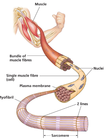
What is a sarcomere?

  • The basic contractile unit of a muscle fiber.

  • It is the region of a myofibril from one Z disc to the next.

  • Shortening of sarcomeres (Z discs moving closer) results in muscle contraction.


3
New cards

Describe the myosin and actin interactions during contraction.

  1. Myosin head is energized (with ADP + Pi) and binds to actin, forming a cross-bridge.

  2. Power stroke: Myosin head pivots, pulling the thin filament toward the center of the sarcomere.

  3. ATP binding: ATP binds to myosin, causing it to release actin.

  4. Recocking: Myosin hydrolyzes ATP to ADP + Pi, returning to its high-energy state, ready to bind actin again. The cycle repeats.

4
New cards

Where does the energy for muscle contraction come from?

  • Directly from ATP.

  • ATP is required for:

    1. The power stroke of the myosin head.

    2. Detaching myosin from actin.

    3. Pumping Ca²⁺ back into the sarcoplasmic reticulum for relaxation.

5
New cards

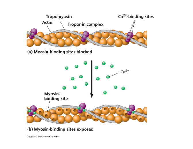
Explain the role of tropomyosin, troponin, and Ca²⁺.

  • At rest, tropomyosin blocks the myosin-binding sites on actin.

  • When an action potential arrives, Ca²⁺ is released from the sarcoplasmic reticulum.

  • Ca²⁺ binds to troponin, which changes shape and pulls tropomyosin away from the myosin-binding sites.

  • This allows myosin to bind to actin and initiate contraction.

<ul><li><p class="ds-markdown-paragraph"><span>At rest, </span><strong><span>tropomyosin</span></strong><span> blocks the myosin-binding sites on actin.</span></p></li><li><p class="ds-markdown-paragraph"><span>When an action potential arrives, </span><strong><span>Ca²⁺</span></strong><span> is released from the sarcoplasmic reticulum.</span></p></li><li><p class="ds-markdown-paragraph"><span>Ca²⁺ binds to </span><strong><span>troponin</span></strong><span>, which changes shape and pulls tropomyosin away from the myosin-binding sites.</span></p></li><li><p class="ds-markdown-paragraph"><span>This allows myosin to bind to actin and initiate contraction.</span></p></li></ul><p></p>
6
New cards

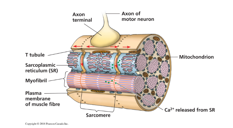
Describe the steps from an action potential to muscle relaxation.

  1. Action potential arrives at the motor neuron terminal, releasing ACh.

  2. ACh triggers an action potential in the muscle cell membrane.

  3. The action potential travels down T-tubules.

  4. This triggers the sarcoplasmic reticulum (SR) to release Ca²⁺.

  5. Ca²⁺ binds to troponin, moving tropomyosin to expose myosin-binding sites on actin.

  6. Cross-bridge cycling and contraction occur.

  7. To relax, Ca²⁺ is actively pumped back into the SR, tropomyosin re-blocks the sites, and the muscle fiber lengthens passively.

<ol><li><p class="ds-markdown-paragraph"><strong><span>Action potential</span></strong><span> arrives at the motor neuron terminal, releasing ACh.</span></p></li><li><p class="ds-markdown-paragraph"><span>ACh triggers an </span><strong><span>action potential</span></strong><span> in the muscle cell membrane.</span></p></li><li><p class="ds-markdown-paragraph"><span>The action potential travels down </span><strong><span>T-tubules</span></strong><span>.</span></p></li><li><p class="ds-markdown-paragraph"><span>This triggers the </span><strong><span>sarcoplasmic reticulum (SR)</span></strong><span> to release </span><strong><span>Ca²⁺</span></strong><span>.</span></p></li><li><p class="ds-markdown-paragraph"><span>Ca²⁺ binds to </span><strong><span>troponin</span></strong><span>, moving </span><strong><span>tropomyosin</span></strong><span> to expose myosin-binding sites on </span><strong><span>actin</span></strong><span>.</span></p></li><li><p class="ds-markdown-paragraph"><strong><span>Cross-bridge cycling</span></strong><span> and </span><strong><span>contraction</span></strong><span> occur.</span></p></li><li><p class="ds-markdown-paragraph"><span>To relax, Ca²⁺ is actively pumped back into the SR, tropomyosin re-blocks the sites, and the muscle fiber lengthens passively.</span></p></li></ol><p></p>
7
New cards

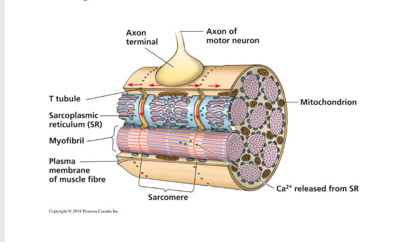
How do the plasma membrane and SR facilitate contraction?

  • Transverse (T) Tubules: Infoldings of the plasma membrane that carry the action potential deep into the muscle fiber.

  • Sarcoplasmic Reticulum (SR): A specialized endoplasmic reticulum that stores and releases Ca²⁺ when triggered by the action potential in the T-tubules.


<ul><li><p class="ds-markdown-paragraph"><strong><span>Transverse (T) Tubules:</span></strong><span> Infoldings of the plasma membrane that carry the action potential deep into the muscle fiber.</span></p></li><li><p class="ds-markdown-paragraph"><strong><span>Sarcoplasmic Reticulum (SR):</span></strong><span> A specialized endoplasmic reticulum that </span><strong><span>stores and releases Ca²⁺</span></strong><span> when triggered by the action potential in the T-tubules.</span></p></li></ul><p><br></p>
8
New cards

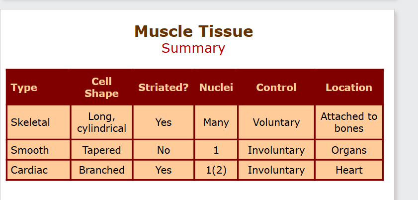
Compare skeletal, cardiac, and smooth muscle.

  • Skeletal:

    • Location: Attached to bones.

    • Structure: Long, striated, multinucleated fibers.

    • Control: Voluntary.

    • Function: Body movement.

  • Cardiac:

    • Location: Heart only.

    • Structure: Branched, striated, usually one nucleus, has intercalated discs.

    • Control: Involuntary.

    • Function: Pump blood.

  • Smooth:

    • Location: Walls of hollow organs (e.g., stomach, intestines, blood vessels).

    • Structure: Spindle-shaped, no striations, one nucleus.

    • Control: Involuntary.

    • Function: Move substances through organs (e.g., peristalsis).

<ul><li><p class="ds-markdown-paragraph"><strong><span>Skeletal:</span></strong></p><ul><li><p class="ds-markdown-paragraph"><strong><span>Location:</span></strong><span> Attached to bones.</span></p></li><li><p class="ds-markdown-paragraph"><strong><span>Structure:</span></strong><span> Long, striated, multinucleated fibers.</span></p></li><li><p class="ds-markdown-paragraph"><strong><span>Control:</span></strong> <strong><span>Voluntary</span></strong><span>.</span></p></li><li><p class="ds-markdown-paragraph"><strong><span>Function:</span></strong><span> Body movement.</span></p></li></ul></li><li><p class="ds-markdown-paragraph"><strong><span>Cardiac:</span></strong></p><ul><li><p class="ds-markdown-paragraph"><strong><span>Location:</span></strong><span> Heart only.</span></p></li><li><p class="ds-markdown-paragraph"><strong><span>Structure:</span></strong><span> Branched, striated, usually one nucleus, has </span><strong><span>intercalated discs</span></strong><span>.</span></p></li><li><p class="ds-markdown-paragraph"><strong><span>Control:</span></strong> <strong><span>Involuntary</span></strong><span>.</span></p></li><li><p class="ds-markdown-paragraph"><strong><span>Function:</span></strong><span> Pump blood.</span></p></li></ul></li><li><p class="ds-markdown-paragraph"><strong><span>Smooth:</span></strong></p><ul><li><p class="ds-markdown-paragraph"><strong><span>Location:</span></strong><span> Walls of hollow organs (e.g., stomach, intestines, blood vessels).</span></p></li><li><p class="ds-markdown-paragraph"><strong><span>Structure:</span></strong><span> Spindle-shaped, </span><strong><span>no striations</span></strong><span>, one nucleus.</span></p></li><li><p class="ds-markdown-paragraph"><strong><span>Control:</span></strong> <strong><span>Involuntary</span></strong><span>.</span></p></li><li><p class="ds-markdown-paragraph"><strong><span>Function:</span></strong><span> Move substances through organs (e.g., peristalsis).</span></p></li></ul></li></ul><p></p>
9
New cards

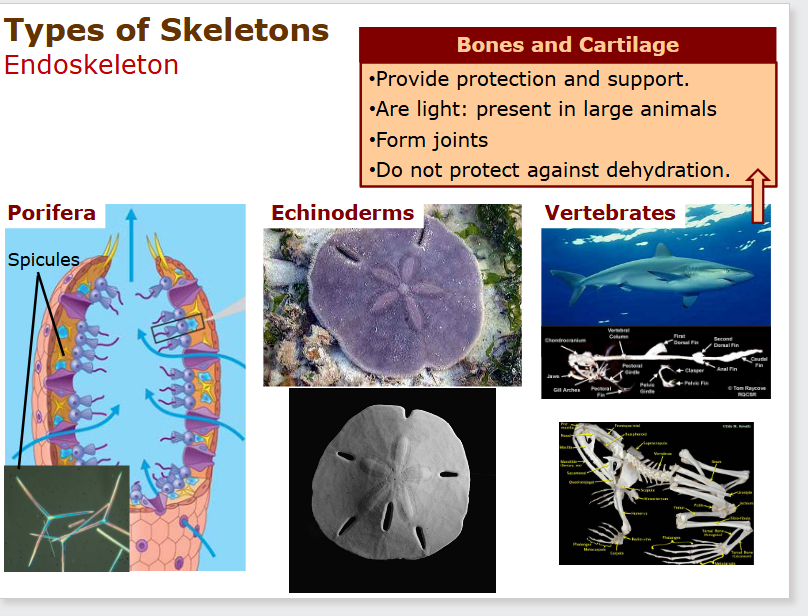
Compare hydrostatic, exoskeletons, and endoskeletons.

  • Hydrostatic: Fluid-filled cavity (coelom) surrounded by muscles (e.g., earthworm). Flexible.

  • Exoskeleton: External, hard encasement (e.g., arthropod chitin shell). Provides protection but must be molted to grow.

  • Endoskeleton: Internal, hard or flexible support (e.g., vertebrate bones, echinoderm plates). Grows with the animal.

10
New cards
  • How does an earthworm use its hydrostatic skeleton to move?


  • Uses muscular antagonism between circular and longitudinal muscles.

  • Contraction of circular muscles makes a segment long and thin, pushing it forward.

  • Contraction of longitudinal muscles makes a segment short and thick, anchoring it.

  • Waves of these contractions create movement. Other examples: Sea anemones, jellyfish.

11
New cards

Compare snail shells and arthropod exoskeletons.

  • Snail Shell: Made primarily of calcium carbonate. Function is mainly protection.

  • Arthropod Exoskeleton: Made of chitin. Function is protection, support, and attachment for muscles for movement.

12
New cards

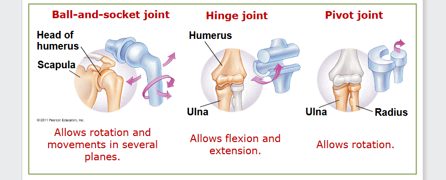
Identify major bones/joints and their movements.

  • Ball-and-Socket Joint (e.g., shoulder, hip): Allows circular movement, rotation.

  • Hinge Joint (e.g., elbow, knee): Allows movement in one plane (flexion/extension).

<ul><li><p class="ds-markdown-paragraph"><strong><span>Ball-and-Socket Joint</span></strong><span> (e.g., shoulder, hip): Allows </span><strong><span>circular movement, rotation</span></strong><span>.</span></p></li><li><p class="ds-markdown-paragraph"><strong><span>Hinge Joint</span></strong><span> (e.g., elbow, knee): Allows movement in </span><strong><span>one plane</span></strong><span> (flexion/extension).</span></p></li></ul><p></p>
13
New cards

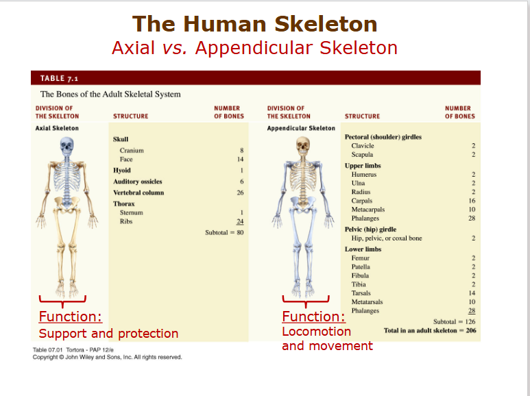
What bones form the shoulder and pelvic girdles, and what is their function?

  • Pectoral (Shoulder) Girdle: Clavicle and scapula. Connects the arms to the axial skeleton.

  • Pelvic Girdle: Fused hip bones (ilium, ischium, pubis). Connects the legs to the axial skeleton.

  • Function: Girdles provide stability and a point of attachment for the limbs.

<ul><li><p class="ds-markdown-paragraph"><strong><span>Pectoral (Shoulder) Girdle:</span></strong> <strong><span>Clavicle and scapula</span></strong><span>. Connects the arms to the axial skeleton.</span></p></li><li><p class="ds-markdown-paragraph"><strong><span>Pelvic Girdle:</span></strong> <strong><span>Fused hip bones (ilium, ischium, pubis)</span></strong><span>. Connects the legs to the axial skeleton.</span></p></li><li><p class="ds-markdown-paragraph"><strong><span>Function:</span></strong><span> Girdles provide stability and a point of attachment for the limbs.</span></p></li></ul><p></p>
14
New cards

Give examples of non-vertebrate animals with endoskeletons.

  • Echinoderms (e.g., sea stars, sea urchins). Their endoskeleton is made of calcareous plates (ossicles) beneath the skin.

<ul><li><p class="ds-markdown-paragraph"><strong><span>Echinoderms</span></strong><span> (e.g., sea stars, sea urchins). Their endoskeleton is made of </span><strong><span>calcareous plates (ossicles)</span></strong><span> beneath the skin.</span></p></li></ul><p></p>
15
New cards

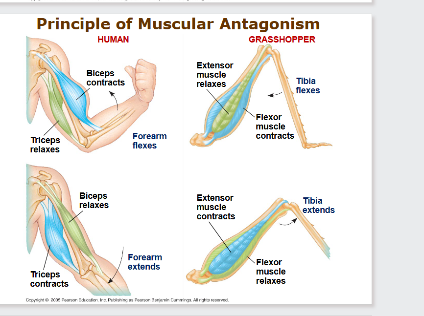
Explain muscular antagonism in the human forearm and grasshopper tibia.

  • Human Forearm:

    • Biceps contracts to flex (bend) the elbow.

    • Triceps contracts to extend (straighten) the elbow.

  • b) Grasshopper Tibia:

    • Flexor muscle contracts to pull the tibia in (fold the leg).

    • Extensor muscle contracts to thrust the tibia out (jump).

<ul><li><p class="ds-markdown-paragraph"><strong><span>Human Forearm:</span></strong></p><ul><li><p class="ds-markdown-paragraph"><strong><span>Biceps</span></strong><span> contracts to </span><strong><span>flex</span></strong><span> (bend) the elbow.</span></p></li><li><p class="ds-markdown-paragraph"><strong><span>Triceps</span></strong><span> contracts to </span><strong><span>extend</span></strong><span> (straighten) the elbow.</span></p></li></ul></li><li><p class="ds-markdown-paragraph"><strong><span>b) Grasshopper Tibia:</span></strong></p><ul><li><p class="ds-markdown-paragraph"><strong><span>Flexor muscle</span></strong><span> contracts to </span><strong><span>pull the tibia in</span></strong><span> (fold the leg).</span></p></li><li><p class="ds-markdown-paragraph"><strong><span>Extensor muscle</span></strong><span> contracts to </span><strong><span>thrust the tibia out</span></strong><span> (jump).</span></p></li></ul></li></ul><p></p>