white light microscopy and staining
0.0(0)
Card Sorting
1/7
There's no tags or description
Looks like no tags are added yet.
Last updated 9:36 PM on 10/21/24
Name | Mastery | Learn | Test | Matching | Spaced | Call with Kai |
|---|
No analytics yet
Send a link to your students to track their progress
8 Terms
1
New cards
different types of light microscopy
brightfield
darkfield
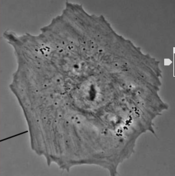
phase contrast
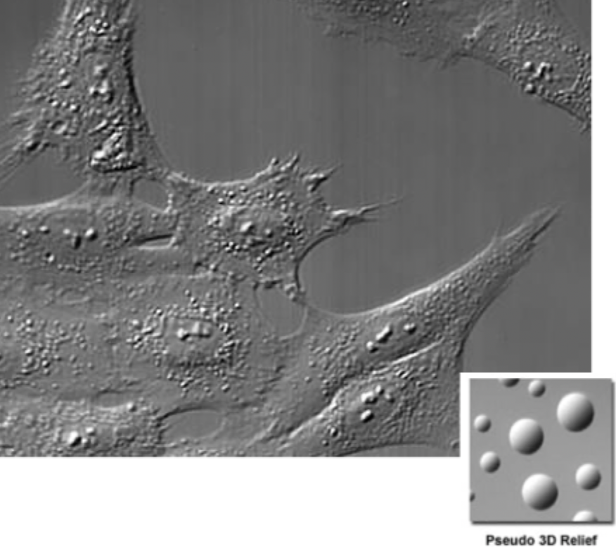
differential interference contrast
2
New cards
feature of phase contrast
white halo around sample
3
New cards
feature of DIC
3D pseudo relief, light and dark sides
4
New cards
what do hematoxylins stain
they stain negatively charged molecules (DNA,RNA)
5
New cards
eosins stain
they stain positively charged molecules (cytoplasm, membrane, ECM)
6
New cards
colour of hematoxylins
purple
7
New cards
colour of eosins
pink
8
New cards
immunohistochemistry (IHC)
enzyme conjugated to antibody targets antigen
substrate is cleaved = leads to stained precipitate
helps us to detect specific proteins in a sample