3.3.2 human gas exchange
1/11
There's no tags or description
Looks like no tags are added yet.
Name | Mastery | Learn | Test | Matching | Spaced | Call with Kai |
|---|
No analytics yet
Send a link to your students to track their progress
12 Terms
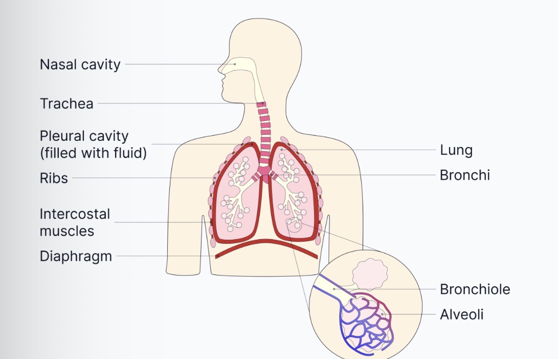
Give the gross structure of the lungs, trachea, bronchi, bronchioles and alveoli- label the picture
Lungs - pair of lobed structures made up of a series of highly branched tubules- called bronchioles which end up in tiny air sacs called alveoli
Trachea- flexible airway supported by rings of cartilage- which prevents the trachea from collapsing in on itself when breathing in
Bronchi - two divisions of the trachea, each leading to one lung. They are similar to trachea structure and also produce mucus to trap dirt particles and cilia that move it towards the throat
Bronchioles - series of Branching subdivisions of the bronchi.
Alveoli- minute air-sacs at the end of the bronchioles where gas exchange occurs
Give the adaptations of the alveoli , what’s the name of thee surface on the alveoli
Alveolar epithelium
large surface area - millions of alveoli increase the surface area and are tightly packed
Thin exchange surface- alveolar epithelium is one cell thick - the epithelial cells are also flattened. Provides a short diffusion pathway
Good blood supply - alveoli are surrounded by a dense capillary network- maintains steep diffusion gradient.
Red blood cells are slowed as they pass through pulmonary capillaries- allowing more time for diffusion
The distance between the red blood cells and alveolar air is reduced as the red blood cells are flattened against the capillary walls
What. Is ventilation
The process of inspiration and expiration to maintain a concentration gradient for gas exchange. It relies on pressure changes in the thoracic cavity due to actions of the diaphragm and the intercostal muscles
Which gases are exchanged in the lungs
Oxygen diffuses down its concentration gradient from the air into the blood
Carbon dioxide diffuses down its concentration gradient from the blood into the air
The concentration gradients required are maintained by the ventilation in the lungs - forcing the air into/out and the continuous flow of blood into the capillaries- carries the blood away from the capillaries at a high rate so there is always a lower concentration gradient/ a higher gradient
Describe eh mechanism of inhalation
External intercostal muscles contract, while the internal intercostal muscles relax
The ribs are pulled UPWARDS and OUTWARDS, increasing the volume of the thorax
The diaphragm muscles contract, causing it to flatten which also increases the volume of the thorax
The increased volume oof the thorax results in the reduction of pressure in the lungs
Atmospheric PRESSURE is now greater than the pulmonary pressure and so air is forced into the lungs
During inhalation, what happens to the ribs
They are pulled upwards and outwards
During inhalation, which intercostal muscles relax relax and which contract
External - contract
Internal- relax
Describe the process of exhalation
internal intercostal muscles contract and the external intercostal muscles relax
The ribs move downwards and inwards, decreasing the volume of the thorax
The diaphragm muscles relax and so it is pushed by the contents of the abdomen that were compressed during inspiration. Volume of the thorax is therefore further decreased
The decreased volume of the thorax increases the pressure in the lungs
The pulmonary pressure is now greater than that of the atmosphere, and s air is forced out of the lungs
What type of relationship does the internal and external intercostal muscles have
An antagonistic interaction - means one does the opposite of the other so one is always contracting and one is always relaxing
(Understanding) where are the external intercostal muscles
Between the ribs and the lungs - and then the contract the ribs are pulled upwards and outwards
Leads to inspiration
(Understanding) where are the intercostal muscles
Located deeper to the external intercostal muscles
They pull the ribcage downwards and inwards- causing expiration