Mastrangelo Lab #10: Spinal Cord (Draft)
1/25
Earn XP
Description and Tags
[INCLUDES REAL CADAVER IMAGES FROM MCGRAW HILL'S A&P REVEALED FOR EDUCATIONAL PURPOSES] Images from A&P Revealed by McGraw Hill & Google. Made for Prof. Michael Mastrangelo's A&P I Lab.
Name | Mastery | Learn | Test | Matching | Spaced | Call with Kai |
|---|
No analytics yet
Send a link to your students to track their progress
26 Terms
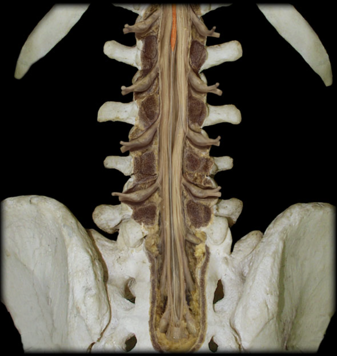
What is the highlighted structure? (orange)
Conus Medullaris
Where is the conus medullaris located?
Upper lumbar vertebral canal
What is the conus medullaris?
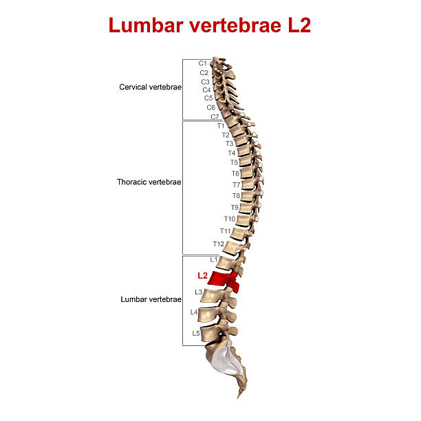
Tapered inferior end of spinal cord (The spinal cord ends at L2 vertebra in adults)
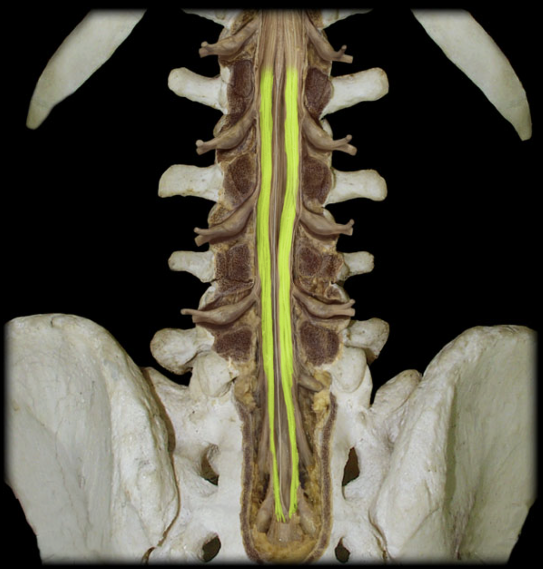
What is the highlighted structure? (yellow)
Cauda equina (horse tail)
Where is the cauda equina located?
Vertebral canal inferior to L2 vertebra level
What is the cauda equina?
A large bundle of dorsal and ventral roots for spinal nerves below L2
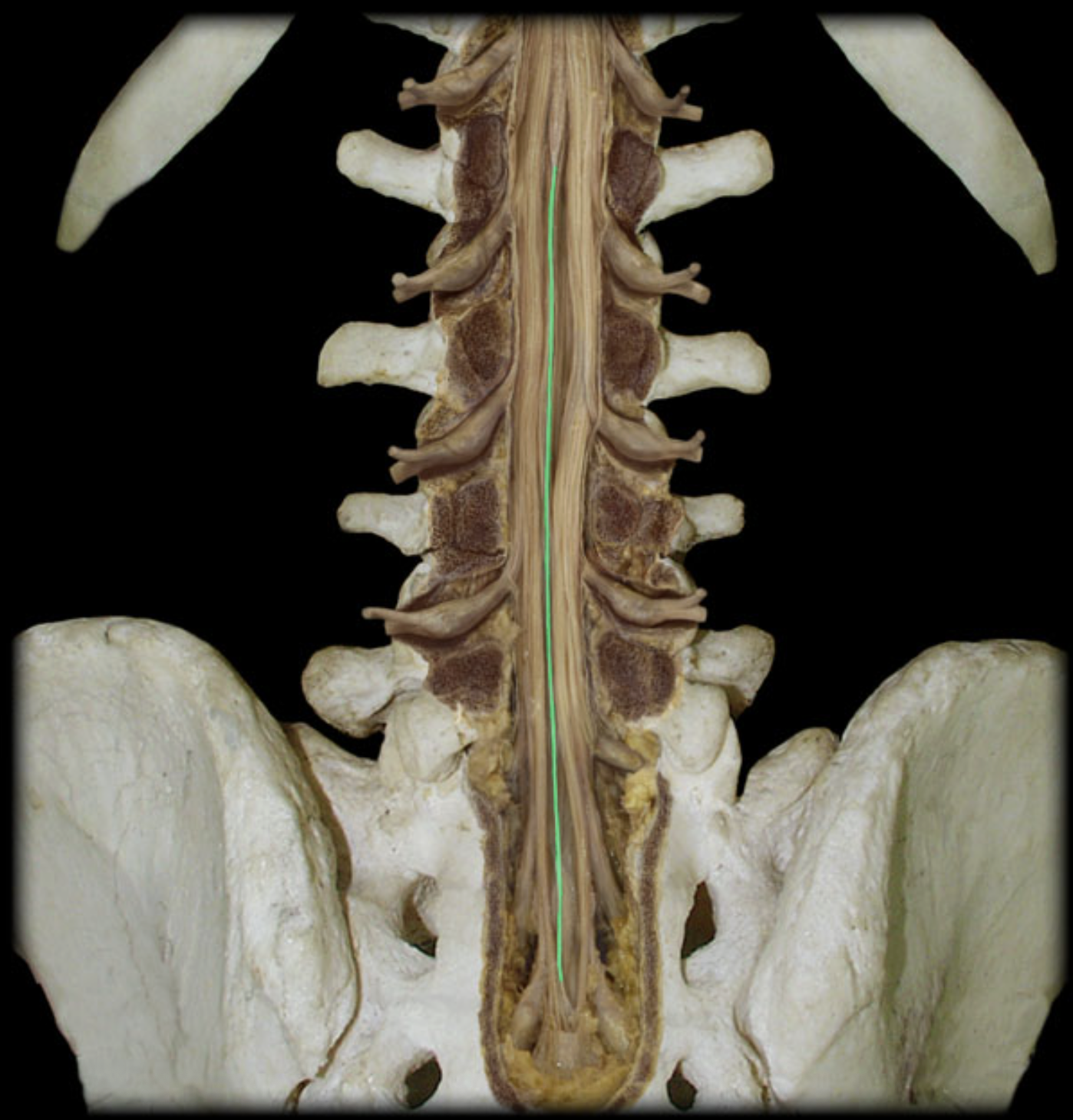
What is the highlighted structure? (green)
Filum terminale
Where is the filum terminale located?
Vertrebral canal (inferior to L2 vertebra)
What is the filum terminale?
A filament of the pia mater which forms at the top of the conus medullaris and passes through the sacral hiatus to attach to the coccyx
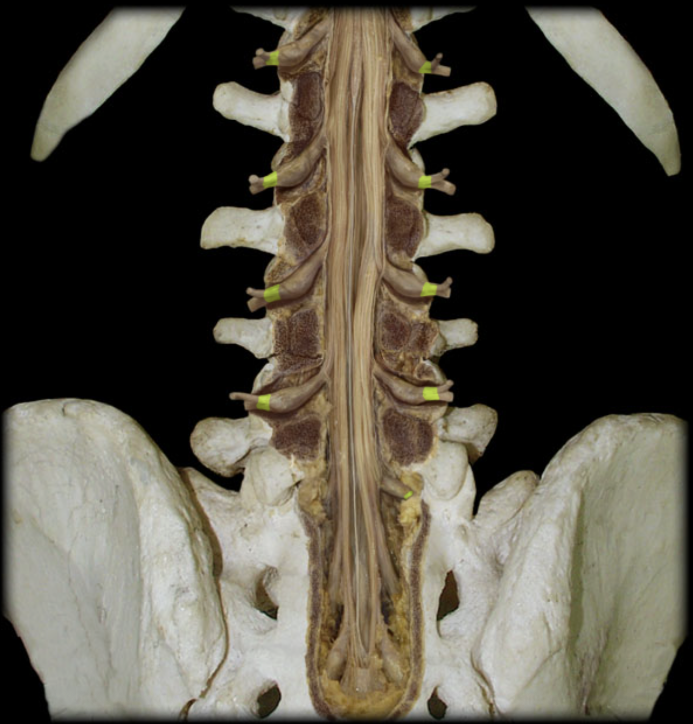
What is the highlighted structure? (yellow)
Spinal nerve
Where is the spinal nerve located?
Intervertebral foramen

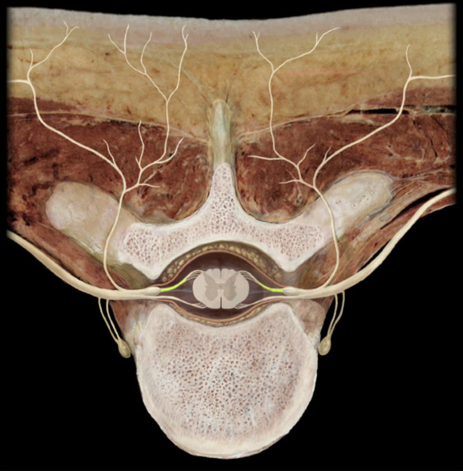
What is the highlighted structure? (yellow)
Dorsal root ganglion
Where is the dorsal root ganglion located?
On the dorsal root
What is the dorsal root ganglion?
A sensory ganglion (contains cell bodies of afferent (sensory) neurons)
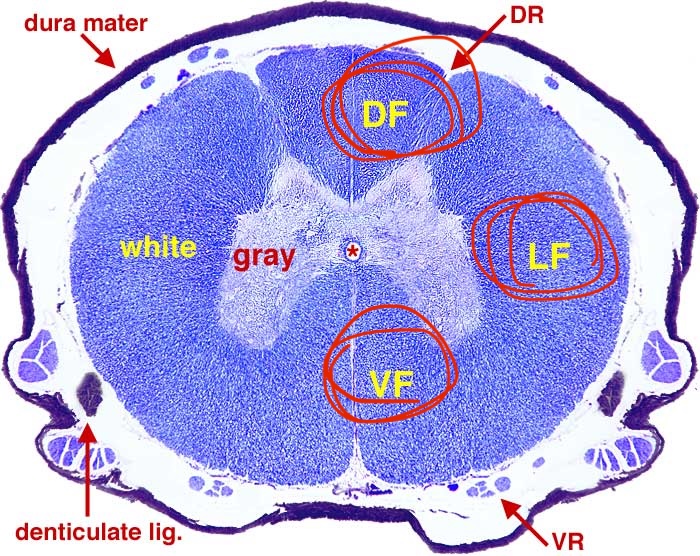
What structures are the labels circled in red indicating?
The white funiculi (3 white columns)
What is the highlighted structure? (yellow)
Dorsal root
What is the highlighted structure?
Ventral root
gray commissure
posterior (dorsal) horn
anterior (ventral) horn
lateral horn
dorsal ramus
ventral ramus
gray matter
white matter
grrrr
bweh