Subluxation Analysis: X-Ray Exam
1/46
There's no tags or description
Looks like no tags are added yet.
Name | Mastery | Learn | Test | Matching | Spaced | Call with Kai |
|---|
No analytics yet
Send a link to your students to track their progress
47 Terms
Chiropractic x-rays:
usually taken weight bearing
most taken A-P and analyzed P-A
Spinography:
Chiropractic analysis of x-rays
Technique specific analysis
Order of patient encounter:
History
Examination: visualization, posture, ROM, leg check, ortho/neuro…
Instrumentation
Static and motion palpation
Diagnostic imaging (if warranted)
Diagnosis and prognosis
Treatment
What are the advantages of X-rays?
Correlate posture findings
Confirm static findings
Identify pathologies
Increases specificity (LOC, SCP, listings, etc.)
What are disadvantages to x-rays?
Exposure
Limited sensitivity to pathologies
Static picture of a dynamic spine (snapshot in time)
Cost
Maintenance
What part of the VSC does x-ray fall under?
Kinesiopathology → relative position
Histopathology → osteological changes
What part of the PART system does x-ray fall under?
A → asymmetry/misalignment
R → range of motion
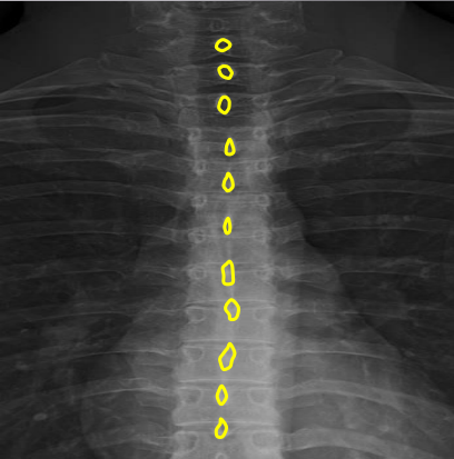
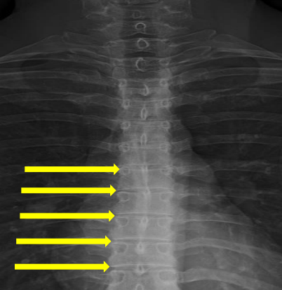
Vertebral bodies
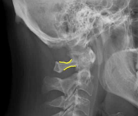
Superior and inferior endplates
Disc space
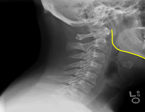
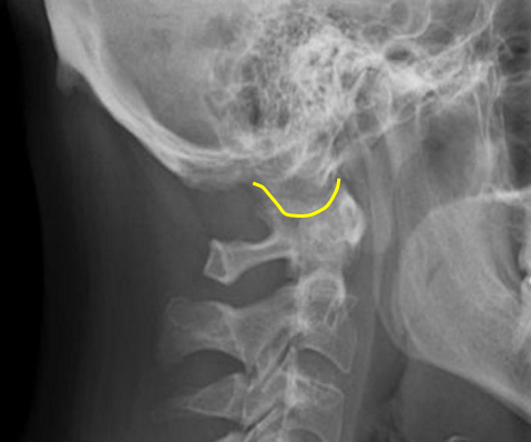
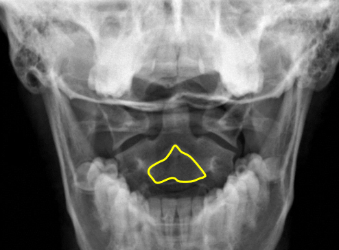
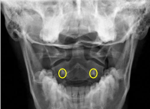
Mandible
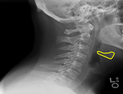
Hyoid bone
Mitchel marker
Occipital condyle

C1 anterior tubercle
C1 posterior arch
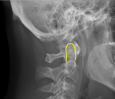
Odontoid process
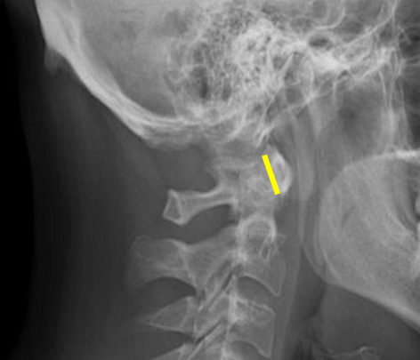
Atlanto-Dental interspace
C1 lateral masses
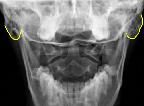
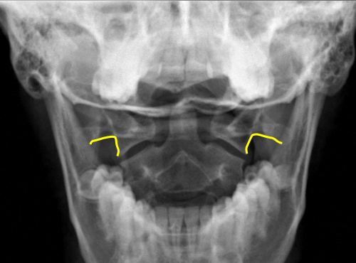
Mastoid process
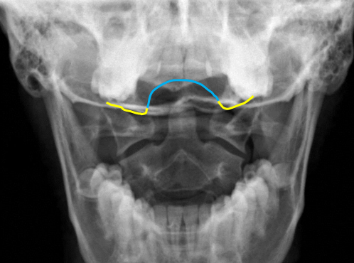
yellow
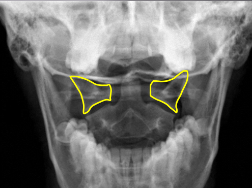
Occipital condyles

blue
Foramen magnum
C1 TVP/ lateral mass junction
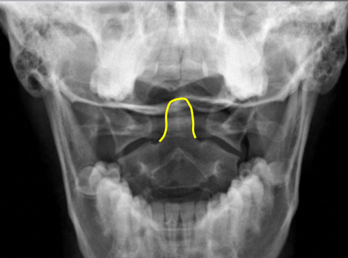
Odontoid process
C2 spinous process
C2 pedicle shadow
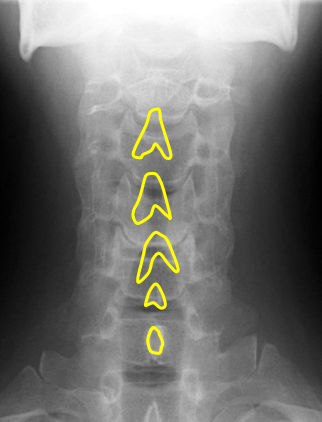
Junction of lamina (top of spinous)
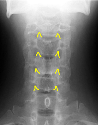
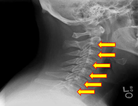
Uncinate processes
Inferior aspect of vertebral bodies
Junction of laminae
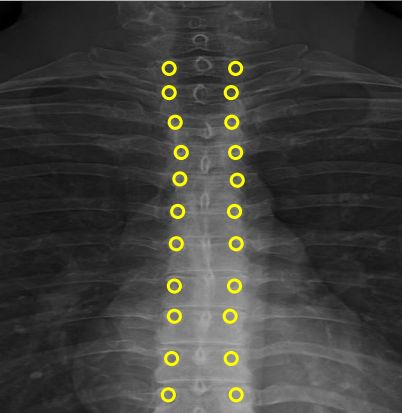
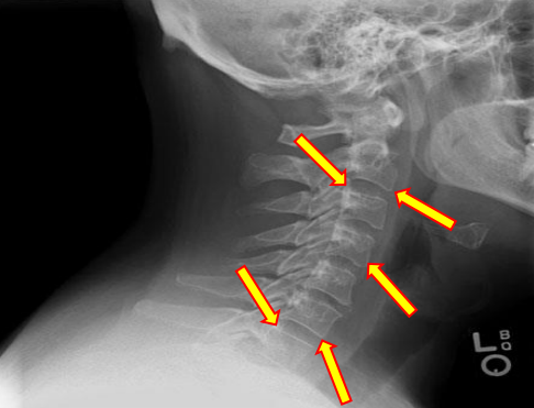
Pedicle shadows
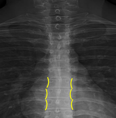
Vertebral waist
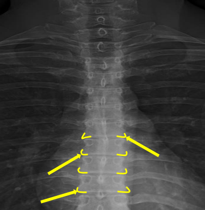
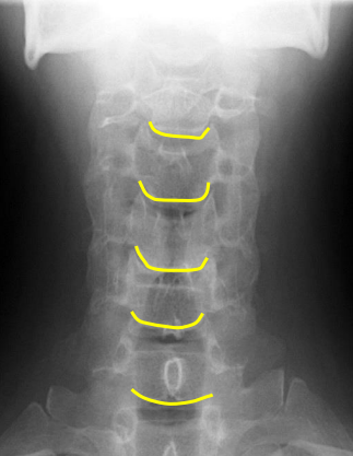
Inferior endplate tips
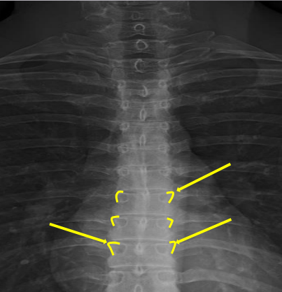
Superior endplate tips
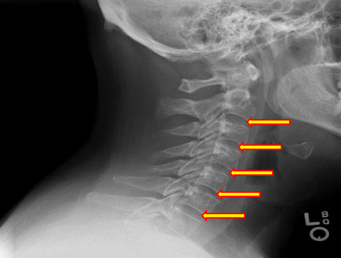
Disc spaces
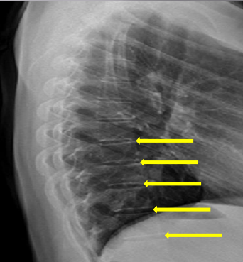
Disc spaces
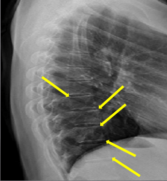
End plate tips
Femur heads
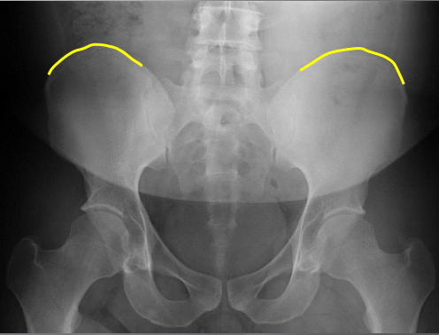
Superior iliac crests
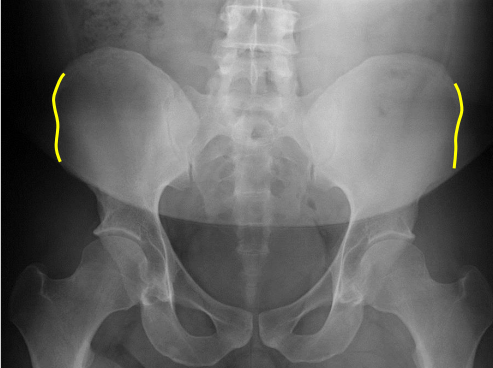
Lateral iliac crests
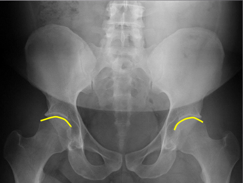
Ischial tuberosities
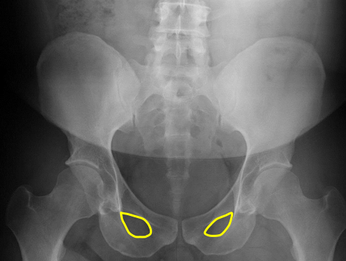
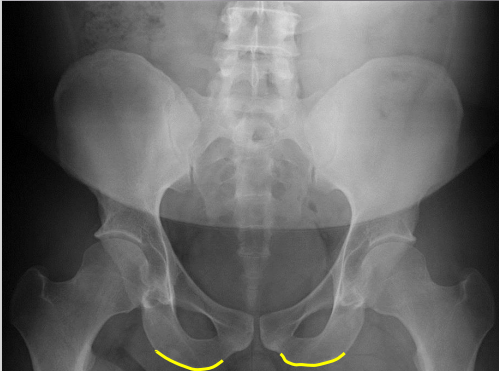
Obturator foramen
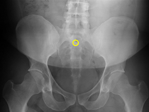
S2 tubercle
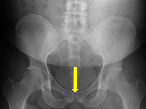
Pubic symphysis
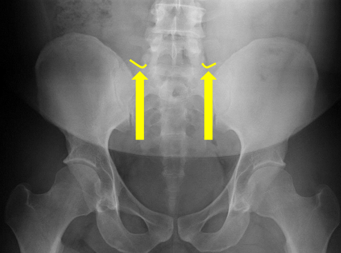
Sacral groove
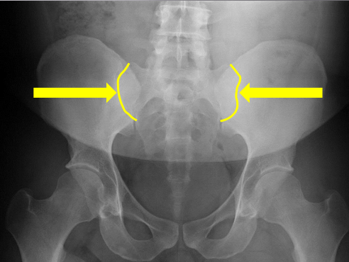
Lateral aspect of sacrum
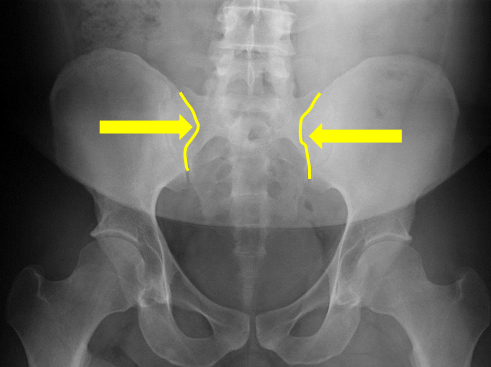
Medial aspect of ilium