How is the oral cavity separated from the nasal cavity?
The palate
What is the oral fissure?
The anterior opening of the oral cavity which bounds it anteriorly.
What are the boundaries of the oral cavity?
Lips/oral fissure (anterior)
Cheeks (lateral)
Soft and hard palate (posterior and anterior)
Floor of mouth (inferior)
Where is the oral vestibule located?
Between the teeth and cheeks (or buccal mucosa)
What is the oral cavity proper?
The region of the mouth that lies internal to the teeth and contains the tongue.
What is the oropharyngeal isthmus?
The posterior aperture of the oral cavity, which opens into the oral part of the pharynx.
What is the epiglottis?
A flap of connective tissue made of elastic cartilage
Present in the posterior floor of the oral cavity to close the trachea during swallowing to prevent food entry into the larynx and trachea
Give some characteristics of the hard palate.
Forms the roof of the anterior portion of the oral cavity.
Formed by fusion of palatine processes of the maxilla and the horizontal plates of the palatine bone.
Lined by layers of oral mucosa.
Give some characteristics of the soft palate.
Forms the roof of the posterior portion of the oral cavity.
Lined by oral mucosa.
No bony support, just muscle and connective tissue.
Can be elevated to separate the nasopharynx from the oropharynx.
Can be depressed to close the oropharyngeal isthmus.
What muscles make up the soft palate? What do they do?
Tensor veli palatini - opens the pharyngotympanic (auditory tube). Forms the palatine aponeurosis to which other muscles attach
Levator veli palatini - elevates the soft palate
Palatopharyngeus - depresses the soft palate and moves the palatopharyngeal arch towards the midline, closing the oropharyngeal isthmus
Palatoglossus - depresses the palate, moves the palatoglossal arch towards the midline, closing the oropharyngeal isthmus
Musculus uvulae - elevates and retracts the uvula
What are the three major salivary glands?
Parotid gland (the largest of these glands)
Submandibular glands
Sublingual glands
Where is the parotid gland located?
Lies anteroinferior to the ear
Lies superficial to the masseter muscle
Lies superior to the angle of the mandible
Where are the submandibular glands located?
Inferior to the mandible
Where are the sublingual glands located?
Inferior to the tongue
How are the salivary glands affected by neural input?
Increased sympathetic input - reduces salivary output via vasoconstriction of blood vessels which supply these glands
Increased parasympathetic input - secretion of the salivary glands is stimulated directly from this
What is the structure of intrinsic muscles of the tongue?
Confined within the tongue
Run in longitudinal, transverse or vertical orientations to change the shape of the tongue
Supplied by CNXII (hypoglossal nerve)
How many extrinsic muscles are there in the tongue and what do they do?
Four: genioglossus, hyoglossus, styloglossus and palatoglossus.
Change the position of the tongue.
How are the extrinsic muscles of the tongue supplied?
Genioglossus, hyoglossus and styloglossus: CNXII (hypoglossal nerve)
Palatoglossus: CNX (vagus nerve)
What provides the arterial supply to the tongue?
The lingual artery, which is a branch of the external carotid artery.
How is the roof of the oral cavity formed?
Formed from the hard and soft palate.
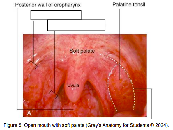
What is the structure of the soft palate?
Extends inferolaterally to form palatoglossal and palatopharyngeal arches, named after the muscles that form them.
Arches lie anterior and posterior to the palatine tonsil respectively.
Bilaterally they form the oropharyngeal isthmus.
Both the palatoglossus and palatopharyngeus are stimulated by CN X (Vagus Nerve)
When is the swallowing reflex initiated?
When the food bolus touches the soft palate.
What does the pharynx lie anterior to?
The first six cervical vertebrae (C1-C6)
Give some characteristics of the oesophagus.
It is a muscular tube that begins in the neck at the lower border of the C6 and descends through the superior and posterior mediastinum of the thorax.
It enters the abdominal cavity by piercing the diaphragm at the oesophageal hiatus at the level of T10.
Inferior to the diaphragm, it opens into the stomach at the cardiac orifice.
The abdominal part of the oesophagus is approx. 2cm in length.
At what certain points can the oesophagus be compressed?
The junction of the oesophagus with the pharynx in the neck
Where the oesophagus is crossed by the arch of the aorta
Where the oesophagus is compressed by the left main bronchus
At the oesophageal hiatus
Where do oesophageal arteries arise from in the posterior mediastinum?
Thoracic aorta
Bronchial arteries
What does the venous drainage of the oesophagus involve?
Involves small vessels returning to the:
Azygos vein
Hemiazygos vein
Oesophageal branches of the left gastric vein in the abdomen
What is the lymphatic drainage of the oesophagus like?
Lymphatic drainage of the oesophagus in the posterior mediastinum returns to posterior mediastinal and left gastric nodes.
Give some characteristics of the innervation of the oesophagus.
It is complex:
Striated muscle fibres in the superior part are innervated by special visceral efferents from the vagus nerves
Smooth muscle fibres in the inferior part are innervated by parasympathetic fibres from vagus nerve visceral efferents.
The sensory innervation involves visceral afferents of the vagus nerves, sympathetic trunks, and splanchnic nerves
The vagus nerves divide into several branches upon reaching the oesophagus, forming the oesophageal plexus
As the oesophagus descends through the diaphragm, the plexus forms two trunks, the anterior vagal trunk formed mainly of left vagus nerve fibres, the posterior vagal trunk, mainly from fibres originally in the right vagus nerve
Briefly describe the stomach.
It is a J-shaped hollow organ that extends from the cardiac orifice to the duodenum distally.
What regions of the abdomen does the stomach occupy?
Left hypochondriac, epigastric and umbilical regions.
Name the two missing structures.
Outer arch: palatoglossal arch
Inner arch: palatopharyngeal arch
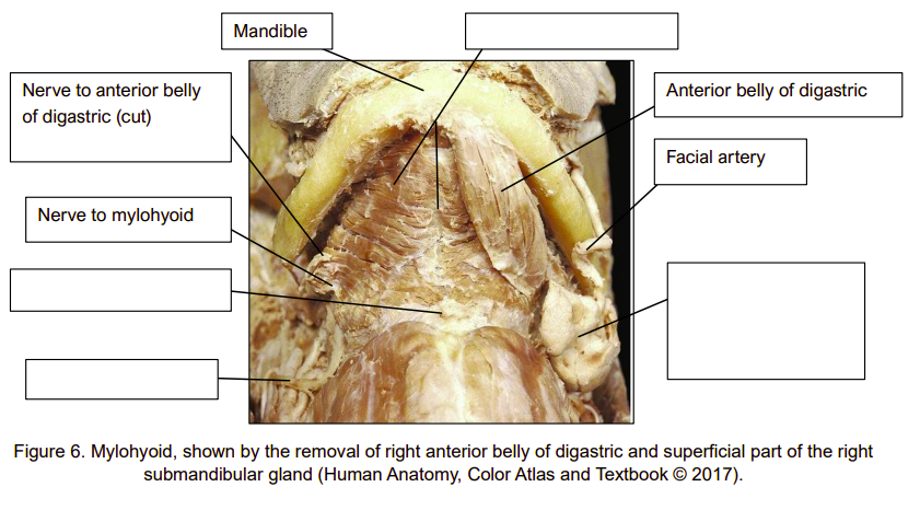
Name the missing structures.
(From left to right clockwise):
Hypoglossal nerve
Hyoid bone
Mylohyoid muscle
Submandibular gland
Which muscle of the cheek is penetrated by the parotid duct?
Buccinator
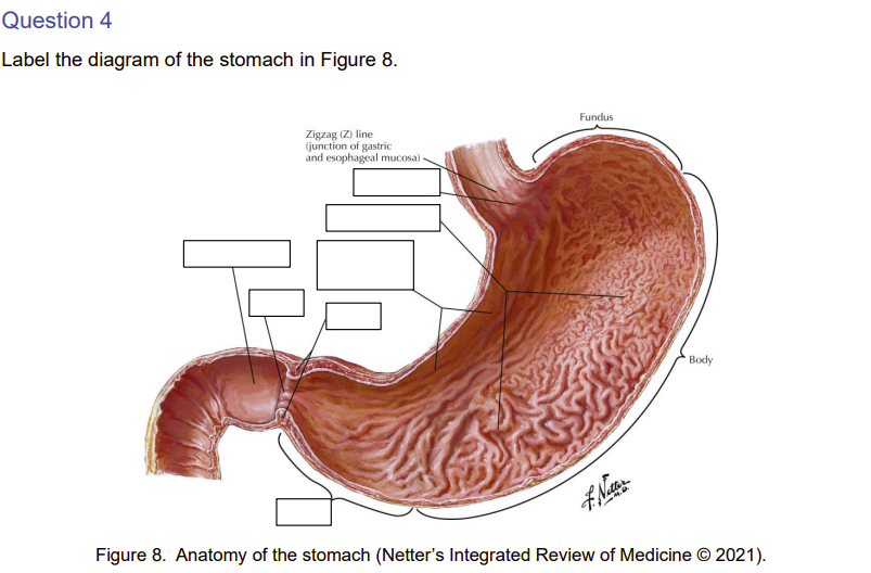
(From right to left anti-clockwise):
Cardiac opening
Longitudinal folds
Rugae
Pyloric sphincter
Pyloric orifice
Duodenum
Antrum
What is the lesser omentum?
A layer of peritoneum which is attached from the lesser curvature of the stomach to the liver.
What is the greater omentum?
A layer of peritoneum which is attached to the greater curve of the stomach and covers most of the intestines.
What is the name of the space posterior to the stomach and lesser omentum?
Lesser sac
Which nerve provides general sensation for the anterior 2/3s of the tongue?
The lingual nerve, which is a branch of the mandibular division of the trigeminal nerve.
Which nerve provides special sensation (taste) to the anterior 2/3s of the tongue?
The chorda tympani nerve, which is a branch of the facial nerve.
Which nerve provides general and special sensation to the posterior 1/3 of the tongue?
CN IX (Glossopharyngeal Nerve)
What nerve provides motor innervation to the palatoglossus?
CN X (Vagus Nerve)
Which nerve provides motor innervation to the intrinsic muscle, genioglossus, hyoglossus and styloglossus?
CN XII (Hypoglossal Nerve)
Which sensory nerve can be overstimulated during intubation to lead to a spasm of the airway (laryngospasm)?
Superior laryngeal nerve - branch of the vagus nerve.
What glands are provided by the branches of the facial nerve?
All minor intra-oral salivary glands, submandibular and sublingual glands.
Is the parotid gland intra-oral or extra-oral?
Extra-oral
What is the parotid gland stimulated by?
Parasympathetic fibres of the glossopharyngeal nerve (CN IX).
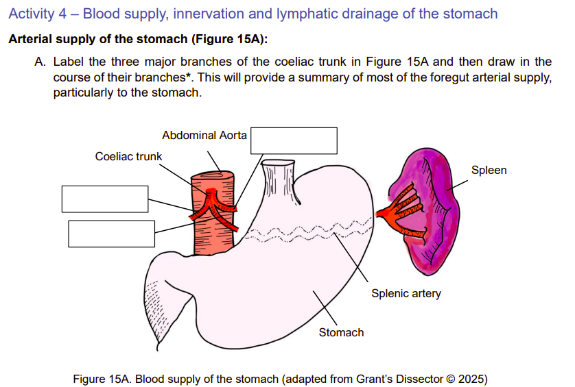
Three major branches (from left to right):
Hepatic proper artery (gives rise to right gastric artery)
Splenic artery
Left gastric artery
All originate from abdominal aorta.
Which arteries supply the lesser curvature of the stomach?
Left and right gastric arteries.
Which arteries supply the greater curvature of the stomach?
Left and right gastro-omental arteries.
What is the drainage pathway of the left and right gastric veins?
Left gastric vein drains into portal vein.
Right gastric vein drains into portal vein.
What is the drainage pathway of the short gastric veins?
Short gastric veins drain into the splenic vein.
What is the drainage pathway of the left and right gastro-omental veins?
Left gastro-omental vein drains into the splenic vein.
Right gastro-omental vein drains into the superior mesenteric vein.
Give some characteristics of the lymphatic drainage to the stomach.
Lymphatics follow the course of the arteries supplying the stomach.
They drain in the opposite direction.
They drain lymph from its posterior and anterior surfaces toward its curvatures, where the gastric (lesser curvature) and gastro-omental (greater curvature) lymph nodes are located.
The efferent lymphatic vessels from these nodes accompany the large arteries to the coeliac lymph nodes.
What is the origin of sympathetic innervation to the stomach?
T6-T9, through pelvic splanchnic nerves and they synapse at the coeliac ganglion.
What is the origin of parasympathetic innervation to the stomach?
Vagus nerve (CN X) from the brain then through anterior and posterior vagal trunks.
What are the effects of sympathetic innervation to the stomach?
Reduces secretions and peristalsis.
What are the effects of parasympathetic innervation to the stomach?
Increases secretions and peristalsis.