CVA Chapter 10 - Sensory Reception and the Special Senses
1/149
There's no tags or description
Looks like no tags are added yet.
Name | Mastery | Learn | Test | Matching | Spaced | Call with Kai |
|---|
No analytics yet
Send a link to your students to track their progress
150 Terms
What are the two main categories of sensory receptors?
Visceral and Somatic Senses
What do visceral receptors monitor?
The internal environment of the body
What are interoceptors?
Another term for visceral receptors
What sensations are included in visceral senses?
Nausea and hunger
What do somatic senses monitor?
The external environment and the external body
What are exteroceptors?
Receptors that detect external stimuli. Type of somatic sense.
What are proprioceptors?
Receptors that provide information about body position and movement. Type of somatic sense.
What are general senses?
Senses with receptors spread out over a large area, such as touch and temperature
What are special senses?
Senses with receptors concentrated in a small area, such as sight and hearing
How is the location of a stimulus determined in mammals?
By the sensory cortex
What are dendritic endings?
Branching tips of sensory neurons that detect stimuli and invade tissues during development. All senses detected by them are general senses.
What are encapsulated dendritic endings?
Dendritic endings encased in a connective tissue capsule to increase surface area for increased sense perception
What are free dendritic endings?
Dendritic endings that lack connective tissue capsules and are 'naked'
What are complete cell receptor cells?
Cells that utilize the entire cell to detect stimuli, associated with special senses. Can be neurons or neuroepithelial cells.
What are neuroepithelial cells?
Specialized epithelial cells derived from neural crest ectoderm that can conduct impulses to a sensory neuron.
What are stereocilia?
Hair-like structures on neuroepithelial cells that detect stimuli
What are neuromast organs?
Structures in fishes and aquatic amphibians that detect mechanical stimuli from water
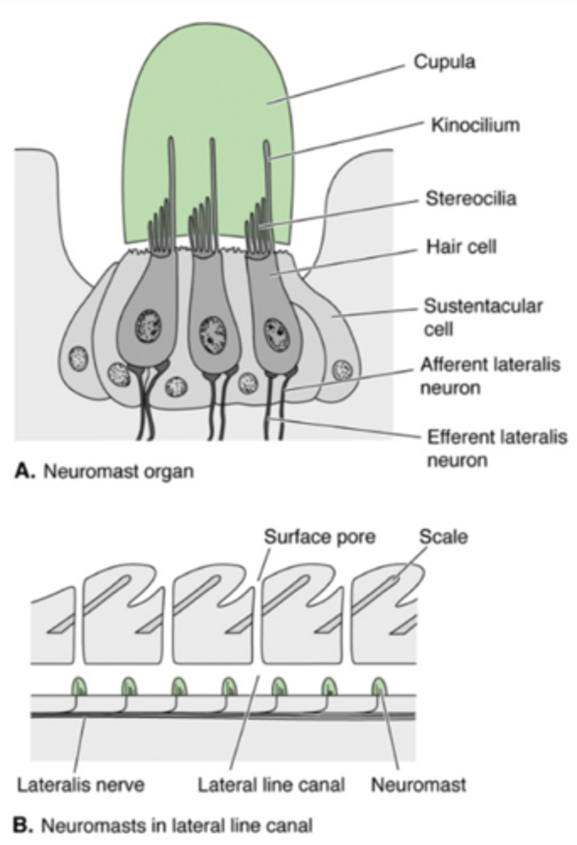
What are sustentacular cells?
Columnar epithelial cells that surround and protect neuromast cells. Also known as "Supporting Cells."
What is a cupola?
An acellular glycoprotein in which stereocilia are embedded
What are external neuromasts?
Neuromasts located in shallow pits or grooves on the body surface with the cupola projecting into the water. Found in agnathans, larval amphibians, and aquatic urodeles.
What are internal neuromasts?
More specialized structures with cupola embedded within the body. The most specialized ones are found in the membranous labyrinth of the inner ear in all gnathostomes.
What are pit organs?
Fluid-filled pits beneath the epidermis that contain internal neuromasts. Open onto the surface by way of pore.
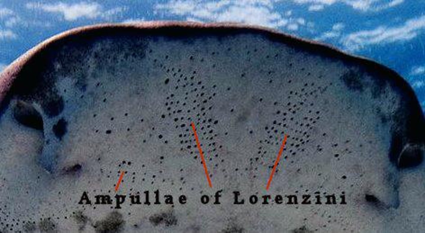
What are ampullae of Lorenzini?
Internal neuromast electroreceptive organs in sharks that detect electrical signals. Open to the surface by means of a long duct. Assists with hunting prey.
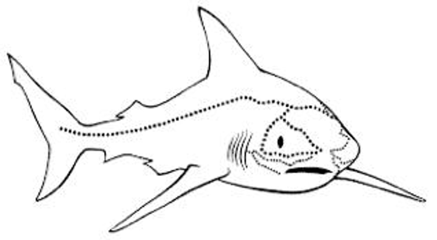
What are the lateral line and cephalic canal systems?
A system in fish that detects mechanical forces such as water currents. Shallow grooves on the head and a singular groove extending along the lateral line to the tail on both sides. Primitively external.
What happens to the lateral line and cephalic canal systems in larval amphibians?
It is lost when they metamorphosize into the adult, terrestrial form.
What group has internal neuromast lateral line and cephalic canal systems?
Jawed fishes.
What has happened to the lateral line canal in most teleosts?
It has sunk below the surface and is now embedded in the dermal bone.
In what groups are the neuromasts innervated by CN 8, CN ALL, and CN PLL?
Fish, larval aquatic amphibians, and adult aquatic amphibians.
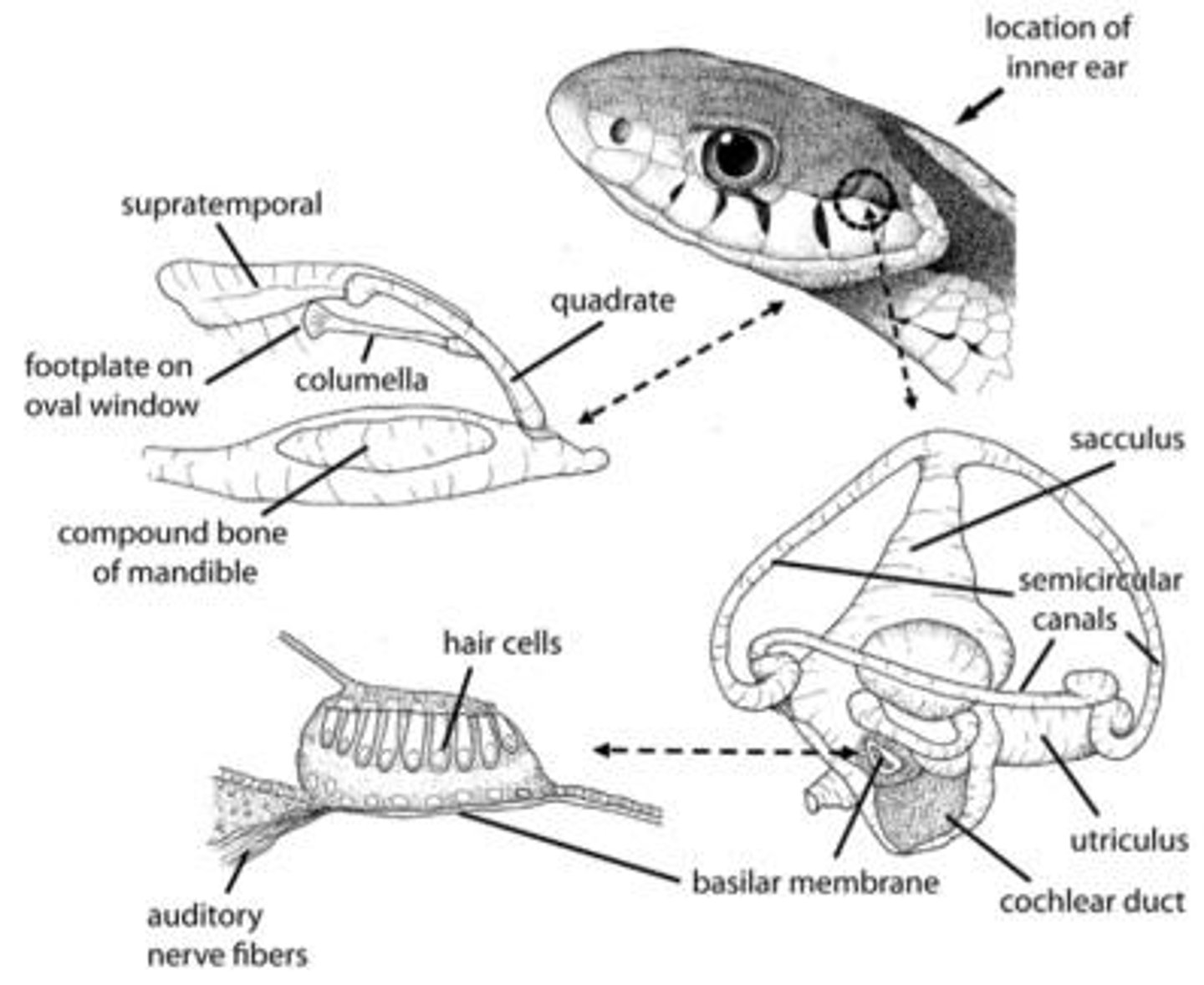
What is the membranous labyrinth?
A structure in all vertebrates that detects equilibrium and audition.
What is the role of the skeletal labyrinth?
It houses the membranous labyrinth and is composed of cartilage or bone
What is the membranous labyrinth located within?
A skeletal labyrinth composed of either cartilage or bone.
What are the two portions of the skeletal labyrinth found in all gnathostomes?
Semicircular Canals and the Vestibule.
Which additional portion do mammals and birds have in their skeletal labyrinth?
The Cochlea.
From what did the cochlea develop in fish and amphibians?
An outpocketing of the vestibule called the Lagena.
What fluid is found between the skeletal and membranous labyrinths?
Perilymph.
What is the space that encloses the perilymph called?
The Perilymphatic Space.
How many regions does the membranous labyrinth have in most gnathostomes? What about in mammals?
Two regions in gnathostomes, three in mammals.
How many semicircular ducts and canals do gnathostomes have?
Three. One for each plane of motion.
What are the two connected portions of the membranous labyrinth found in the vestibule?
Utricle and Saccule.
Where is the cochlear duct located?
In the cochlea of birds and mammals.
What is the fluid that fills the membranous labyrinth?
Endolymph.
What structure extends from the vestibule in most vertebrates?
The Endolymphatic Duct.
Where are the endolymphatic ducts located in elasmobranchs?
On the surface of the chondrocranium in depressions called Endolymphatic Fossae.
What accompanies the Endolymphatic sacs in the subarachnoid space in mammals?
A set of perilymphatic sacs.
What is the role of the receptors in the utricle and saccule?
To provide sensory information regarding the orientation of the head in space.
What is the name of the structure where the receptors of static equilibrium are located?
The Macula on the wall of the utricle and saccule
What do the hair cells in the macula extend into?
An overlying acellular membrane called the Otolith Membrane.
What are otoliths composed of in amniotes and some fishes?
Calcium carbonate crystals.
What happens to the otoliths when the head moves?
They move due to gravitational forces, triggering a nerve impulse that is transmitted along the vestibular branch of CN 8 to the cerebral cortex.
Where are the receptors of dynamic equilibrium located?
In the Ampulla of the semicircular canal portion of the bony labyrinth.
How many semicircular ducts do mammals and birds have?
Three, positioned at right angles to one another.
What is the sensory organ for dynamic equilibrium called?
Crista Ampullaris.
What are the two classes of Crista Ampullaris epithelial cells?
Supporting hair/receptor cells.
What does the bending of the Crista Ampullaris stereocilia cause?
Stimulation and an impluse that travels to the vestibular branch of CN 8 and to the cortex.
What is the cochlea's shape and position?
Spiral-shaped portion of the bony labyrinth located anterior to the vestibule.
What are the three canals internally divided in the cochlea?
Scala Vestibuli, Scala Tympani, and Scala Media.
What is housed in the Scala Media?
The sensory structure responsible for hearing, known as the Cochlear Duct.
What separates the scala media from the scala vestibuli?
Vestibular membrane.
What separates the scala media from the scala tympani?
Basilar membrane.
What is the organ of hearing located on the basilar membrane?
The Organ of Corti (aka Spiral Organ)
What does the organ of Corti consist of?
A series of epithelial cells located on the inner surface of the basilar membrane.
What types of neuroepithelial hair cells are found in the Organ of Corti?
Outer and Inner Hair Cells.
What do the Outer and Inner hair cells extend into and what do they contact?
Extend into the acellular membranous structure called the Tectorial Membrane. They contact the cochlear branch of CN 8.
What causes the production of an impulse in the cochlea?
Vibrations from sound waves traveling through endolymph and perilymph.
What mechanism do some fishes use for hearing?
Cypriniformes use Weberian ossicles to transmit sound waves to the lagena. Clupeiformes use a swim bladder extension.
What are Weberian ossicles?
Modifications of the transverse processes of the first 3 or 4 vertebrae that aid in hearing.
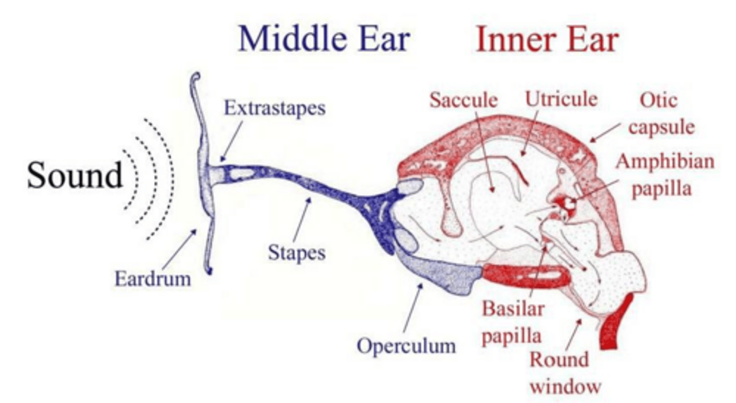
What mechanism(s) of hearing do amphibians and reptiles have?
In addition to the normal macula, there is a modified macula located in the sacculus, near the lagena. Amphibians have two special maculae in the sacculus.
What are the two special maculae in the amphibian sacculus?
The amphibian and basal papillae. They have tectorial membranes instead of otolith membrane.
What is the function of the tectorial membrane in amphibians?
It stimulates the stereocilia in response to sound waves.
How is the lagena modified in reptiles?
Reptiles have an expanded lagena, and their basal papilla has grown into it.
What is the Organ of Corti?
It is formed when the basal papilla becomes fully engulfed within the lagena in crocodilians, birds, and monotreme mammals.
What is the structure of the external ear in mammals?
It consists of the pinna, external auditory canal, and tympanic membrane.
What is the role of the pinna?
It captures and concentrates sound waves.
Where is the external auditory canal located?
Within the petrous portion of the temporal bone.
What is the function of the tympanic membrane?
It separates the external auditory canal from the middle ear and is involved in sound wave transmission.
What is the Eustachian Tube?
It connects the middle ear to the pharynx and helps equalize air pressure.
What are the auditory ossicles in mammals?
The three small bones are Malleus, Incus, and Stapes, which transmit sound waves from the tympanic membrane to the oval window.
What is the function of the Tensor Tympani Muscle?
It increases tension on the tympanic membrane to prevent damage from loud sounds.
What is echolocation?
The ability to project sound waves and form an image based on the reflection of these sound waves, found in certain mammals.
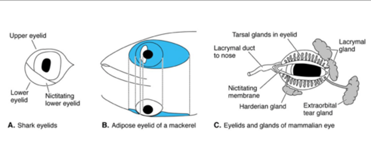
What are eyelids, or palpebrae, in amniotes?
Structures that shade the eyes from excessive light, protect the eyes when sleeping, and distribute lubricating secretions.
What are nictitating membranes?
A third, transparent eyelid found in many vertebrates that protects the eye.
What is the function of eyelashes?
They help protect the eye from particulate matter.
What are the Glands of Zeis?
Glands at the base of each eyelash follicle that secrete lubricant for the eyelashes.
What is the lacrimal apparatus?
A structure found in tetrapods that is involved in tear production and eye lubrication.
How do amphibians and reptiles differ in their auditory mechanisms?
Amphibians have two special maculae in the sacculus, while reptiles have an expanded lagena with a modified basal papilla.
What is the role of the middle ear?
It receives sound waves conducted from the tympanic membrane and transmits them to the inner ear.
What is the significance of the round and oval windows in the middle ear?
They are openings that separate the middle ear from the inner ear and allow sound wave transmission.
What is the function of the Stapedius Muscle?
It protects the inner ear from loud sounds by drawing the stapes posteriorly.
What evolutionary adaptation allows the Eustachian Tube to function in tetrapods?
It evolved from the spiracle of fishes.
What is the auditory mechanism in reptiles?
It includes a tympanic membrane and an expanded lagena with a basal papilla for hearing.
What is the primary function of the outer ear?
To collect sounds and direct them inward.
How do sound waves reach the inner ear in vertebrates?
They are conveyed across the middle ear from the tympanic membrane to the oval window by ossicles.
What is the function of the lacrimal apparatus?
To manufacture, distribute, and drain tears.
What produces lacrimal fluid?
The Lacrimal Gland.
What is the role of lacrimal fluid?
To lubricate the eye and help remove particulate matter.
Where are the lacrimal sacs located?
In the lacrimal sulci of the lacrimal bones.
What does the nasolacrimal duct do?
Carries lacrimal fluid into the posterior nasal cavity.
How is lacrimal fluid spread over the eye?
By the blinking of the palpebrae.
What is the Harderian Gland?
An extra component in species with a nictitating membrane that lubricates it.
How many extrinsic ocular muscles are there?
Six skeletal muscles.