Unit 4 - Spinal Cord
1/17
There's no tags or description
Looks like no tags are added yet.
Name | Mastery | Learn | Test | Matching | Spaced | Call with Kai |
|---|
No analytics yet
Send a link to your students to track their progress
18 Terms
Spinal/skull foramen labeling
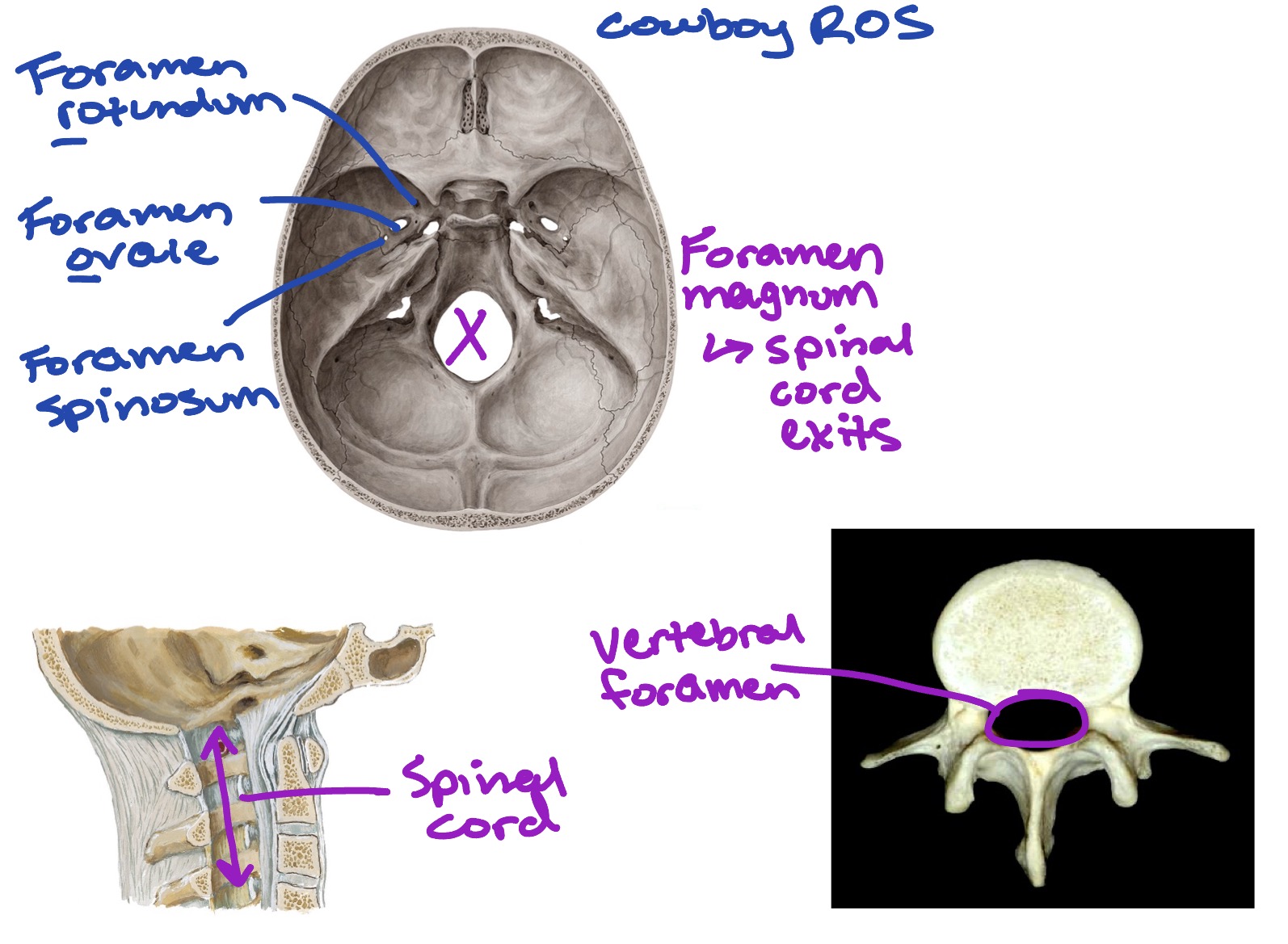
Cowboy R.O.S.:
Foramen rotundum
Foramen ovale
Foramen spinosum
Foramen magnum: where spinal cord exits
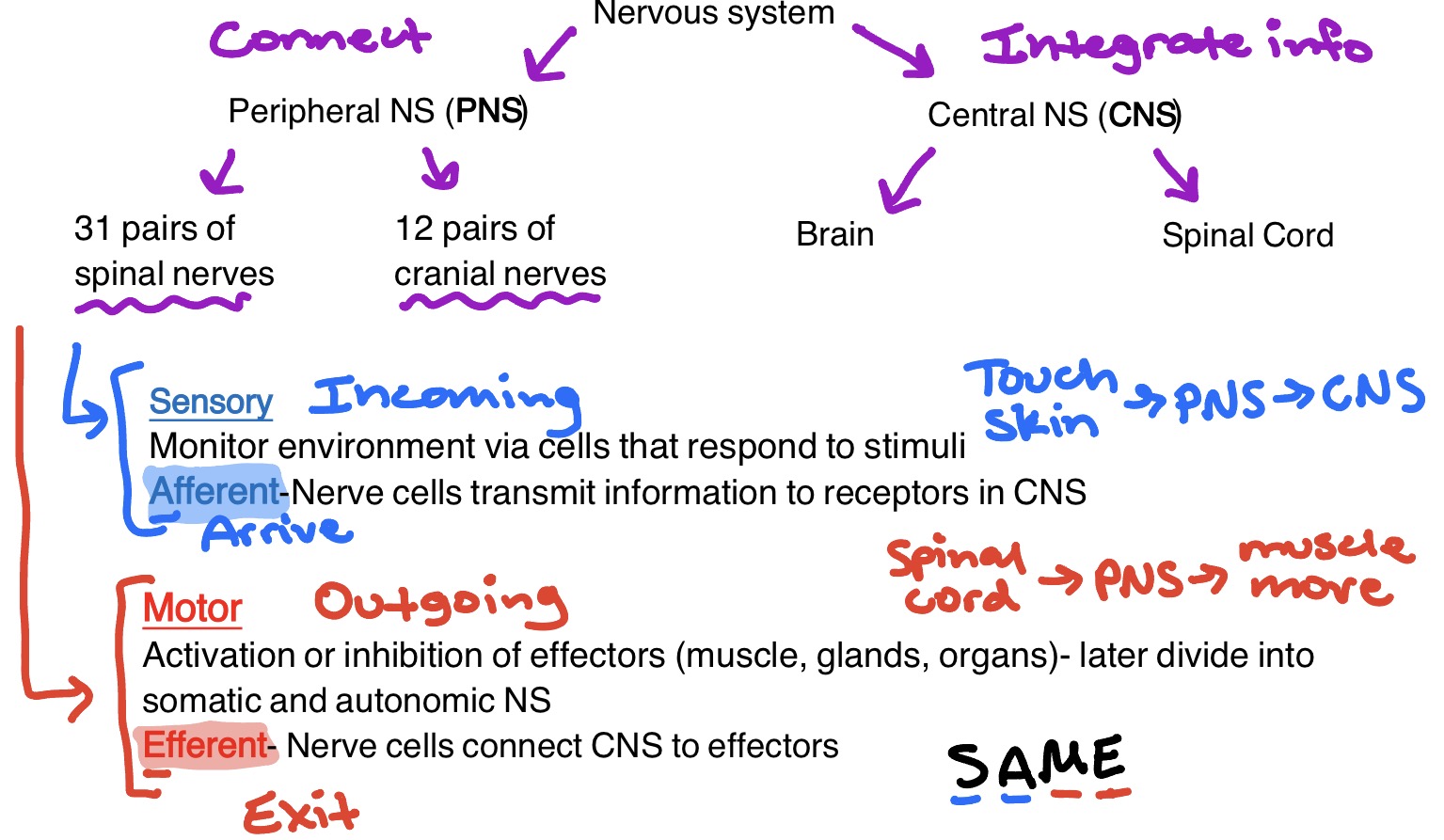
Divisions of the Nervous System
Peripheral Nervous System (PNS)
31 pairs of spinal nerves
12 pairs of cranial nerves
Sensory (incoming):
Monitor environment via cells that respond to stimuli
Afferent (arrive) nerve cells transmit info to receptor in the CNS
Touch skin → PNS → CNS
Motor (outgoing):
Activation or inhibition of effectors (muscles, glands, organs) - later divide into somatic & autonomic NS
Efferent (exit) nerve cells connect the CNS to effectors
S.A.M.E. → Sensory - Afferent/Arrive; Motor - Efferent/Exit
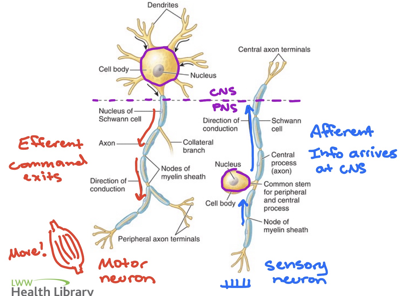
Motor & Sensory neuron visuals
Motor: sends signals from CNS to effectors
Sensory: sends signals from effectors to CNS
Spinal cord
Serves as communication b/w the brain & PNS
Reflex center
Extends from the foramen magnum to approximately the L1 or L2 vertebrae level
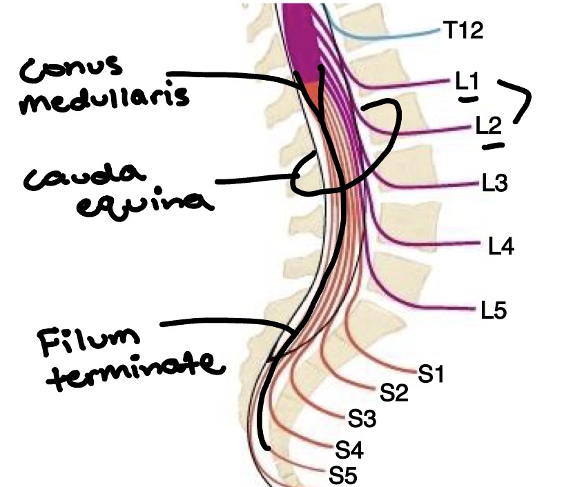
Spinal cord gross anatomy
Conus medullaris = Distal tapered end of spinal cord
Cauda equina = Collection of spinal nerve roots that extend inferior to spinal cord → also called “Horse’s tail”
Filum terminale = Extension of pia mater from the conus medullaris, anchoring to the posterior surface of the coccyx
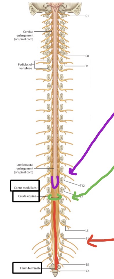
Distal spinal cord
Conus medullaris
Cauda equina
Filum terminale
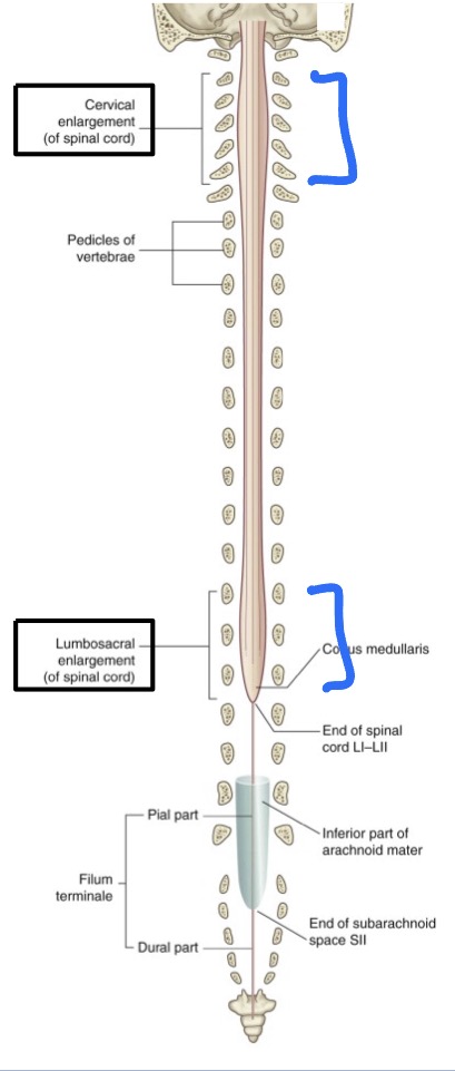
2 major enlargements
Cervical enlargement
C5-T1
Brachial plexus → upper limb
Lumbosacral enlargement
L1-S3
→ lower limb
Spinal cord protected by…
Bones
Meninges
CSF
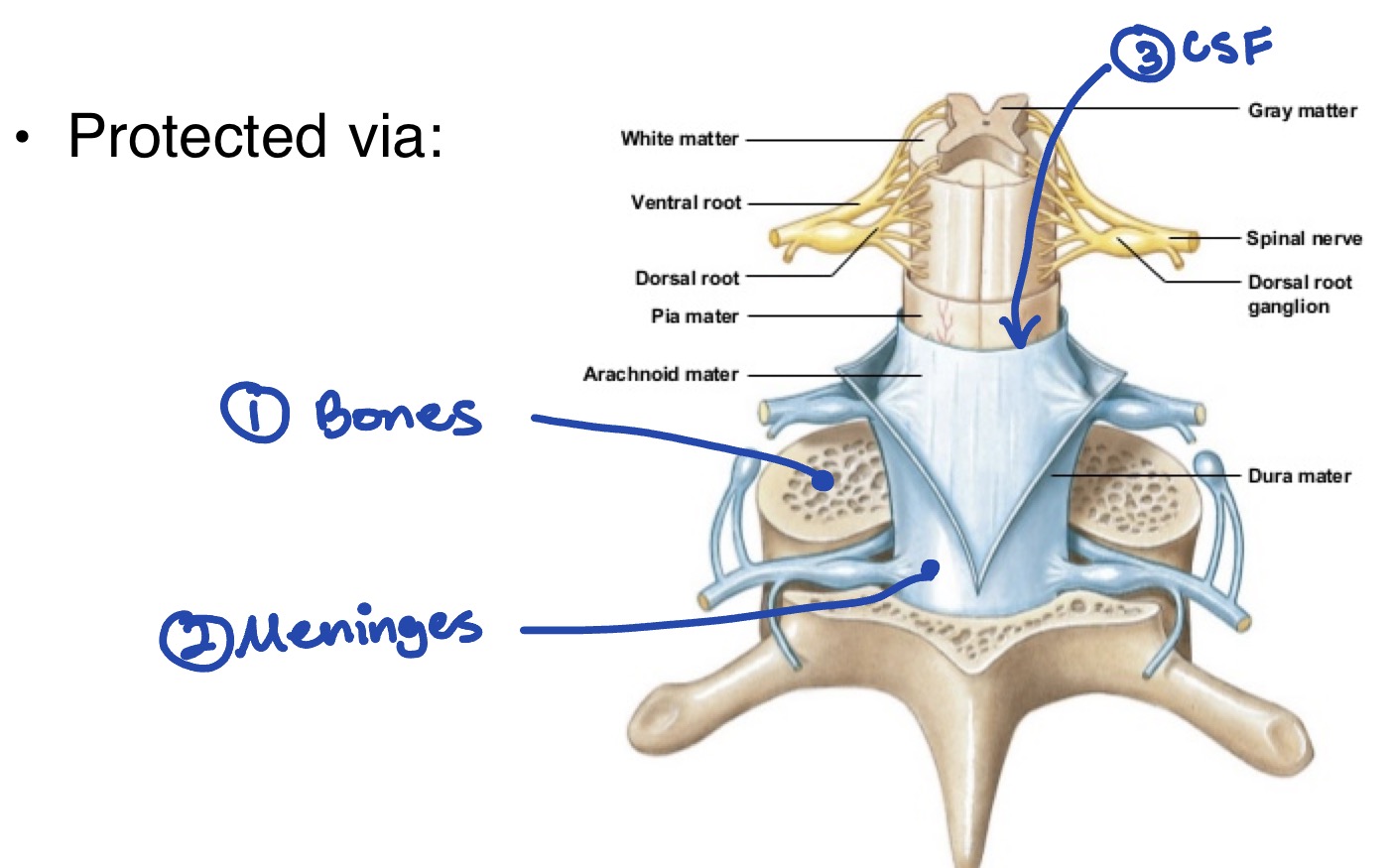
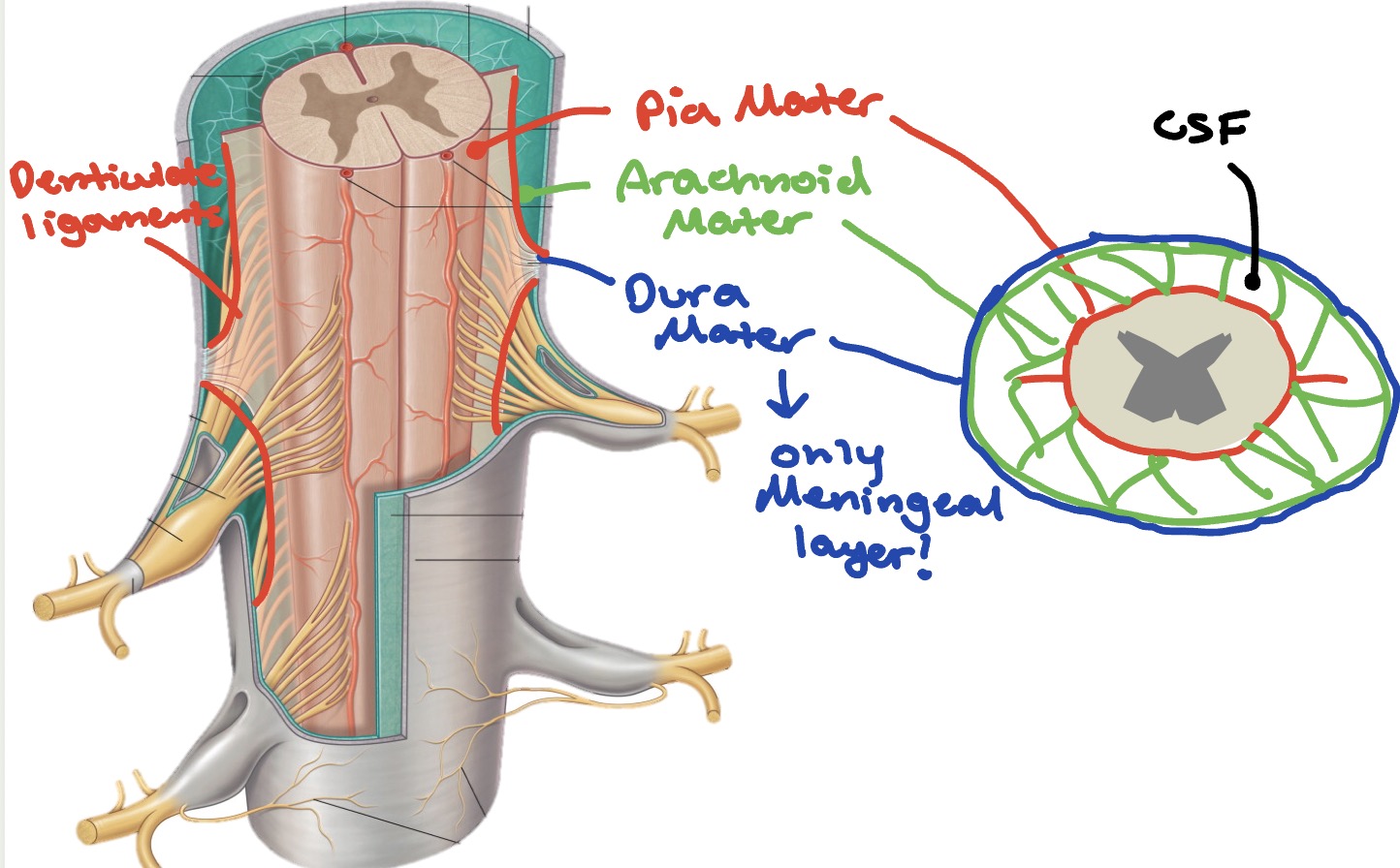
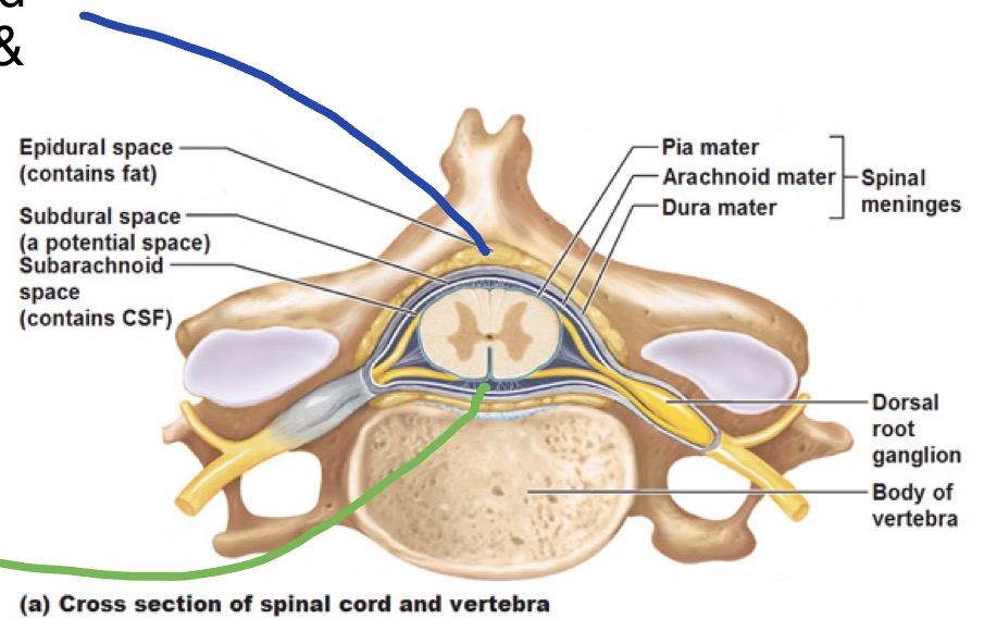
Spinal cord meninges
Pia Mater
Arachnoid Mater
Dura Mater
**Only meningeal layer b/c periosteal layer is absent in the vertebral column!
Meningeal Space
Epidural (extradural) space
Actual space; filled w/ epidural fat & venous plexus
Subdural space
Potential space
Subarachnoid space
Actual space; location of CSF
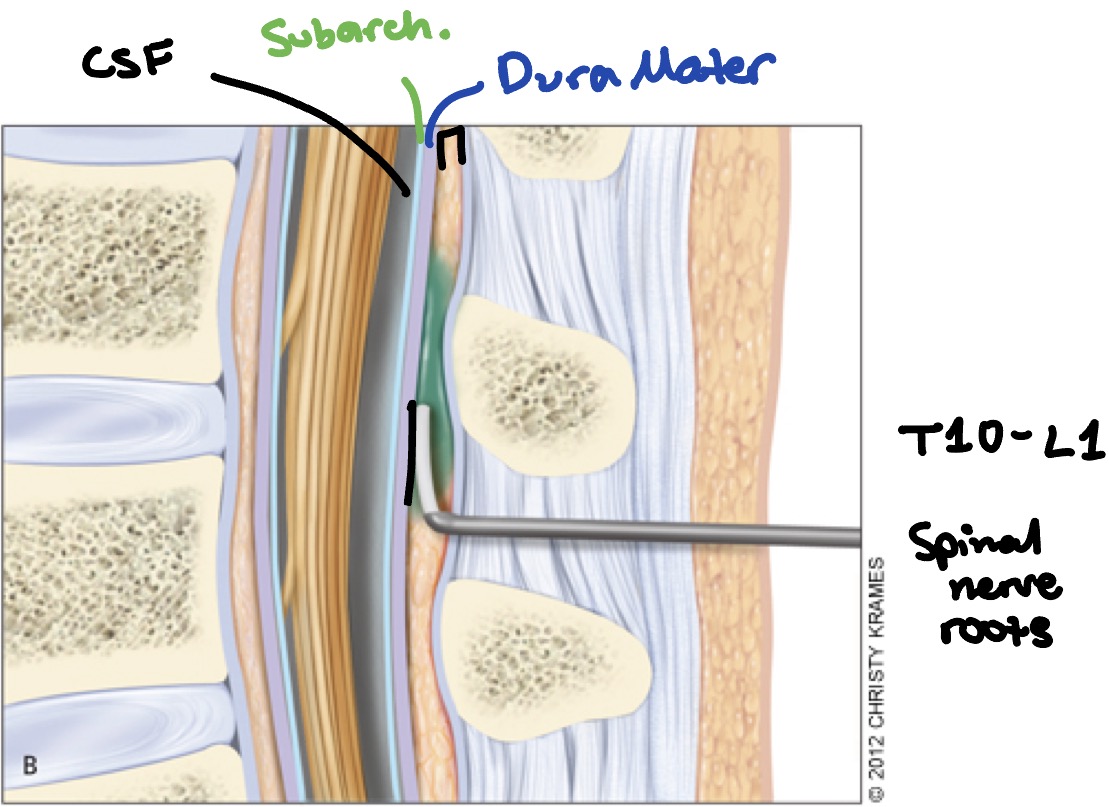
Epidural
T10-L1
Spinal nerve roots
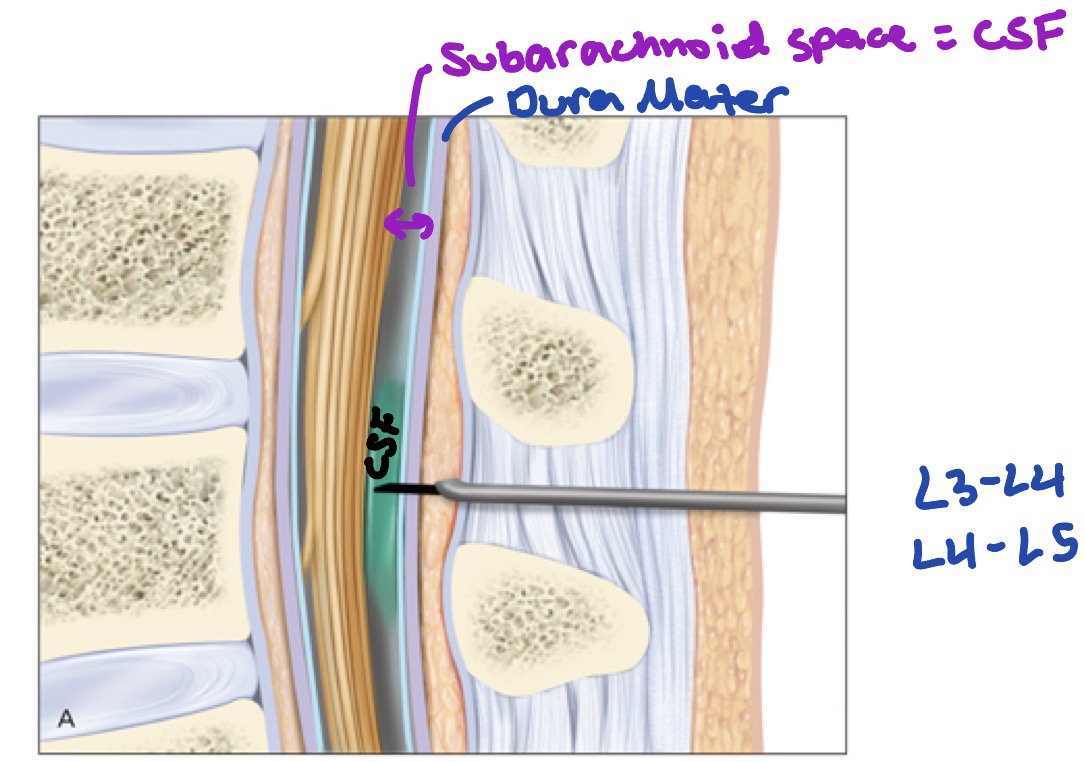
Spinal tap
L3-L4
L4-L5
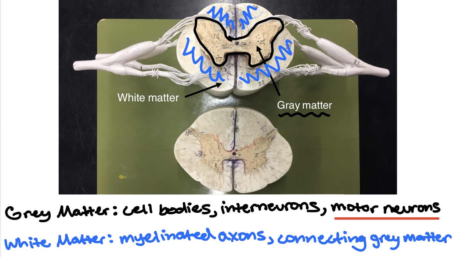
Review of White vs. Gray matter
Gray matter: cell bodies, interneurons, motor neurons
White matter: myelinated axons, connecting grey matter
Anatomy of Spinal Nerves - visual
Motor/efferent
Sensory/afferent
Interneurons
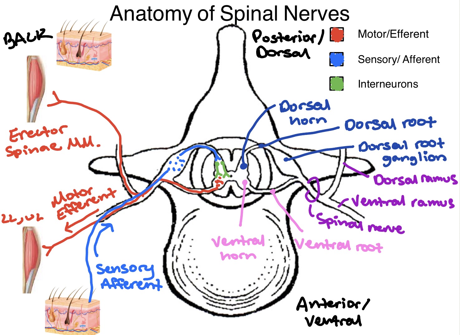
Summary: the typical spinal nerve
Motor/efferent:
Ventral horn (gray matter)
Lower motor neuron cells bodies
Ventral roots
Lower motor axons
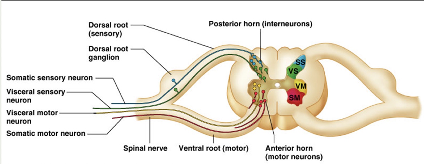
Sensory/afferent:
Dorsal root ganglion
Sensory neuron cells bodies
Dorsal roots
Sensory axons
Dorsal horn (gray matter)
Sensory axons, interneurons
Mixed:
Spinal nerve
Mixed motor & sensory axons
Ventral rami
Motor & sensory axons to muscles & skin of anterior trunk & limbs
Dorsal rami
Motor & sensory axons to muscles & skin of the back
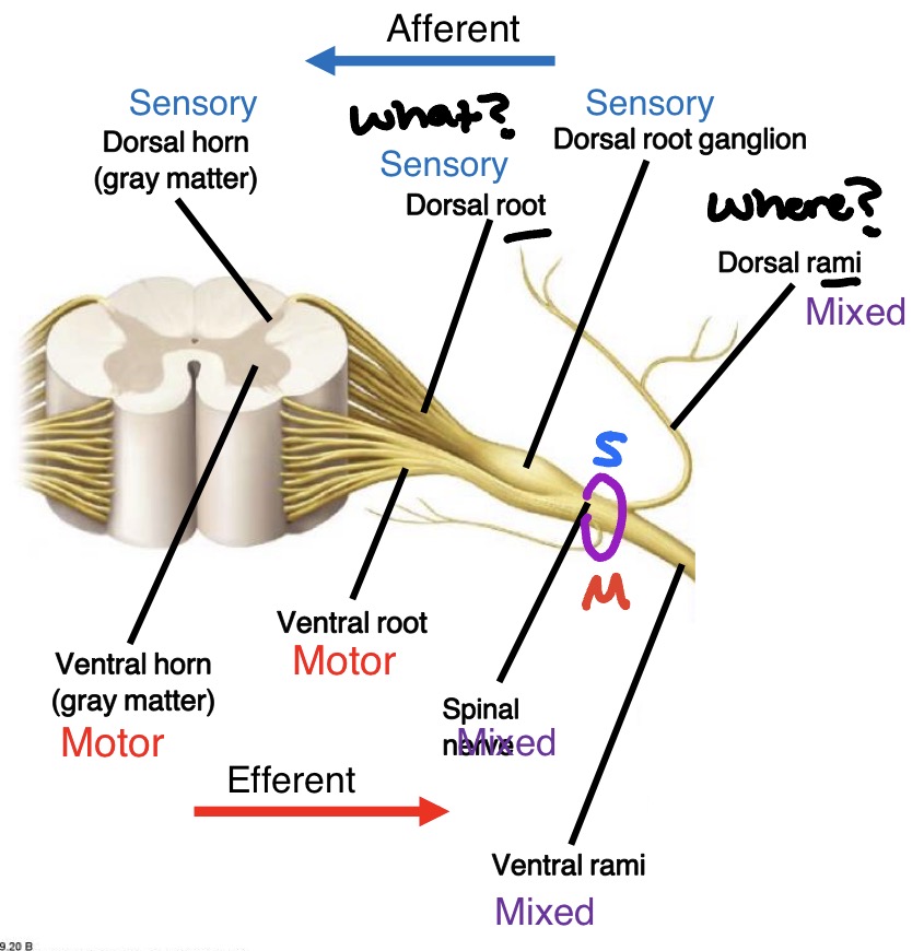
Cross-section of spinal cord
Cell bodies of motor neurons = located in ventral horn
Axons of motor neurons exit spinal cord through ventral root → spinal nerve → ventral OR dorsal ramus → through named nerves to effector
Cells bodies of sensory neurons = located in dorsal root ganglia
Axons of sensory neurons enter through dorsal OR ventral ramus → spinal nerve → dorsal root → synapse with interneuron in dorsal horn
White matter of spinal cord consists largely of organized myelinated axons organized into tracts or fasciculi
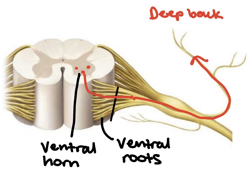
What type of signal would be interrupted here?
Motor - efferent
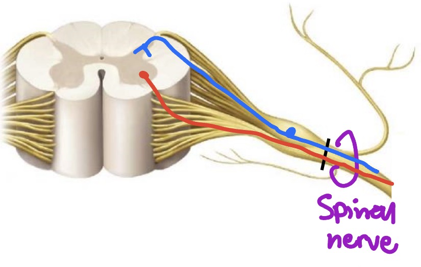
What type of signal would be interrupted here?
Mixed