Immunity 3.2.4
 Call Kai
 Learn
Learn Practice Test
 Spaced Repetition
 Match
Match Flashcards
 Knowt Play
1/20

Earn XP
Description and Tags
Name | Mastery | Learn | Test | Matching | Spaced | Call with Kai | Chat |
|---|
No analytics yet
Send a link to your students to track their progress
21 Terms
Defence mechanisms
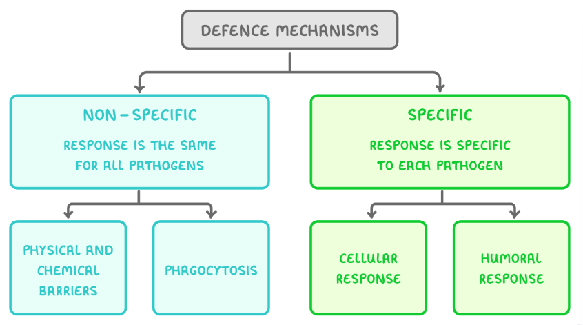
Physical and chemical barriers
Non-specific defence mechanisms that prevent entry of pathogens. Examples:
Skin is both a physical and chemical (produces sebum which lowers pH and inhibits growth of pathogens) barrier.
Mucous membranes line the ears, nose, throat and digestive tract. Mucus traps and lysozymes destroy.
Expulsive reflexes - coughing/sneezing remove pathogens from gas exchange system and vomiting/diarrhoea expel pathogens from the gut.
Blood clotting/wound repair seals possible pathogen entry points with a scab.
Inflammation causes heat which prevents pathogens from reproducing.
Antigens
Molecules found on the surface of cells to signpost them as self or non-self. Antigens allow the immune system to identify various cells including pathogens, cancerous cells and toxins.
Phagocytosis
A type of non-specific defence mechanism that uses phagocytes (a type of white blood cell) that engulf and destroy pathogens during phagocytosis.
The phagocyte is attracted to chemicals released by the pathogen and recognises the pathogen’s antigens as non-self, causing the phagocyte to bind to and engulf the pathogen into a phagosome. The phagosome fuses with a lysosome to form a phagolysosome, where the hydrolytic enzymes (lysozymes) digest and destroy the pathogen. The phagocyte presents the pathogen’s antigens on its surface, becoming an antigen-presenting cell, to activate other cells in the immune system.
The cellular response
A specific defence mechanism that uses T lymphocytes/cells (which mature in the thymus gland) to respond to antigens presented on body cells.
1. Phagocytosis → APC.
2. T helper cells with complementary receptors bind to APCs.
3. The T cell is activated to divide by mitosis to form genetically identical clones which can: Develop into memory cells, stimulate phagocytosis, stimulate division of B cells, or activate cytotoxic T cells.
Cells involved in the cellular response
T helper cells - bind to complementary antigens or APCs to form memory cells, stimulate B cells or phagocytes, and activate cytotoxic T cells.
Cytotoxic T cells - kill abnormal/foreign cells by producing a protein called perforin which makes holes in the cell-surface membrane, becoming permeable and the cell to die.
Memory T cells - provide long-term immunity.
The humoral response
A specific defence mechanism that uses B lymphocytes/cells (which mature in the bone marrow) to produce antibodies in body fluids/humours.
1. A B cell with a complementary antibody binds to the antigens on a pathogen.
2. The B cell engulfs the pathogen and becomes an APC.
3. Clonal selection - Activated T helper cells bind to the B cell, activating the B cell.
4. Clonal expansion - The activated B cell divides by mitosis to form plasma and memory cell clones.
Cells involved in the humoral response
B cells - have antibodies on their surface that bind to complementary antigens causing them to engulf and display the antigens (becoming APCs). Once activated, B cells can divide into plasma or memory cells.
Plasma cells - a type of B cell that can produce and secrete antibodies.
Memory cells - a type of B cell that provides long-term immunity by rapidly dividing into plasma cells if the body is re-infected by the same pathogen.
Helper T cells - bind to APCs to activate the division of B cells.
Primary vs secondary immune response
Primary immune response occurs when the body is exposed to a pathogen for the first time. The response is slow and the infected individual experiences symptoms of the disease.
Secondary immune response occurs when the body has been exposed to the same pathogen before. This response is much faster and stronger and pathogens are destroyed before any symptoms appear, because of memory cells. A higher concentration of antibodies is produced.
Antibody structure
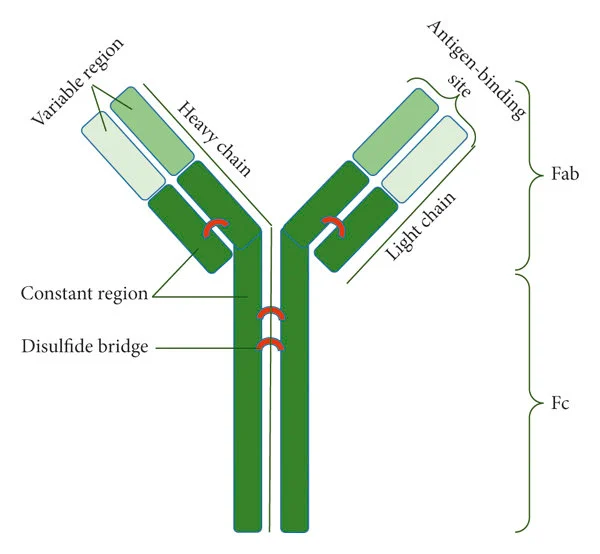
Y-shaped glycoproteins made of 4 polypeptide chains (2 heavy and 2 light), held together by disulphide bridges. The constant region is the same for all antibodies and binds to receptors on B cells. The variable region is complementary to a specific antigen.
Antibody functions
When an antibody binds to an antigen, they form an antigen-antibody complex and carry out three roles to destroy pathogens:
Agglutination of pathogens - antibodies cause pathogens to clump together, making it easier for phagocytes to locate them and also allows them to engulf multiple pathogens at once.
Neutralisation of toxins - antibodies bind to toxins produced by pathogens, neutralising/inactivating them to prevent damage to body cells.
Preventing pathogens from binding to cells - by binding to a pathogen’s antigens, antibodies block the cell-surface receptors needed to bind to host cells.
Uses of monoclonal antibodies
Diagnosis of disease
Treatment of disease
Pregnancy testing
Detecting certain cancers
ELISA test
The Enzyme-Linked ImmunoSorbant Assay test uses monoclonal antibodies to detect the presence and quantity of protein/antigen in a sample. Steps of the indirect ELISA test:
1. Add sample (containing target protein) to a well plate.
2. Add specific antibodies to target protein, these will bind.
3. Wash to remove unbound antibodies.
4. Add a second antibody that is attached to a colour-changing enzyme that will bind to the first.
5. Wash to remove unbound antibodies.
6. Add a solution containing the substrate to the enzyme causing a colour change. The intensity of the colour indicates the quantity of the protein.
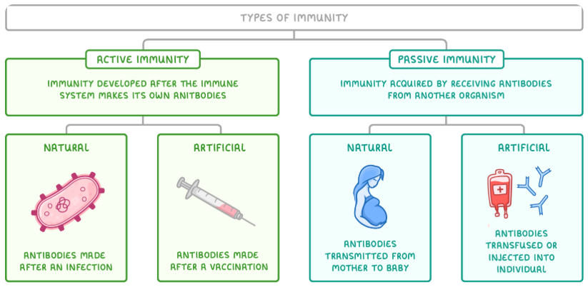
Types of immunity
Vaccination
The introduction of a (inactivated/attenuated) pathogen’s antigens into the body to stimulate the production of antibodies (artificial active immunity).
The antigens in the vaccine enter the blood stimulating the primary immune response to produce antibodies and memory cells. These memory cells remain in the body to rapidly divide into plasma cells if the pathogen is encountered in the future, destroying the pathogen before symptoms can be experienced.
Herd immunity
When a large proportion of a population is vaccinated, it provides some protection for the unvaccinated individuals because less people can spread the disease.
Antigenic variability
Some pathogens can change their antigens (in a process called antigenic variability), making it difficult to develop vaccines against the pathogen as the antigens will not be recognised. As a result, vaccines need to be changed frequently to provide protection against recent pathogenic strains.
HIV structure
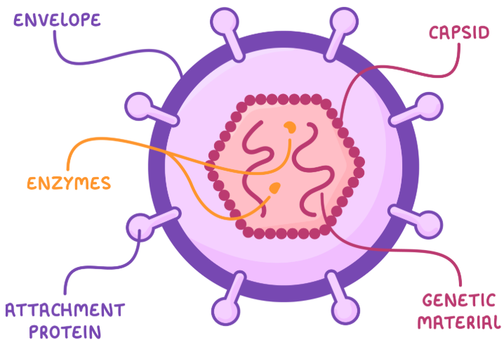
The genetic material (two single strands of RNA) and enzymes (e.g. reverse transcriptase converts RNA to DNA) are surrounded by a capsid (a layer of protein molecules). The outer layer is called the envelope (made of phospholipids) and contains glycoproteins/attachment proteins (help HIV to bind to host cells).
HIV replication
The attachment proteins on HIV attach to receptors on a helper T cell (HIV uses T cells as host cells, therefore damaging the immune system of the infected individual). HIV releases its viral RNA into the helper T cell. Reverse transcriptase converts this RNA into DNA. the viral DNA is inserted into the helper T cell’s genome so when its DNA is translated, the viral DNA is also translated to make viral proteins. These proteins assemble new HIV particles which burst out the cell (causing damage) in order to infect other cells.
Antibiotics
Drugs that kill/inhibit the growth of bacteria by:
Preventing synthesis of bacterial cell walls, disrupting protein activity in the membrane, disrupting enzyme action, preventing DNA synthesis and preventing protein synthesis.
Antibiotic resistance
Random genetic mutations occur, making some bacteria resistant to an antibiotic. When an infection is treated with antibiotics, resistant bacteria will survive and reproduce, passing on the allele for antibiotic resistance to their offspring.