Anterior Abd Wall + Inguinal Ligament + Descent of the Testes + Inguinal Ligament
1/18
Earn XP
Description and Tags
Name | Mastery | Learn | Test | Matching | Spaced |
|---|
No study sessions yet.
19 Terms
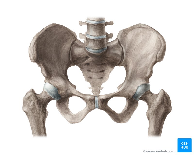
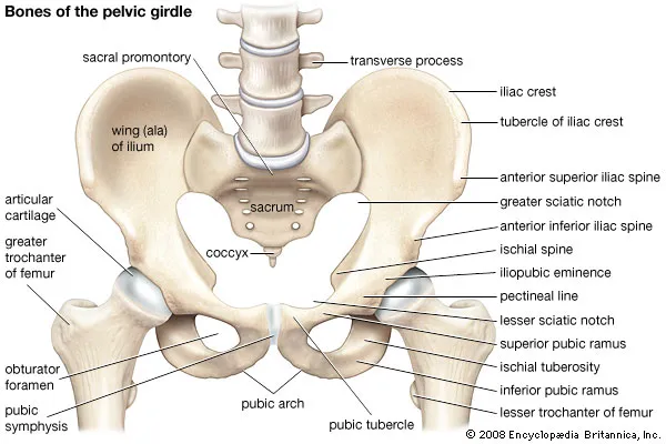
Label the pelvis
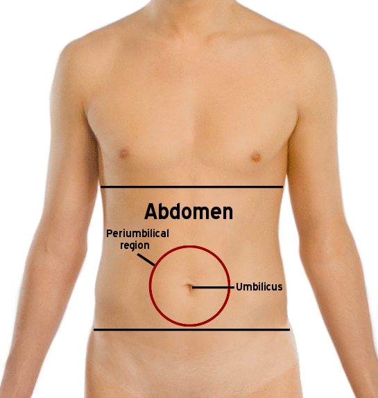
Where is the umbilicus?
Level of L3 to L4.
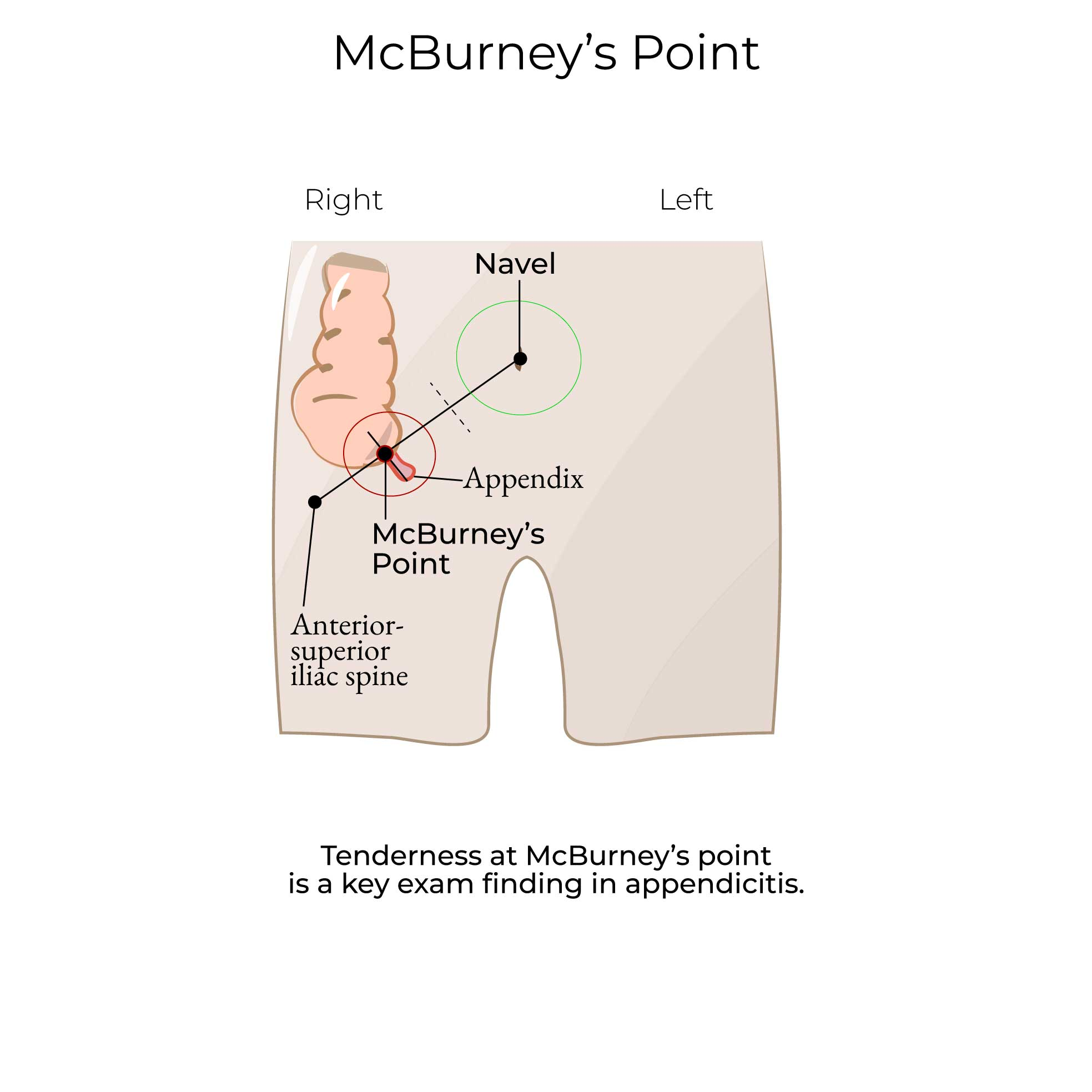
Where is McBurney’s point? What is it used for?
Location on the lower right side of the abdomen that is 1/3rd of the way between the anterior superior iliac spine and the umbilicus.
It is a key landmark in the physical examination for appendicitis, as tenderness at this point is a strong indicator of inflammation of the appendix.
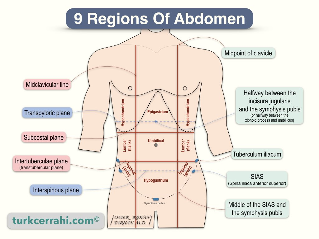
What are the 9 parts the abdomen is divided into?
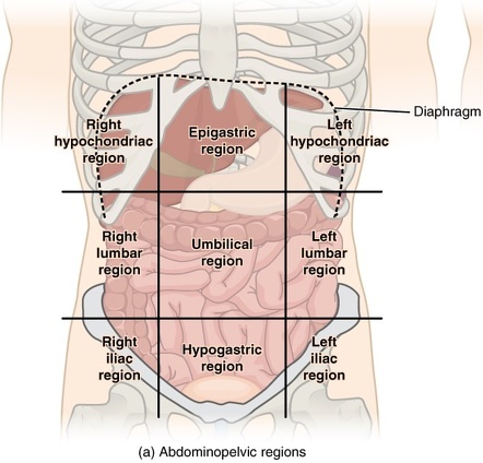
What are the planes the abdomen is divided by?
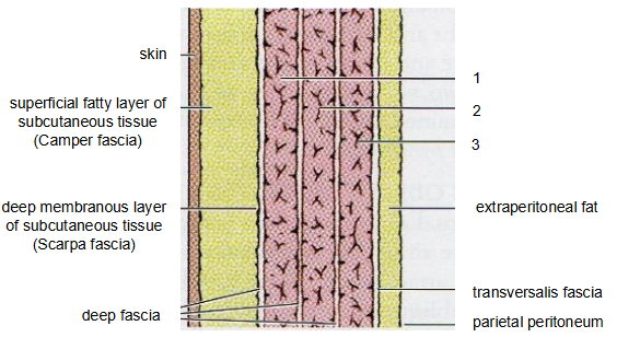
What are the layers of the abdominal wall from outwards to inwards?
Skin
Superficial fascia
Camper’s fascia - fatty layer of sub cut tissue
Scarpa’s fascia - membranous layer
Deep fascia between muscles
Muscular layer
External oblique
Internal oblique
Transversus abdominis
Transversalis fascia
Extraperitoneal fat
Parietal peritoneum
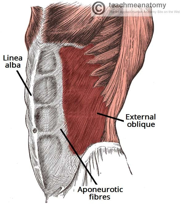
Describe the external oblique - origin, insertion, function and innervation.
Location: The largest and most superficial of the three flat abdominal muscles. Fibers run downward and medially (outwards).
Origin: Lower eight ribs. From outer surfaces of ribs 5 to 12.
Insertion: Linea alba (fibrous structure running down midline of abdomen), pubic tubercle, and iliac crest (anterior).
Function: Compresses the abdominal cavity, aids in trunk flexion, and rotates the trunk.
Innervation: Primarily innervated by T7-T12, lower thoracic nerves and subcostal nerve.
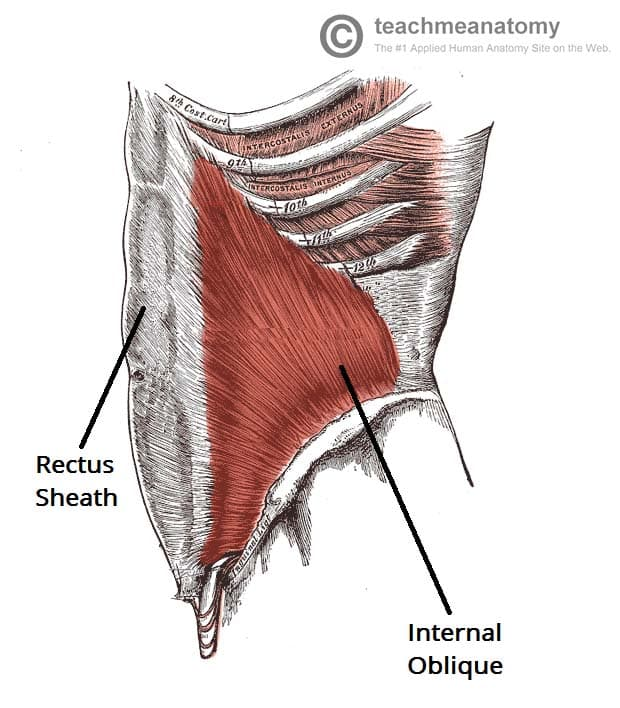
Describe the internal oblique - origin, insertion, function and innervation.
Location: Lies just beneath the external oblique. Fibers run upwards and medially – opposite to the external oblique.
- Aponeurosis – transitions to this as approaching insertion.
Origin: Thoracolumbar fascia (connective tissue which supports the lower back), iliac crest, and inguinal ligament.
Insertion: Ribs 10-12, linea alba via aponeurosis, and pubic crest.
Function: Compresses the abdominal cavity, aids in trunk flexion, and rotates the trunk (opposite side of the external oblique).
Innervation: T7-T11 thoracic nerves, T12 subcostal, L1 nerve.
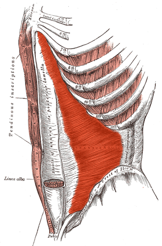
Describe the transversus abdominis - origin, insertion, function and innervation.
Location: The innermost of the three flat abdominal muscles. Underneath internal obliques. Muscle fibers lie horizontal.
Origin: Thoracolumbar fascia, iliac crest, inguinal ligament, and lower six ribs.
Insertion: Linea alba and pubic crest.
Function: Compresses the abdominal contents, providing stability to the trunk.
Innervation: T7-T11 thoracic nerves and L1 nerve.
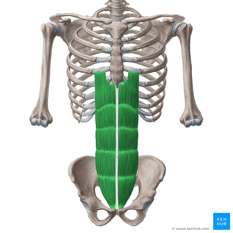
Describe the rectus abdominis - origin, insertion, function and innervation.
Location: Runs vertically on each side of the abdomen. Positioned centrally, running from the pubis to the xiphoid process, this muscle is responsible for flexing the trunk and is what we commonly refer to as the “abs.
Origin: Pubic symphysis and pubic crest.
Insertion: Xiphoid process and costal cartilages of ribs 5-7.
Function: Flexes the vertebral column, tenses the abdominal wall, and assists in forced expiration.
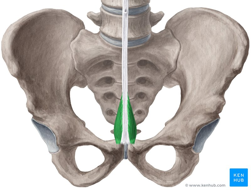
Describe the pyramidalis - origin, insertion, function and innervation.
Location: Small triangular muscle located in the lower abdomen, in front of rectus abdominus.
Vestigial muscle – meaning it has diminished in function due to evolution.
Origin: Pubic symphysis.
Insertion: Linea alba.
Function: Tenses the Linea alba.
Innervation: T12.
What is the inguinal ligament?
The inguinal ligament is a strong, fibrous band of tissue that runs from the anterior superior iliac spine (ASIS) to the pubic tubercle.
It's essentially the thickened lower edge of the aponeurosis of the external oblique muscle. The ligament forms the inferior (lower) border of the inguinal canal and is a crucial landmark in the groin area.
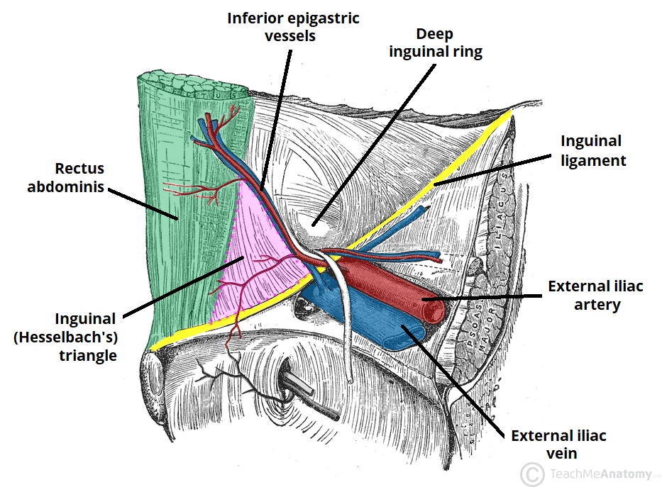
What passes through the inguinal canal?
Round ligament of the uterus - female
Spermatic cord - male
What is the rectus sheath?
The rectus sheath is a tendon sheath (aponeurosis) which encloses the rectus abdominis and pyramidalis muscles.
It is an extension of the tendons of the external abdominal oblique, internal abdominal oblique, and transversus abdominis muscles.
In addition to these muscles, the rectus sheath also contains neurovasculature of the anterior abdominal wall. Its function is to protect the contents it encloses.
Describe the rectus sheath composition
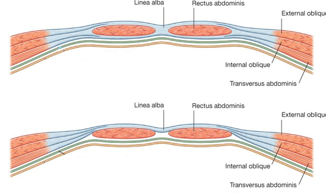
Superior to the costal margin, the anterior wall is made of aponeurosis of the external abdominal oblique muscle only, and there is no posterior wall at all.
From the costal margin to the lower quarter of the rectus abdominis muscle, both walls completely wrap the upper three-quarters of the rectus abdominis muscle. Here, the anterior wall is comprised of aponeuroses of the external abdominal oblique and internal abdominal oblique muscles, whereas the posterior wall is made of aponeuroses of the internal abdominal oblique and transversus abdominis muscles.
Inferior to the arcuate line, the lower quarter of the rectus abdominis muscle is covered by the rectus sheath on its anterior surface only, while the posterior surface is in direct contact with the transversalis fascia. The rectus sheath here is made of aponeuroses of all three above mentioned muscles – external and internal abdominal oblique muscles and the transversus abdominis muscle
Composition of the rectus sheath
Walls of upper three-quarters | Anterior wall: |
Walls of lower quarter | Anterior wall: |
What 2 arteries supply the abdomen?-
Superior and Inferior epigastric arteries
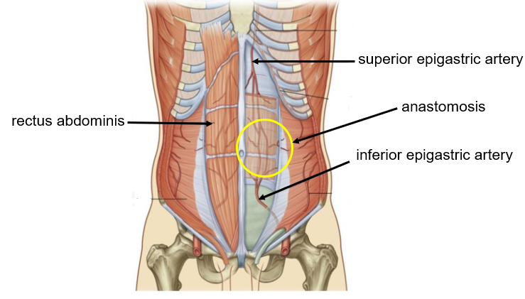
Where does the Superior Epigastric artery arise from?
Internal thoracic (mammary) artery
Where does the Inferior Epigastric artery arise from?-
External Iliac artery