11_Signal transduction
1/46
There's no tags or description
Looks like no tags are added yet.
Name | Mastery | Learn | Test | Matching | Spaced | Call with Kai |
|---|
No analytics yet
Send a link to your students to track their progress
47 Terms
signaling cells
Signaling molecules are released by
Ligand
the chemical signal molecule
binds to its specific receptor on a target cell
primary chemical messenger/molecules which comes from a distant or nearby source; it binds with the receptor causing production of additional molecules within the cell that received the signal.
conformational or shape-change in the receptor
Ligand binds to its specific receptor on a target cell. This ligand-receptor interaction induces a ___
Extracellular Signaling
Produces a specific response - The cellular response can include a vast array of compounds
Cells interact with their environment by interpreting extracellular signals via proteins that span their plasma membrane called receptors
Extracellular domain
relays information about the outside world to the intracellular domain
Intracellular domain
then interacts with other intracellular signaling proteins
further relay the message to one or more effector proteins
Effector proteins
mediate the appropriate response
Cellular communication
is vital in living systems and important to produce response from a certain stimulus coming from the environment
Use of chemical transmitters (neurons)
Regulatory chemical messengers to local receptors on cellular surface and distant from secreting cells
Signal can be transferred from one cell to another by:
The Nervous System
It collects information from the environment.
It processes that information.
It elicits responses to that information by triggering specific effectors.
Membrane potential
is a fundamental property of the cells
Resting membrane potential (observed in cells at rest)
Negative resting membrane potential (intracellularly)
Electrical excitability
A property of specialized cells triggered by certain types of stimuli that allow rapid changes in membrane potential called action potential
action potential
A property of specialized cells triggered by certain types of stimuli that allow rapid changes in membrane potential called ____
Differences in ion concentration in cytosol (ICF) and ECF.
Anions & smaller cations - usually are concentrated in ICF
Cations mostly – concentrated in ECF
How is Membrane potential achieved?
Electroneutrality
K+ counterion for trapped cytosolic anions; Cl- counterion for Na+ in ECF
Potassium ions (K+)
are more concentrated in the cytosol and have a tendency to move out of the cell, leaving behind trapped anions. Membrane potential becomes more negative.
Sodium ions (Na+)
are much more concentrated outside the cell than inside and tend to enter the cell. As Na+ ions enter, they neutralize some excess negative charge in the cytosol, and membrane potential becomes more positive.
Chloride ions (CI)
usually crosses the membrane together with a permeable cation (normally K+). As CI enters a cell, it tends to make the membrane potential more negative.
voltage sensor and part of the gating mechanism
The fourth transmembrane helix, S4, is a ____
Channel gating
The channel is regulated by a gate, which can open or close, depending on the state of voltage sensor domains in the channel. The channel gate opens and closes depending on the conformational state of channel subunits.
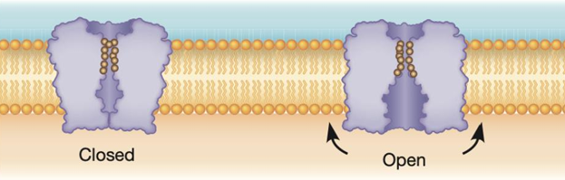
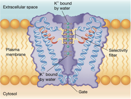
Pore structure
The transmembrane region of a potassium channel in the closed position. This diagram is based on the bacterial KcsA channel, but vertebrate potassium channels are similar.
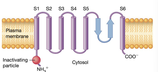
Voltage-gated channels
Domain structure. ___ for sodium, potassium, and calcium ions all share the same basic structural themes.
The channel is essentially a rectangular tube whose four walls are formed from either four subunits (for example, potassium channels) or four domains of a single polypeptide (for example, sodium channels).
Each subunit or domain contains six transmembrane helices, labeled S1–S6. The fourth transmembrane helix, S4, is a voltage sensor and part of the gating mechanism. For voltage-gated sodium channels
And some types of potassium channels, a region near the N-terminus protrudes into the cytosol and forms an inactivating particle.
Transmission of an Action Potential Along a Nonmyelinated Axon
A nonmyelinated axon can be viewed as a string of points, each capable of undergoing an action potential. Notice that no backward propagation occurs near sites where action potentials form because sodium channels are in an inactivated state and the membrane is hyperpolarized.
nonmyelinated axon
can be viewed as a string of points, each capable of undergoing an action potential.
Prokaryotes
have membrane-bound receptor molecules on the cell surface to respond to substances in their environment
Eukaryotic cells
produce signals may be thru display of molecules on their surfaces that are recognized by receptors on the surfaces of other cells
Multicellular organisms
often control the activities of specialized cells through the release of chemical messengers
Hormones
endocrine signals
Local mediators
growth factors diffuse to nearby tissues (paracrine), or require physical contact between nearby source & target cells (juxtacrine), and, act on the same cell that produces them (autocrine)
Chemical Signal Transduction
Cellular ability to translate a receptor-ligand interaction to changes in its behavior or gene expression
Effector tissues/cells
– target structures which contain the surface receptor initiating the signaling process
Second messenger
messenger molecules produced within the effector/target tissue by the effect of the primary messenger/ligand
Hydrophilic form second messenger
more common type; it has the ability to bind with one or more specific receptors on target cell.
Hydrophobic form second messenger
acts on receptors in the nucleus or cytosol that regulate transcription of particular genes (i.e. steroid hormones, retinoids cholesterol derivatives)
RECEPTOR AFFINITY and RECEPTOR DOWN-REGULATION
Ligand-Receptor Interactions
RECEPTOR AFFINITY
Ligand-Receptor Interactions:
at low concentration of ligand and most of receptors are already occupied = High receptor affinity
is described by Kd – it tells us the concentration of ligand is enough to produce a response
RECEPTOR DOWN-REGULATION
Ligand-Receptor Interactions:
Cells can sense changes in ligand concentrations. Cells adapt to these changes and hence called down-regulation.
Cellular change in the receptor density on its surface – Receptor-mediated endocytosis
Desensitization – alter receptor affinity by lowering its affinity for ligand & render inability to initiate changes in cellular function
down-regulation
Cells adapt to these changes and hence called ___
Receptor-mediated endocytosis
Cellular change in the receptor density on its surface – ___
Desensitization
alter receptor affinity by lowering its affinity for ligand & render inability to initiate changes in cellular function
G-protein linked and Protein kinase linked
TWO FAMILIES OF RECEPTORS:
The G Protein-Linked Receptors
Family of receptor:
A ligand binds to the extracellular portion of the receptor, causing an intracellular portion of the receptor to bind and activate a G protein.
Specific amino acids in the cytosolic region are also targets for phosphorylation by G protein linked receptor kinases (GRKs) and protein kinase A.
C-GMP
Nitric oxide couples G-protein linked receptor stimulation in endothelial cells to vasodilation by relaxation of tunica media in blood vessels
Protein kinases
are enzymes that add a phosphate group from ATP onto a substrate protein; this reaction is called phosphorylation
kinases
Activated receptors frequently transmit signals through through intracellular signaling proteins called _____
are often themselves activated by other kinases via phosphorylation and can organize into phosphorylation cascades
phosphorylation
Protein kinases are enzymes that add a phosphate group from ATP onto a substrate protein; this reaction is called ___
frequently serves to activate the substrate of the kinase, but can also target the substrate for degradation
NF-κβ (nuclear factor kappa-light-chain enhancer of activated B cells)
a key regulator of inflammatory reaction, is sequestered in the cytoplasm (in a transcriptionally inactive form) by members of the IκB (inhibitor of NF-κβ) family of proteins.