PSY10060-Brain and Behavior: Lecture 2: Human Nervous System
1/56
There's no tags or description
Looks like no tags are added yet.
Name | Mastery | Learn | Test | Matching | Spaced | Call with Kai |
|---|
No analytics yet
Send a link to your students to track their progress
57 Terms
Central nervous system (CNS) content
Brain and spinal cord
Peripheral nervous system (PNS) content
Sensory and motor connections
What are the major structures of the brain?
Cerebrum (forebrain)
-Hemispheres
Brainstem
Cerebellum
What is the cerebrum?
Forebrain, two hemispheres
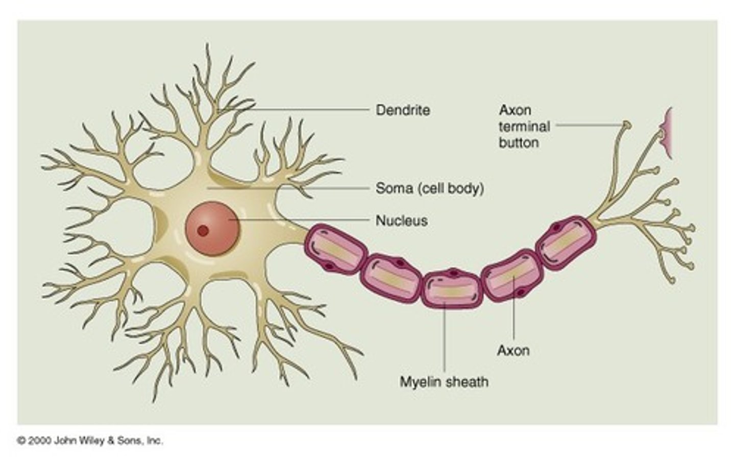
What are the different parts of the neuron and where are they located?
Dendrites
Cell body
Nucleus
Axon
Myelin sheath
Axon terminals
What does the role of a neuron depend on?
The inputs and outputs of the neuron
What are the two manners in which a wide variety of methods used to trace the connections of nuerons?
1. To and from a neuron
2. To and from a region of the brain
What do we need to know in order for methods of connectional measurement to work?
The approx functions (what the regions do) of the input and output regions
What is a DTI? What does it do?
Diffusion Tensor Imaging (DTI)
-Uses MRI scanner
-Non-invasive
-Produces detailed maps of the diffusion of water within neural tissue
What do correlational methods involve?
Making observations of brain activity WHILE an individual is performing some type of behavior
What is an invasive way to study correlational methods?
Record electrical activity of neurons via microelectrodes implanted in the brain
What are 4 less-invasive correlational methods?
1. Electroencephalography (EEG)
2. Magnetoencephalography (MEG)
3. Positron emission tomography (PET)
4. Functional magnetic resonance imaging (fMRI)
What an MRI?
Magnetic resonance imaging
Why does an MRI work?
-The differences in the properties of tissue create major differences in how protons in the tissue behave when place in a strong magnetic field
What does every MRI have? Why is this essential?
-Every MRI contains a superconducting magnet
-In order to collect an image the MRI must partially magnetize the body
Structural Analysis: How does an MRI work? What are characteristics of the images?
-Strong magnetic field passed through the brain, followed by a radio wave
-Very clear 3D images of various types of tissues within the body
-Images have excellent spatial resolution (less than a mm)
What is an fMRI?
Functional magnetic resonance imaging
Why does an fMRI work?
-When neurons become more active they need oxygen
-fMRI can detect these changes in the magnetic properties of blood because the magnetic properties of oxygen rich blood differ from oxygen poor blood
What does an fMRI measure? What is it dependent on?
Measures the ratio of oxygenated to deoxygenated blood
-Blood oxygen level-dependent (BOLD) response
What is different between what an MRI studies and an fMRI studies?
MRI studies the brain structure, while an fMRI studies brain function
How do we study the damage to the brain? What does it study?
Neuropsychological testing
-The effects of brain damage on specific cognitive functions
What would neurological testing on a temporal lobe lesion likely discover?
Temporal lobe lesion is associated with memory disturbance as it plays a large role in memory
Is it likely that a lesion to the temporal lobe would affect all forms of memory? Can we test all at once?
No, memory is not a single function (semantic, procedural, episodic...) thus its rare to be impaired in all forms of memory
-Each must be tested separately
Can we use correlational studies alone to prove theories? What is an example of something supportive?
No, we need to back up our findings from correlational studies with causation studies such as lesion studies
What are lesion studies?
Studies the effects of brain damage (lesions)
What wide variety of events can cause lesions (5)?
-TBI -traumatic brain injury
-Stroke -bleeding or blockage of blood supply into brain region
-Tumors
-Degenerative diseases
-Infections
What role do single and double dissociations play in studying the damaged brain?
-A single dissociation is able to compare a lesion in one region a brain to a control, while double dissociation is able to compare a patient group with a lesion in for example the temporal lobe to another patient group with a lesion in the frontal lobe, and then both of those to a control.
What is a single dissociation in lesion studies?
Patient vs control
-Patient group will likely only show impairment on one task and not the other
What is double dissociation in lesion studies?
Patient X vs Patient Y vs. Control
-Patient groups will show different levels of impairment on different tasks
What are two methods of functional & structional brain stimulus
1. Deep brain stimulus (DBS)
2. Transcranial magnetic stimulation (TMS
What is DBS?
Deep-brain stimulation (DBS)
How does DBS work?
Implant electrodes in the brain to stimulate a targeted area with a low voltage electrical current to facilitate behavior
-Pacemaker for the brain, provides regular set of electrical impulses that drowns at noise and let natural signals go through
-Often done awake
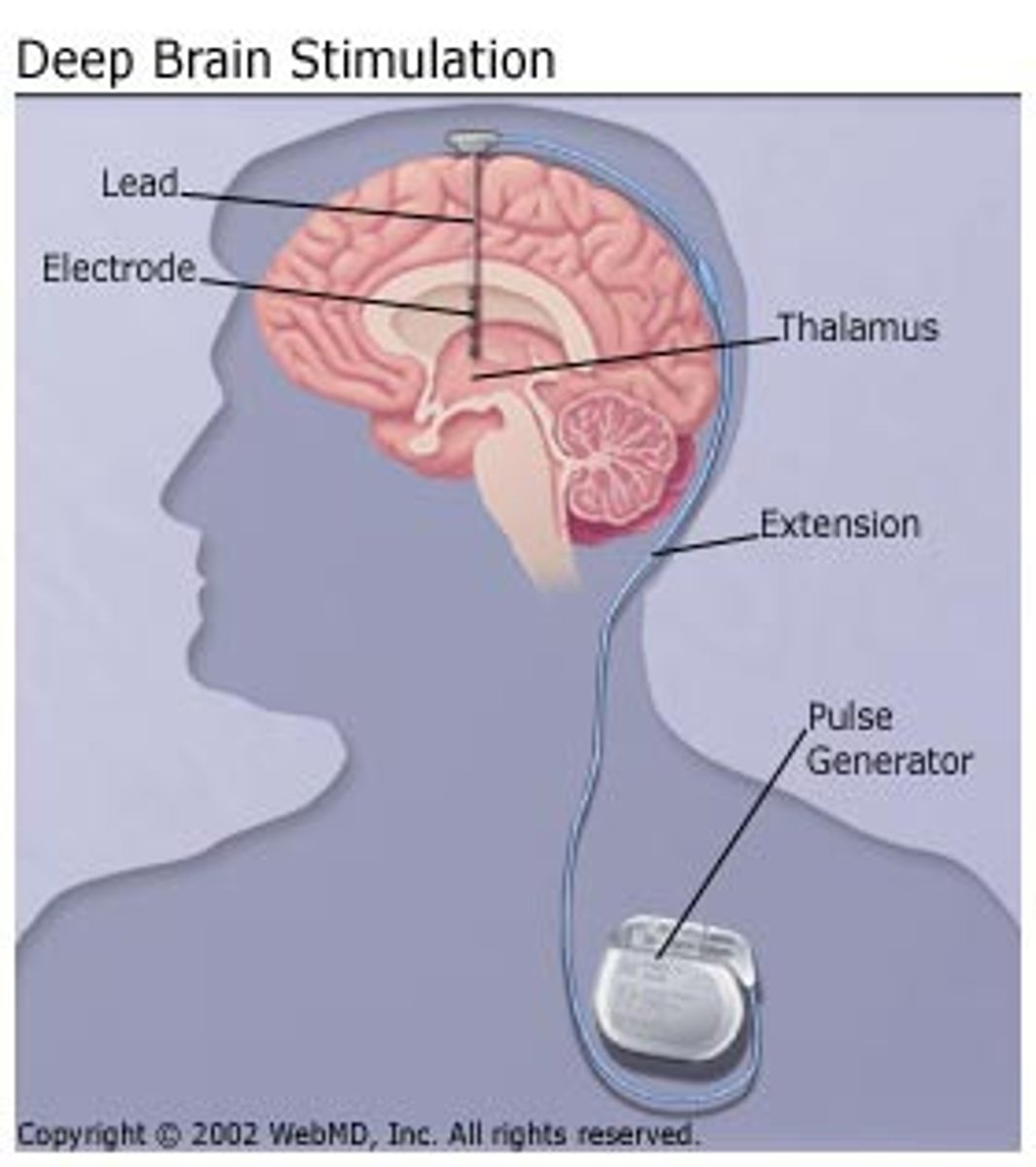
What are therapeutic applications of DBS?
-Parkinsons disease
-Depression
-OCD
What is TMS?
Transcranial magnetic stimulation
How does TMS work?
-Magnetic coil is placed over the skull stimulating the underlying brain area
-Wire in coil carries an electric current, rapid change in current creates a magnetic field
-This induces a current in the nearby neurons, causing them to fire (generate action potentials)
-Can be used to disrupt ongoing cognitive or motor function (creating a virtual lesion)
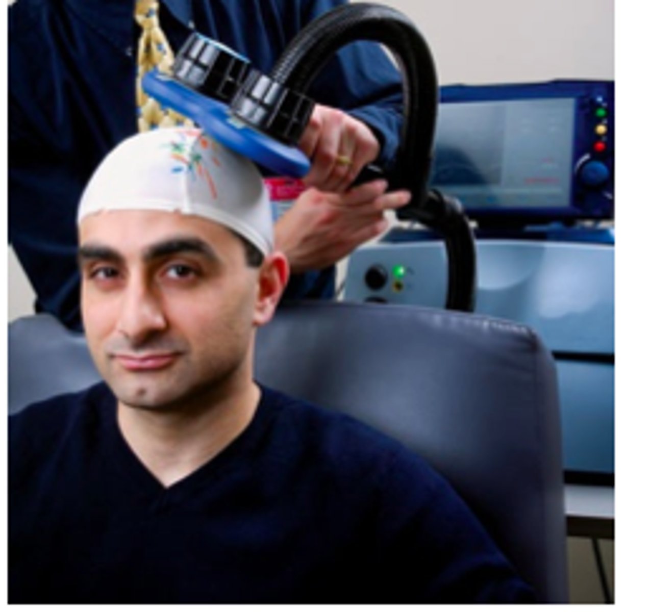
How strong is the electrical current in TMS?
-TMS is relatively mild, many natural situations stimulate the brain
-However not used on people with epilepsy
-Number/rate of pulses regulated by ethical guidelines
Potential therapeutic applications of TMS?
-Depression
What are the 4 major techniques that use electrical activity to study brain function?
1. Single-cell recording
2. Electroencephalography (EEG)
3. Event related potentials (ERP)
4. Magnetoencephalography (MEG)
What do single cell recordings do?
-Measure single-neuron action potentials with fine electrodes
In single cell recordings what is the difference between extracellular electrodes and intracellular electrodes?
-Electrodes placed next to cells (extracellular) or inside (intracellular)
-Extracellular recording -distinguish activity of up to 40 neurons
-Intracellular recording -study of a single neuron's electrical activity
Can single cell recordings be used on humans?
Yes, when electrodes are implanted for clinical reasons (such as DBS surgery)
What is an EEG?
Electroencephalography (EEG)
What do EEGs do?
-Record thousands of cells as opposed to single neurons
-Reveals features of the brains electrical acitvity
-Is able to signal changes in behavior
-Recordings from the cortex show an array of patterns, some rythmical
-Electrical activity continues during sleep or coma
What can an EEG be used to measure?
Ongoing brain activity or changes in response to a particular event/stimulus
What are clinical applications of EEGs?
-Can detect abnormalities in the electrical activity of your brain
-can diagnose epilepsy (seizures: rapid spiking waves)
-sleeping disorders
What are alpha waves and beta waves in EEG recordings?
Alpha-occur only in parietal and occipital lobes
Beta-frontal and parietal lobes
What is characteristic of an EEG recording of someone who is awake and excited?
High frequency low amplitude waves, as if someone is super focused
What is characteristic of an EEG recording of someone who is relaxed or have their eyes closed? What type of waves?
-Alpha rythmas generated
-Pretty high frequency, pretty high amplitude
What is characteristic of an EEG recording of someone who is drowsy?
-Slowed frequency, higher amplitude than awake/excited but lower amplitude than relaxed eyes closed
What is characteristic of an EEG recording of someone who is asleep?
-frequecy is slower, high amplitude waves
What is characteristic of an EEG recording of someone who is in deep sleep?
Frequency is even slower and amplitude is even higher, in comparison to asleep
What is characteristic of an EEG recording of someone who is in coma?
Even further slowing from deep sleep
What is an ERP?
Event-related potential
-activity that occurs in association with an event
What is the purpose of an ERP?
To see the effect of a certain stimulus on brain waves in the EEG
-Complex EEGs waveforms are related in time to a specific sensory event
-This is used to counter noise effects, the stimulus is presented repeatedly & recorded responses are averaged
How do we detect ERPs?
-Playing for example a tone or auditory stimulus at a specific time (for example time=0 ms), EEG response is recorded, and then after many successive presentations the EEG wave sequence develops distinctive shape
Why are ERPS useful?
-ERPs provide a precise measure of when brain activity occurs in response to a specific event or stimulus. -By averaging multiple EEG recordings time-locked to an event, the background "noise" of unrelated brain activity is reduced, revealing the specific brain responses linked to cognitive or sensory processes.
Does ERP stimulus have to be auditory?
No, ERPs can be visual (VEP), auditory (AEP), motor (MEP), or somatosensory (SEP)